Anesthesia | Eklavya Medical
vaccines
medical_services Main Specialty Domain lock Subscription Required

Anesthesia

Clinical anesthesiology, local and general anesthesia delivery systems, monitoring, and perioperative care.

verified Verified Medical Faculty menu_book 337 Q&A Modules
lock Protected Clinical Document • Pulse App • Screenshots Strictly Prohibited
QUESTION 331
bookmark_add

MD DNB Questions 1 Describe the cell based model of coagulation and how it differs from classical cascade.Explain the principals of TEG and ROTEM and their clinical parameters.Outline a goal directed massive transfusion protocol using viscoelastic testing. 2 Revised Cardiac Risk index 3 CPET principals and parameters for surgical risk stratification 4 Perioperative Beta blockade latest recommendations 5 Physiological effects of CO2 pneumoperitoneum on CVS and RS system 6 Epidural haematoma incidence risk factors and time critical management

collections Question Diagrams & Reference Images (7)
Question Reference Diagram
zoom_in View Image
Question Reference Diagram
zoom_in View Image
Question Reference Diagram
zoom_in View Image
Question Reference Diagram
zoom_in View Image
Question Reference Diagram
zoom_in View Image
Question Reference Diagram
zoom_in View Image
Question Reference Diagram
zoom_in View Image
picture_as_pdf Clinical Reference Documents & PDFs (1)
Protected Inline Viewer
description X7Xlpsr85Ds8tisoI9lfVuSzy7A2GUtW7SCwmH5K.pdf
lock Protected Clinical Document • Pulse App • Screenshots Strictly Prohibited
QUESTION 332
bookmark_add

Aspiration pneumonia in Anesthesia and critical .

collections Question Diagrams & Reference Images (1)
Question Reference Diagram
zoom_in View Image
picture_as_pdf Clinical Reference Documents & PDFs (1)
Protected Inline Viewer
description z5OKWPkEpQ24C4oeWuj2SdDuGJzmJatgKCBh5AN7.pdf
lock Protected Clinical Document • Pulse App • Screenshots Strictly Prohibited
QUESTION 333
bookmark_add

CHRONIC PAIN 1Classify opioids according to their action on opioid receptors. Compare and contrast fentanyl and remifentanyl. 2What is eutectic mixture? Describe the principle of transdermal drug delivery. Describe the advantages and disadvantages of transdermal fentanyl patch. 3 CRPS 4 Definition and classification all details of Osteoarthritis knee.Give all treatment and modalities of non surgical treatment of OA knee as a pain physician latest guidelines and modalities 5 Radio frequency Ablation in Chronic Pain Management

collections Question Diagrams & Reference Images (7)
Question Reference Diagram
zoom_in View Image
Question Reference Diagram
zoom_in View Image
Question Reference Diagram
zoom_in View Image
Question Reference Diagram
zoom_in View Image
Question Reference Diagram
zoom_in View Image
Question Reference Diagram
zoom_in View Image
Question Reference Diagram
zoom_in View Image
picture_as_pdf Clinical Reference Documents & PDFs (1)
Protected Inline Viewer
description jV4Xfu0Nq8A3dXjgDoSSEBiBJZApPYeKhd3FmBKo.pdf
lock Protected Clinical Document • Pulse App • Screenshots Strictly Prohibited
QUESTION 334
bookmark_add

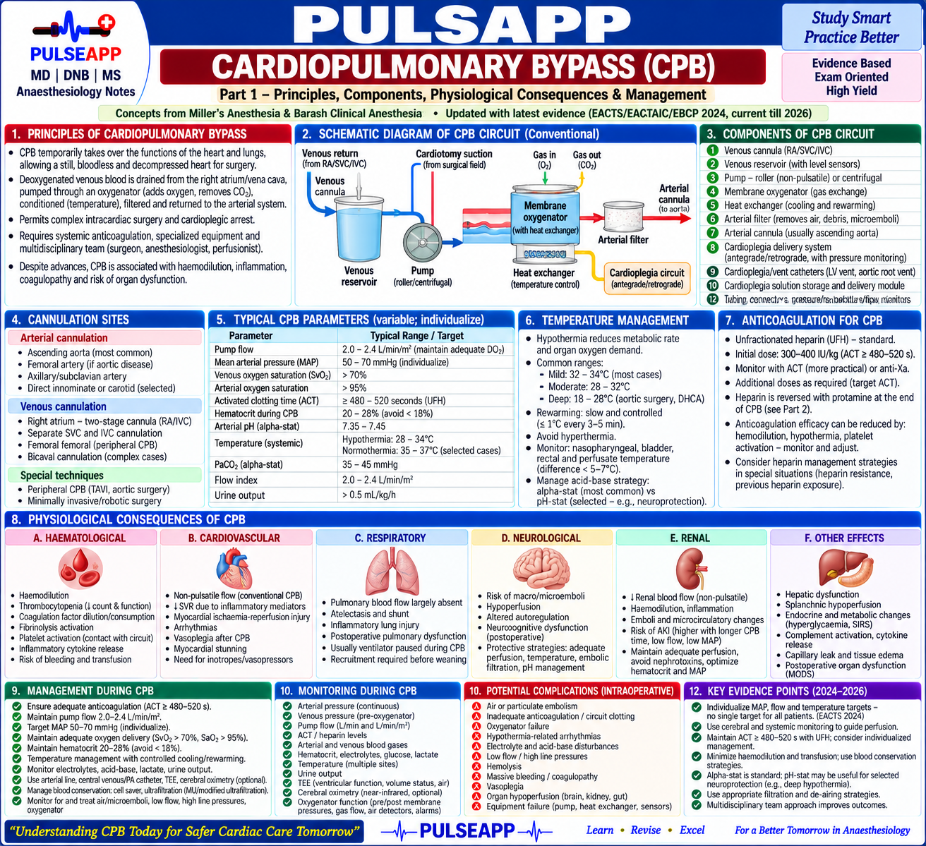
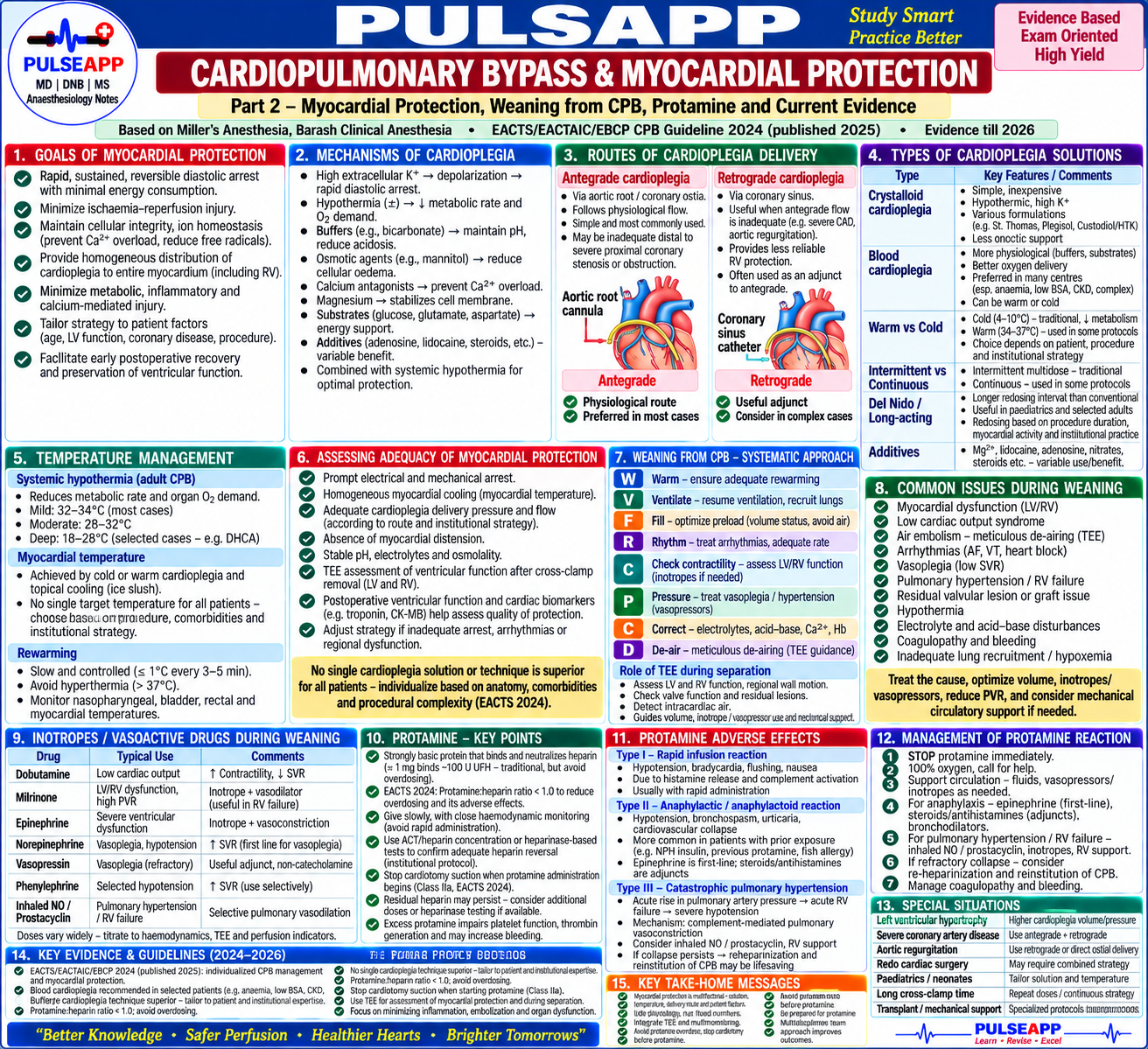
Cardiopulmonary Bypass and Myocardial Protection Principles of CPB — Components, Physiological Consequences & Management, Myocardial Protection Strategies Including Cardioplegia, Haemodynamic Management of Protamine Administration & Its Adverse Effects ✦ A Note on the Most Current Evidence

collections Question Diagrams & Reference Images (2)
Question Reference Diagram
zoom_in View Image
Question Reference Diagram
zoom_in View Image
picture_as_pdf Clinical Reference Documents & PDFs (1)
Protected Inline Viewer
description PpLha3J0OhQl0OhDogETcxo7WukCrH1JVCFyuacA.pdf
lock Protected Clinical Document • Pulse App • Screenshots Strictly Prohibited
QUESTION 335 person Asked by Dr. Ananya S.
bookmark_add

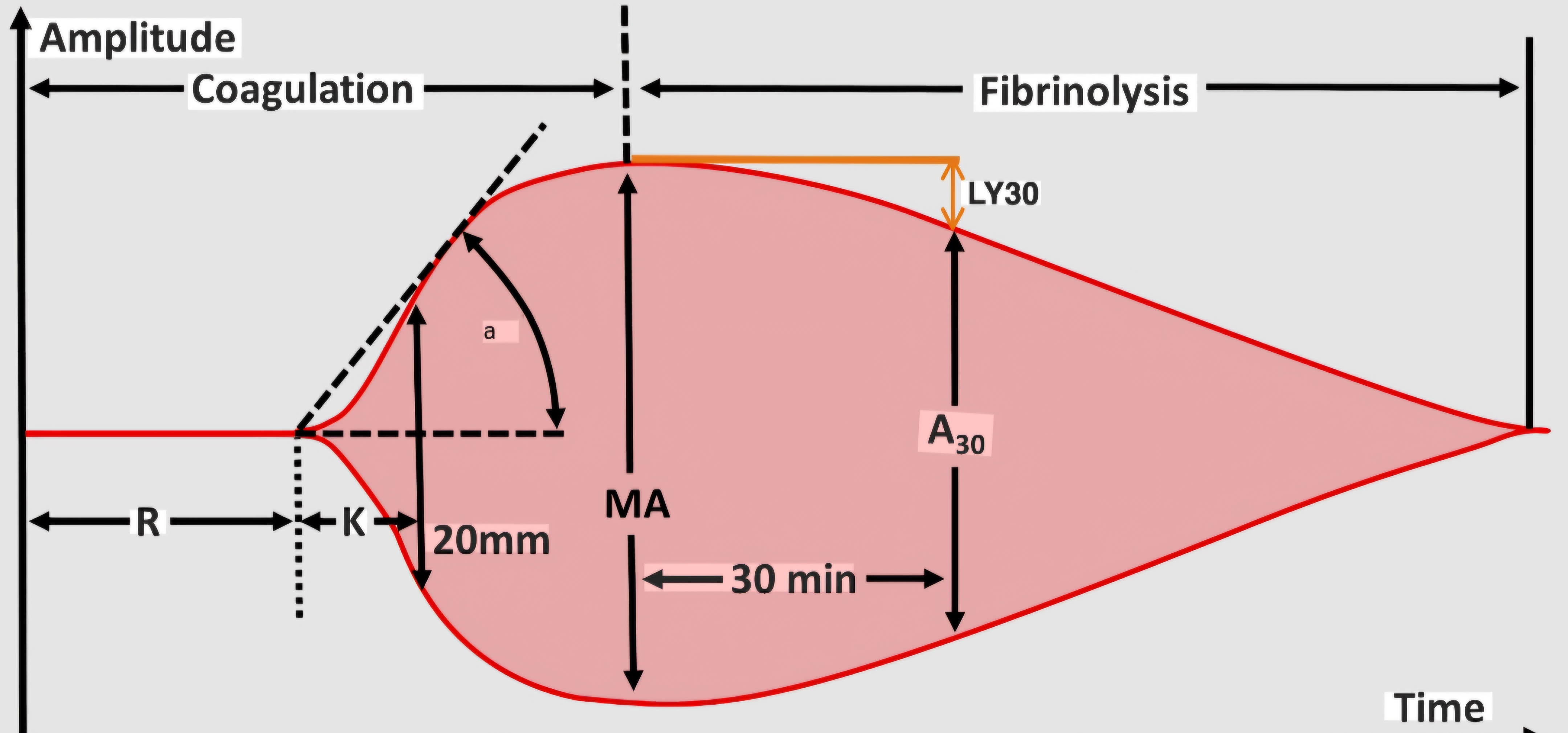
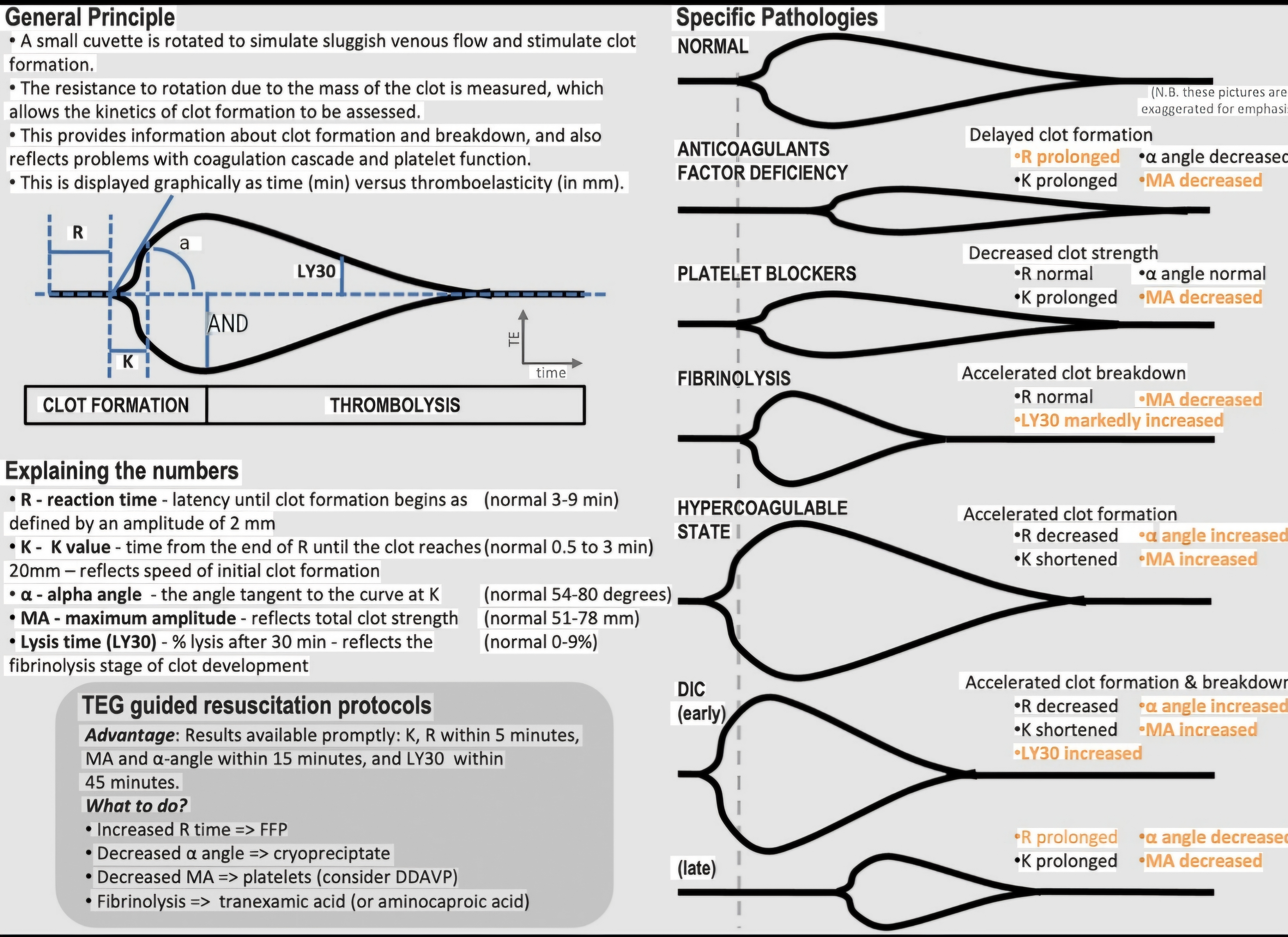
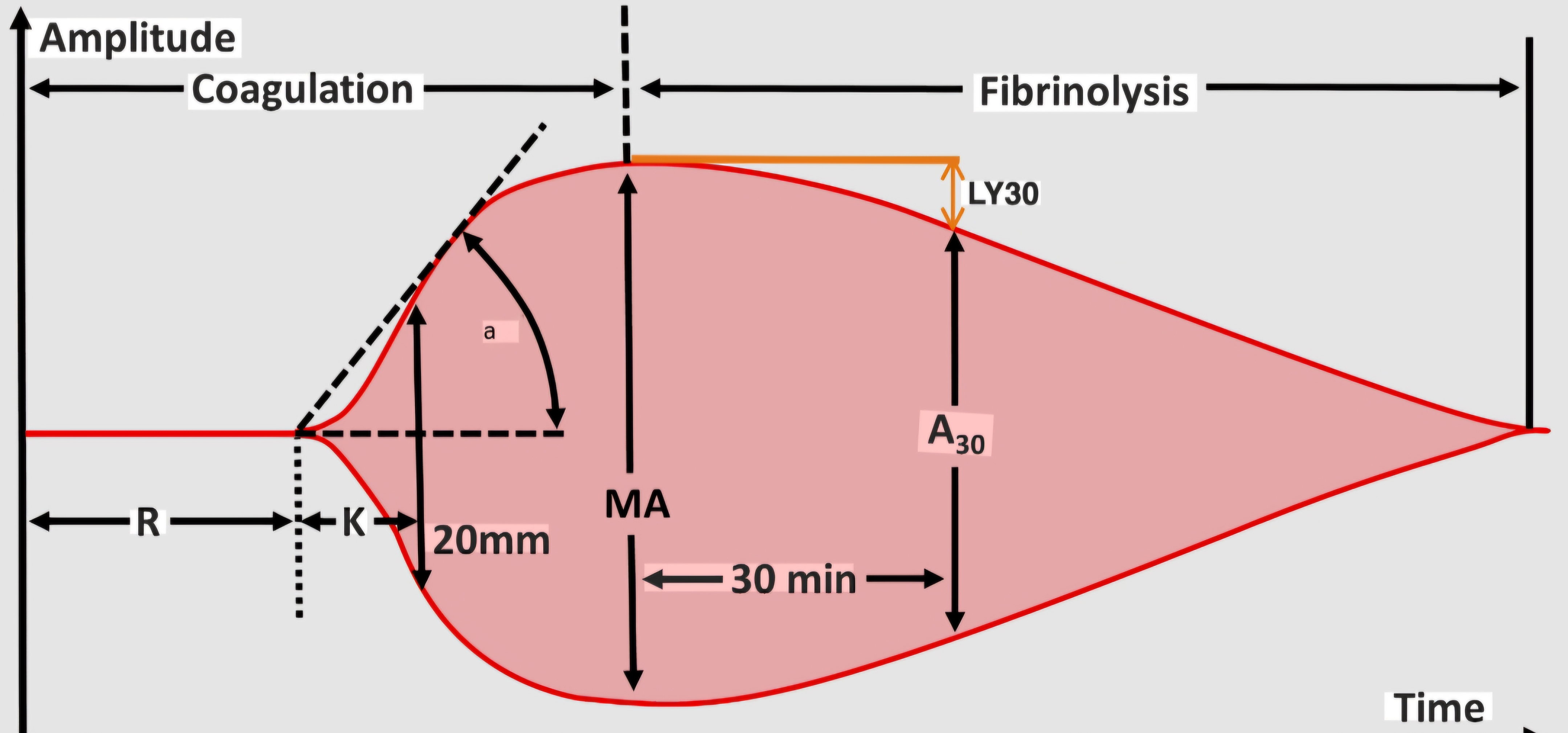
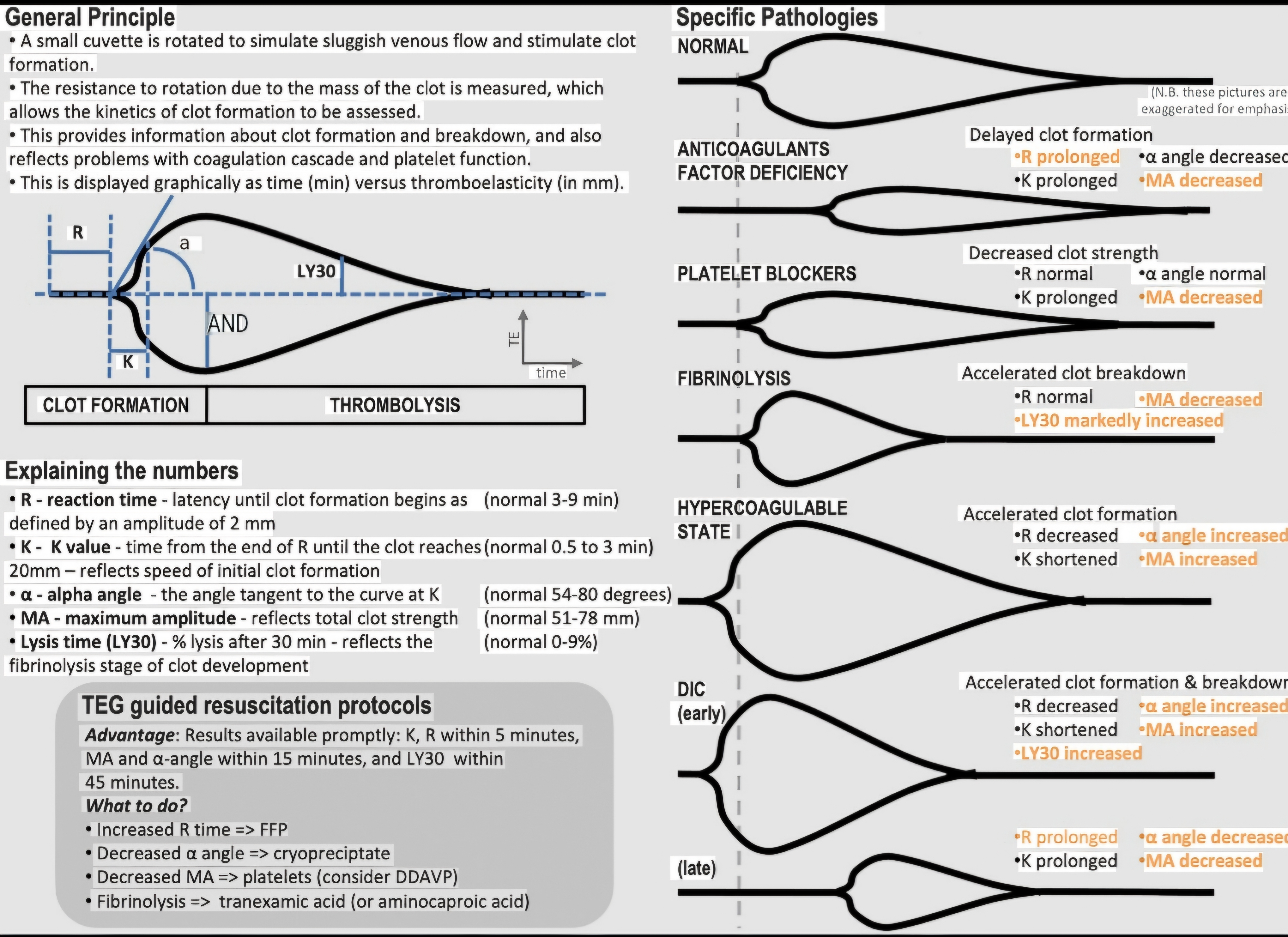
Describe the principle and technique of thromboelastography (TEG). Define each parameter (R, K, Alpha angle, MA, LY30) and its clinical significance. Compare TEG with standard coagulation tests. Outline TEG-guided management of coagulopathy in major haemorrhage.

collections Question Diagrams & Reference Images (4)
Question Reference Diagram
zoom_in View Image
Question Reference Diagram
zoom_in View Image
Question Reference Diagram
zoom_in View Image
Question Reference Diagram
zoom_in View Image
description Clinical Response
settings Core Concept

Standard laboratory coagulation tests (PT, APTT, fibrinogen, platelet count) are performed on plasma at 37°C in test tubes — they measure individual components in isolation, take 30–60 minutes, and fail to assess platelet function, clot strength, or fibrinolysis. TEG, by contrast, measures the entire coagulation process in whole blood in real time — providing a dynamic, holistic picture of haemostasis. This allows targeted component therapy (FFP, cryoprecipitate, platelets, TXA) based on the failing phase of the cascade.

A. Principle and Technique 2 marks

  • Physical principle: A small sample of whole blood (~0.36 mL) is placed in an oscillating cylindrical cup. A pin suspended by a torsion wire is lowered into the blood. As the blood clots, fibrin strands bind the cup and pin, transmitting the cup's rotation to the pin. This displacement is measured and plotted as the TEG waveform.
  • Modern systems: TEG (Haemonetics) uses an oscillating cup; ROTEM uses a rotating pin in a stationary cup. The two systems are not directly interchangeable (different parameters and reference ranges).
  • Activators used: Kaolin (intrinsic pathway); tissue factor/TF (extrinsic pathway — Rapid TEG); heparinase (neutralises heparin to measure true coagulation status); functional fibrinogen (inhibits platelets to measure fibrinogen only).

B. TEG Parameters — Normal Values & Clinical Significance 4 marks

Parameter What It Measures Normal Range Abnormal → Interpretation → Action
R (Reaction time) Time from placement to first detectable fibrin formation (2mm amplitude). Reflects clotting factors. 5–10 minutes (kaolin activated) ↑R (prolonged): clotting factor deficiency or anticoagulant effect (heparin, warfarin) → Give FFP (or protamine if heparin).
K (Kinetics time) Time from R to when clot amplitude reaches 20mm. Reflects speed of clot formation (fibrinogen & platelets). 1–3 minutes ↑K: low fibrinogen or thrombocytopenia → Give cryoprecipitate (or platelets).
Alpha angle (α) The angle of the tangent to the TEG curve at 2mm. Represents rate of fibrin build-up. 53–72 degrees ↓Alpha (flat): low fibrinogen or low platelets → Give cryoprecipitate + platelets.
MA (Maximum Amplitude) Maximum width of the waveform. Reflects total clot strength (primarily 80% platelets, 20% fibrinogen). 55–73 mm ↓MA: thrombocytopenia or platelet dysfunction → Give platelets (or desmopressin).
LY30 (Lysis at 30 min) Percentage decrease in clot amplitude 30 mins after MA. Reflects fibrinolysis status. <8% lysis ↑LY30 (>8%): hyperfibrinolysis → Give Tranexamic Acid (TXA).

C. TEG Waveform Patterns — Quick Recognition 2 marks

Pattern Appearance Interpretation Treatment
Normal Classic "footprint" shape, mild narrowing after MA. All parameters normal. None required.
Factor deficiency Elongated R time; normal or slightly reduced MA. Slow initial clot formation. FFP (or factor concentrate).
Fibrinogen deficiency Normal R; prolonged K; reduced Alpha; reduced MA. Fibrin formation slow and weak; thin/fragile clot. Cryoprecipitate 10 units or fibrinogen concentrate.
Platelet dysfunction Normal R; normal K and Alpha; markedly reduced MA. Poor platelet contribution to clot strength. Platelet transfusion; desmopressin.
Hyperfibrinolysis Normal R/K/Alpha/MA initially; then dramatic narrowing ("onion peel" shape). Clot forms normally but is rapidly lysed (↑LY30 >8%). Tranexamic acid 1g IV immediately.

D. TEG vs Standard Coagulation Tests 1 mark

Feature TEG / ROTEM Standard Tests (PT, APTT, Platelets)
Sample Whole blood (includes platelets, RBCs, WBCs — holistic). Plasma (platelets removed — function NOT assessed).
Result time 10–30 minutes for actionable results. 45–90 minutes (laboratory processing time).
Fibrinolysis YES — directly measured via LY30. NO — PT/APTT cannot detect fibrinolysis (D-dimer is indirect).
forum Viva Corner

Q. Why can a patient have a completely normal PT and APTT yet still have a clinically significant coagulopathy that TEG would detect?

The PT and APTT are performed on cell-free plasma and only measure the time to initial fibrin formation. They provide no information about platelet function, clot strength, or fibrinolysis. For example, a patient on dual antiplatelet therapy (aspirin + clopidogrel) or a patient with severe hyperfibrinolysis will have completely normal PT and APTT, yet their TEG will show a severely reduced MA or elevated LY30, respectively, indicating a high risk of surgical hemorrhage.

grade Examiner's Pearl

Reproduce the five parameters (R/K/Alpha/MA/LY30) with exact normal ranges in a table. The link between abnormal parameters and specific clinical actions (prolonged R → FFP; low Alpha/K → cryo; low MA → platelets; high LY30 → TXA) is the key grading milestone.

lock Protected Clinical Document • Pulse App • Screenshots Strictly Prohibited
QUESTION 336 person Asked by Dr. Rahul V.
bookmark_add

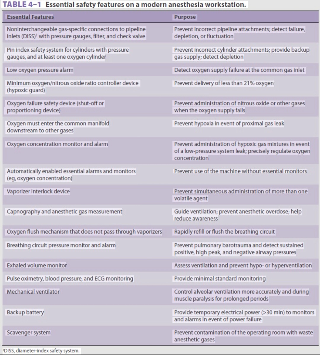
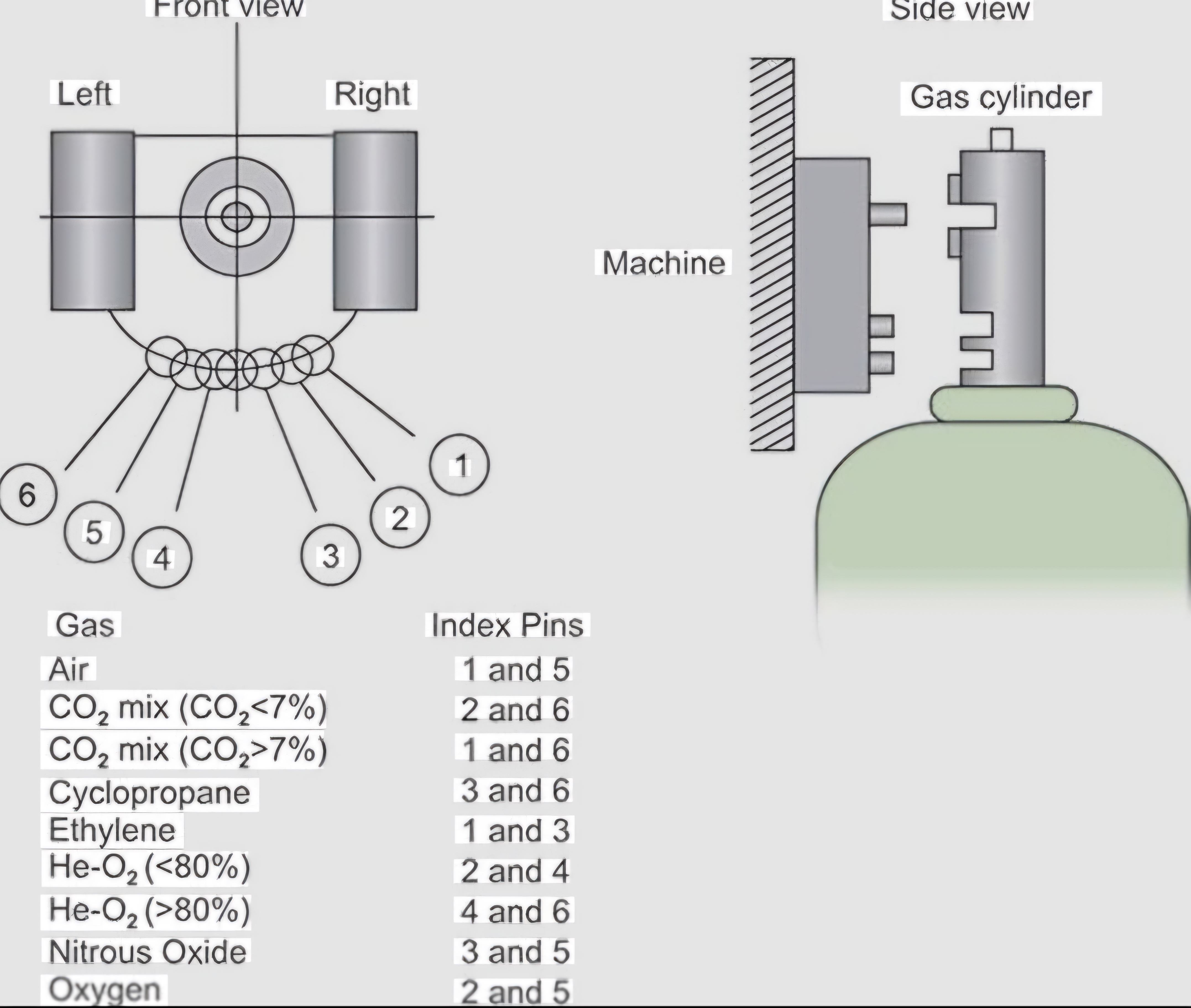
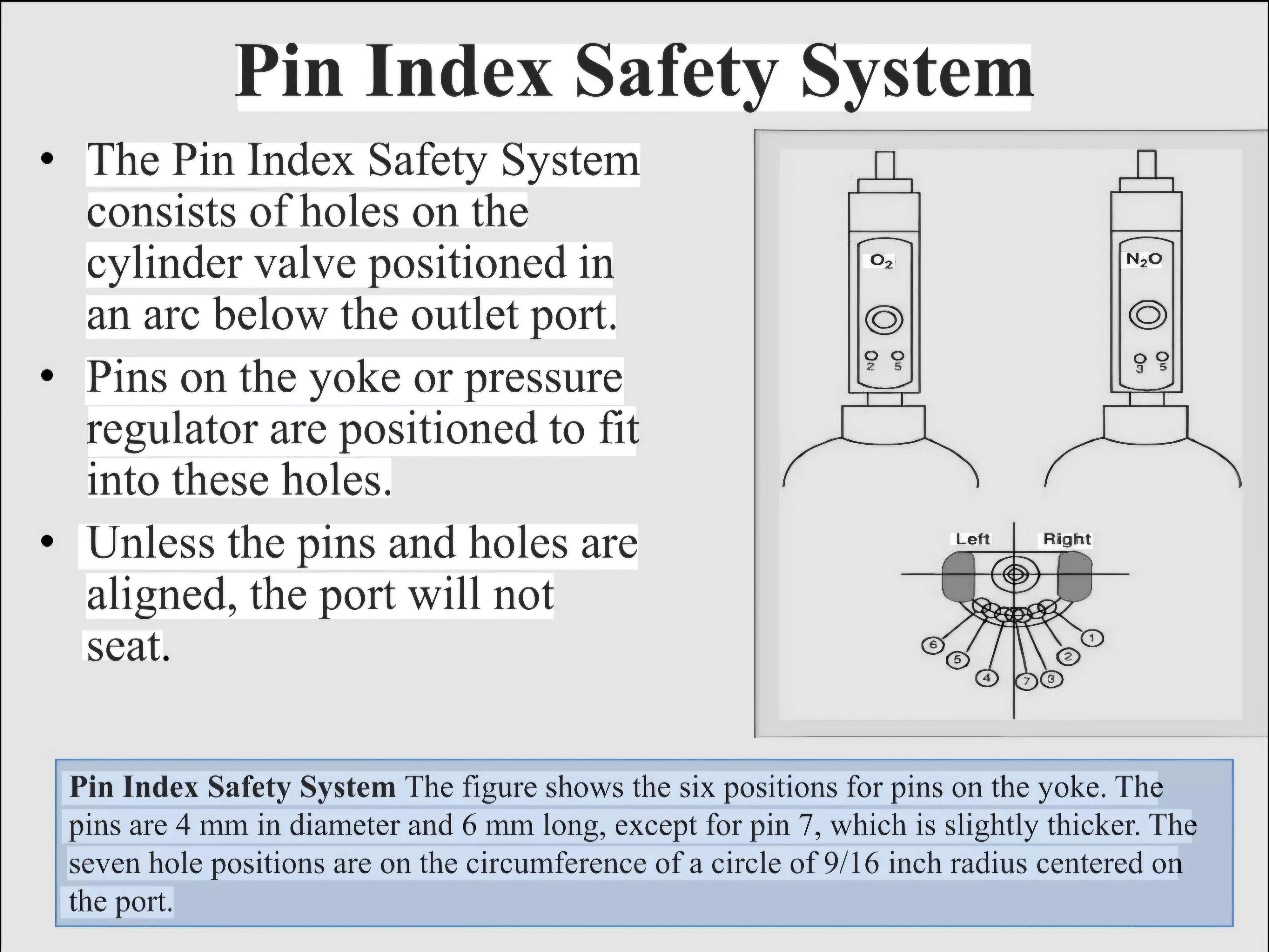
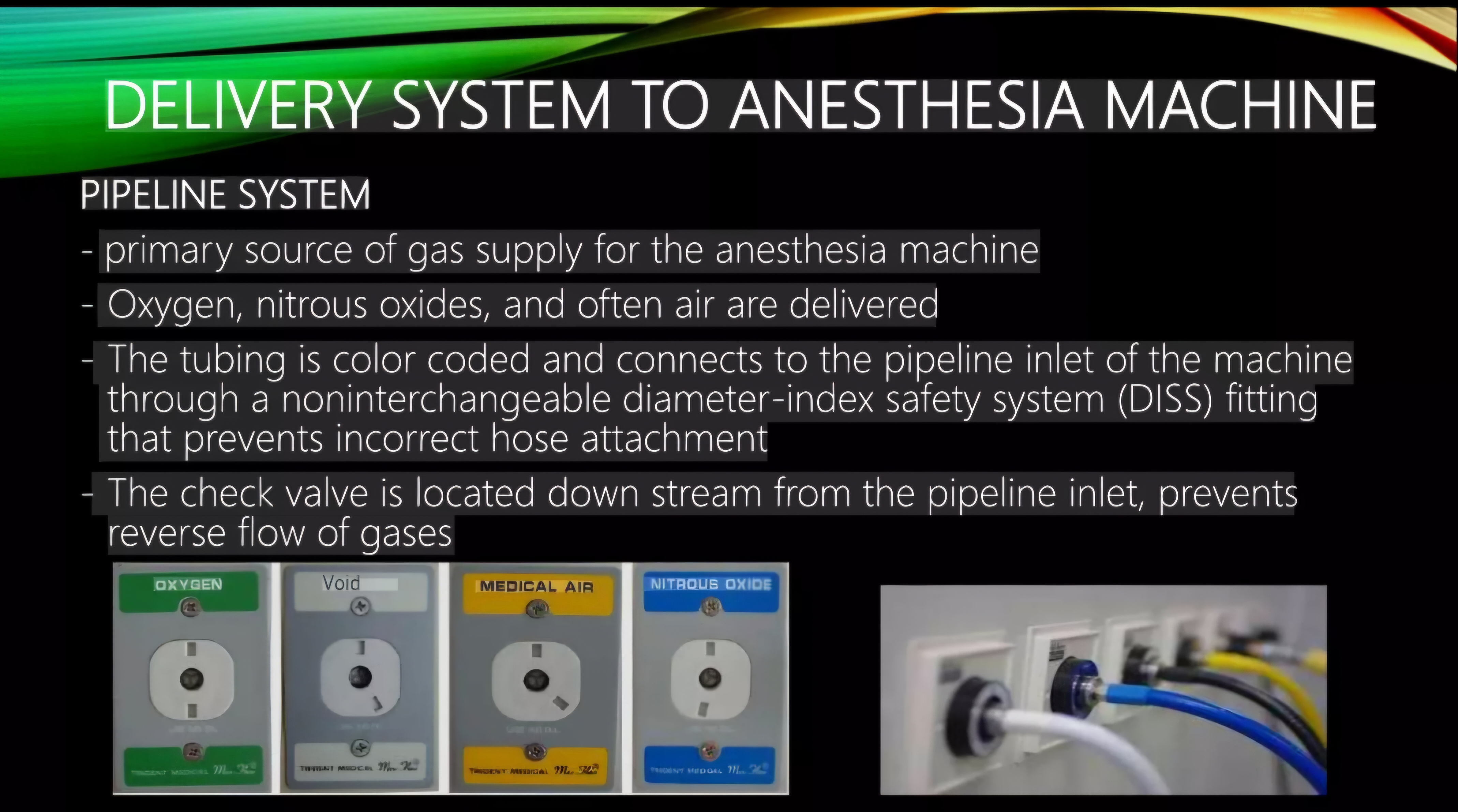
Describe the safety features incorporated in the modern anaesthesia workstation to prevent delivery of a hypoxic or toxic gas mixture. Include: fail-safe valve, oxygen proportioning system, Pin Index Safety System, Diameter Index Safety System, pre-use check, and ventilator alarms.

collections Question Diagrams & Reference Images (4)
Question Reference Diagram
zoom_in View Image
Question Reference Diagram
zoom_in View Image
Question Reference Diagram
zoom_in View Image
Question Reference Diagram
zoom_in View Image
lock Protected Clinical Document • Pulse App • Screenshots Strictly Prohibited
QUESTION 337 person Asked by .
bookmark_add

Fluid Regimes in Diabetic Patient in Anesthesia

picture_as_pdf Clinical Reference Documents & PDFs (1)
Protected Inline Viewer
description Qik9OyeQlhWWQOTjTZjRisjD8XT9KCVE2xuGDjq4.pdf

Showing 331337 of 337 questions

arrow_back Prev 1 32 33 34 Next arrow_forward
account_tree

Subcategory Tree

Explore Anesthesia subcategories

folder_special Anesthesia
Main
lock

Category Subscription

Subscribe to Anesthesia to unlock this module and all nested subcategories.

  • check_circle Access Anesthesia & all subcategories
  • check_circle Detailed, Peer-Reviewed Answers
  • check_circle High-yield visual aids & imaging
Get Category Subscription arrow_forward
Secure 256-bit SSL Connection

Anatomical Models

Explore high-fidelity 3D visualizations included in premium modules.

Case Reviews

Real-world clinical scenarios narrated by senior consultants.

Pulse App