Anesthesia | Eklavya Medical
vaccines
medical_services Main Specialty Domain lock Subscription Required

Anesthesia

Clinical anesthesiology, local and general anesthesia delivery systems, monitoring, and perioperative care.

verified Verified Medical Faculty menu_book 337 Q&A Modules
lock Protected Clinical Document • Pulse App • Screenshots Strictly Prohibited
QUESTION 11 person Asked by .
bookmark_add

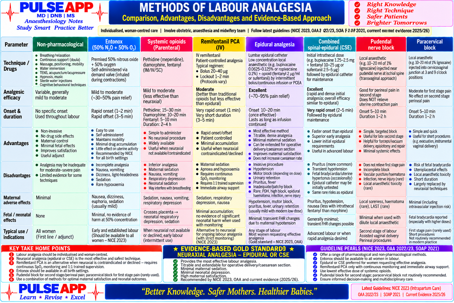
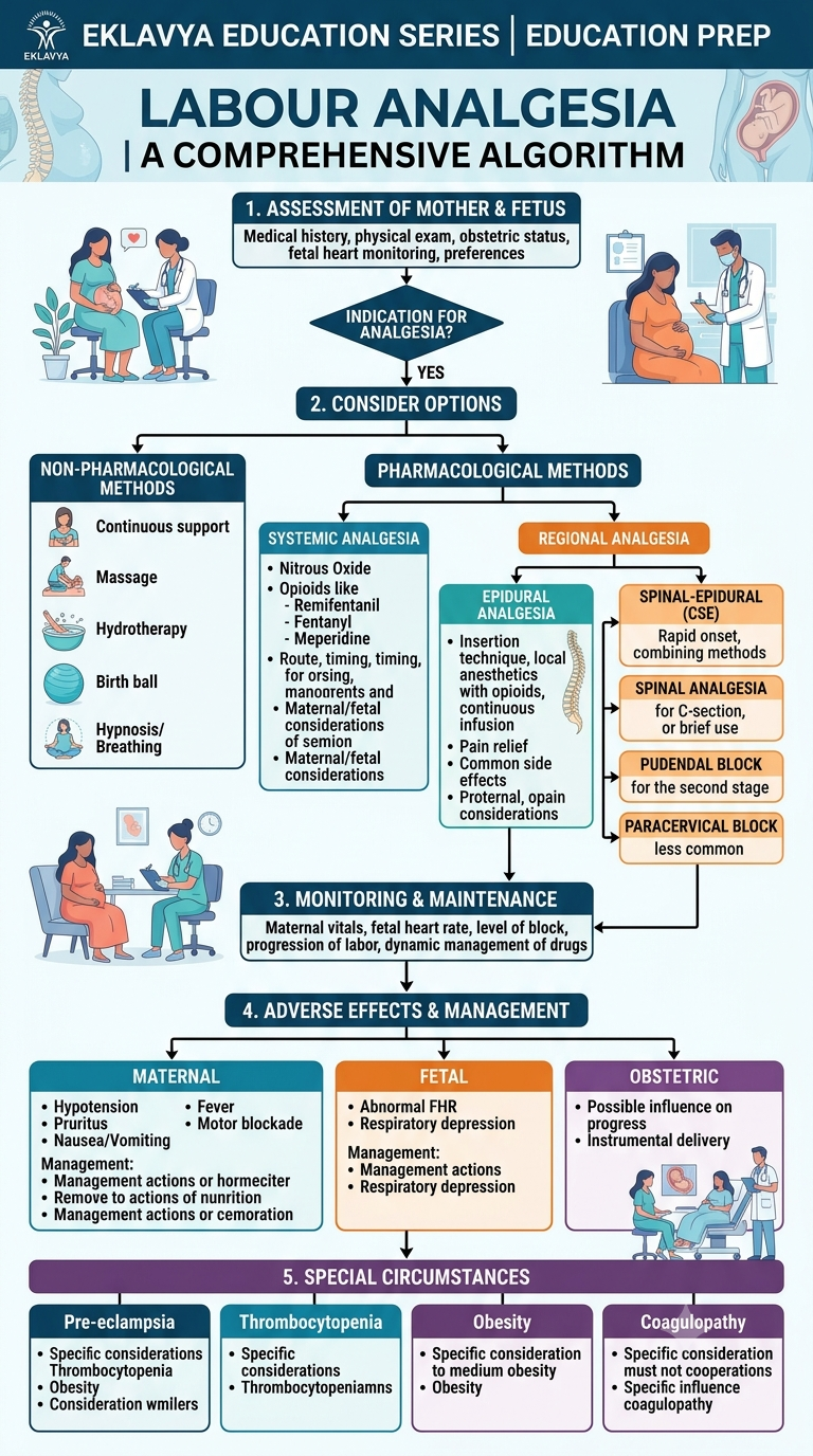
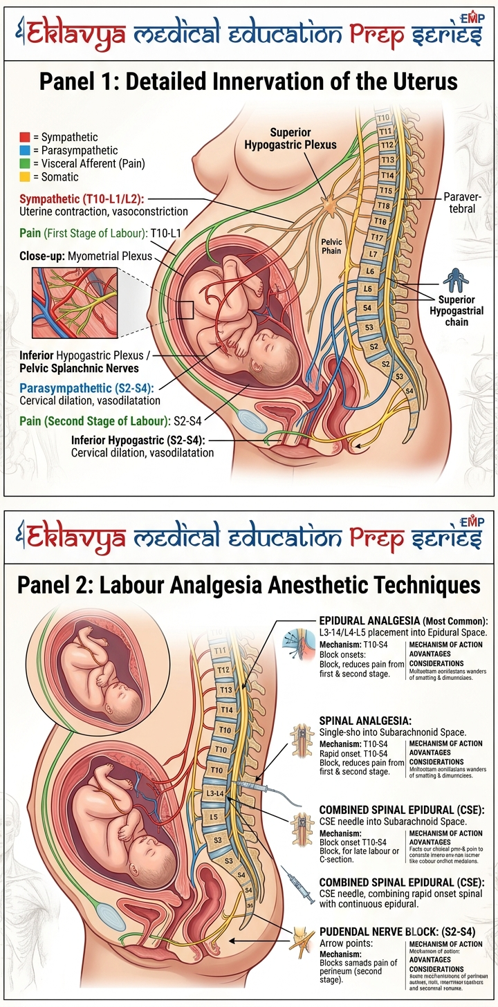
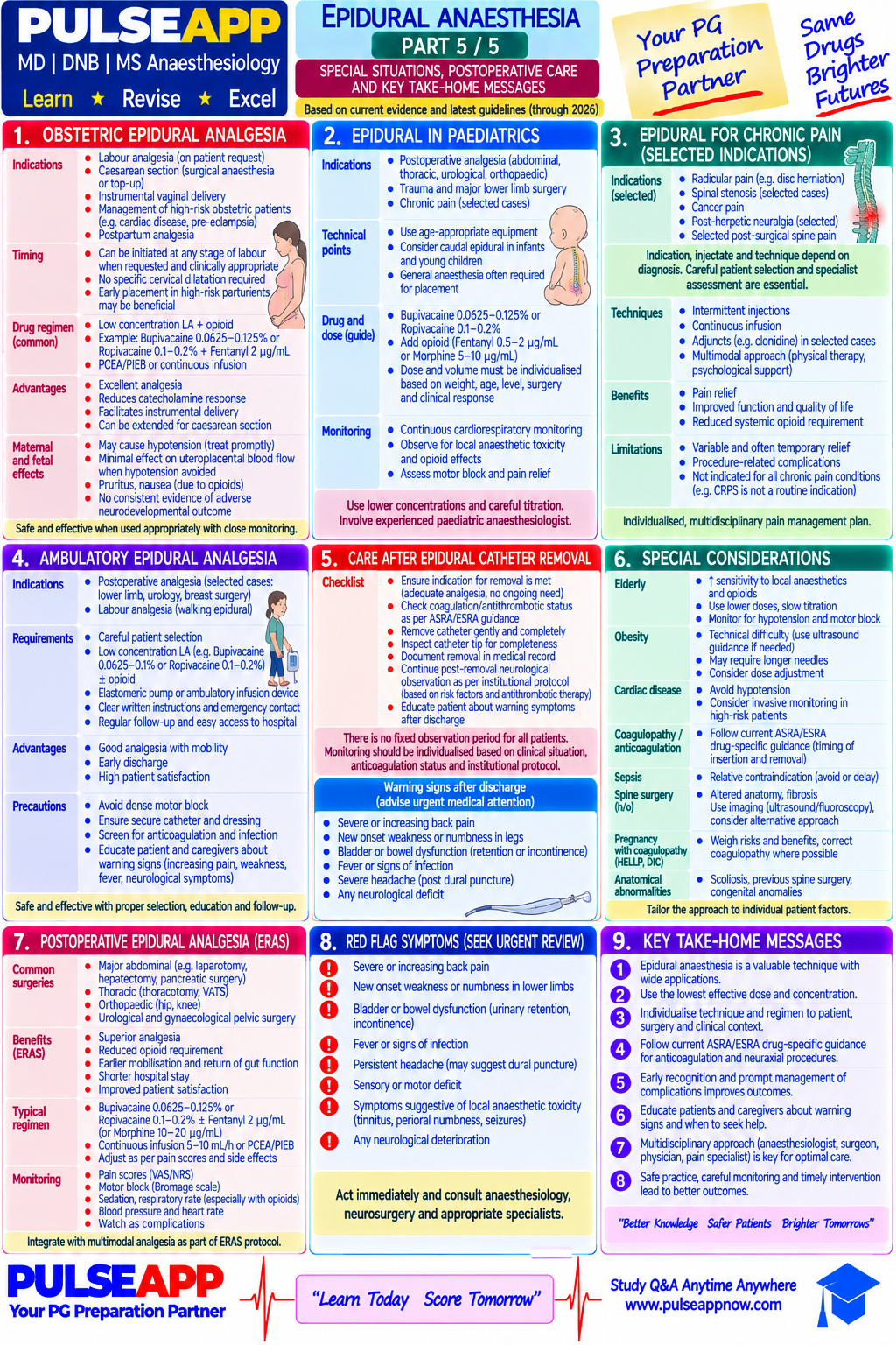
Describe and compare all available methods of labour analgesia including: non-pharmacological, Entonox, systemic opioids (remifentanil PCA), epidural, combined spinal-epidural (CSE), and regional nerve blocks (pudendal, paracervical). State the advantages and disadvantages of each and the evidence-based 'gold standard.'
collections Question Diagrams & Reference Images (2)
Question Reference Diagram
zoom_in View Image
Question Reference Diagram
zoom_in View Image
description Clinical Response
⚙ Core Concept
Labour pain is unique — management must balance maternal comfort against fetal wellbeing and maternal participation in labour. Epidural remains the gold standard, but must be offered among several options, not as the only choice.
A. Non-Pharmacological and Entonox2 marks

Hydrotherapy, TENS, massage/acupuncture/hypnobirthing — modest analgesic effect, no maternal/fetal side effects.

Entonox (50% N₂O/O₂): onset 30–45s, inhaled before contraction peak; ~50–60% find helpful but most progress to epidural; nausea/dizziness common; occupational exposure risk to staff.

B. Remifentanil PCA2 marks

Uniquely suited to labour PCA (onset 30–90s, context-sensitive half-time 3 min) — timed to contraction cycle.

Protocol: 40mcg bolus, 2-min lockout, no background infusion. Superior to pethidine, inferior to epidural.

⚠ Mandatory Safety Requirements
Maternal respiratory depression (SpO₂<94% in 10–15%) requires CONTINUOUS SpO₂ monitoring and 1:1 midwife nursing.
C. Epidural — The Gold Standard4 marks

Achieves complete/near-complete pain relief in >95%; Cochrane review confirms superiority over all other methods; NICE recommends offering to all who request it.

Technique: L2–L3/L3–L4; test dose 3mL 2% lidocaine + adrenaline 1:200,000; loading 10–15mL 0.1% bupivacaine + fentanyl.

'Walking epidural': 0.0625–0.1% bupivacaine + fentanyl — preserves motor function.

✅ Does NOT Increase C-Section Rate
Cochrane and multiple RCTs disprove the historical association — a specifically tested misconception.

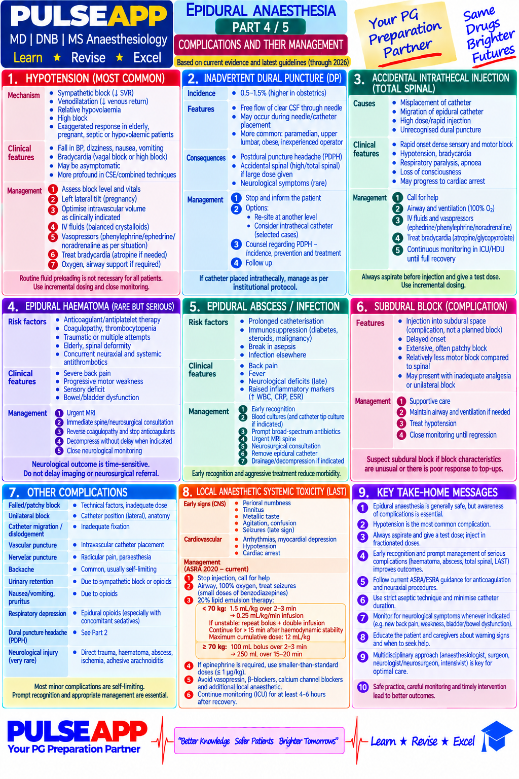
Complications: PDPH (1–2%), inadequate analgesia (5–15%), hypotension, motor block, urinary retention; rare: epidural haematoma/abscess, total spinal.

D. CSE and Regional Nerve Blocks2 marks

CSE: intrathecal bupivacaine 2.5mg + fentanyl 25mcg gives immediate dense analgesia within 5 min; catheter for maintenance — faster onset than epidural alone.

Pudendal block: perineal analgesia for delivery/instrumental delivery only, no uterine pain relief. Paracervical block: largely abandoned (fetal bradycardia risk).
🎤 Viva Corner
Q. A labouring woman on remifentanil PCA has SpO₂ 87% during a contraction. What has happened and immediate management?
Opioid-induced respiratory depression. Immediate: alert midwife, apply supplemental O₂ 10–15L/min, stimulate the patient, press emergency call. If no improvement >60s: naloxone 100mcg IV titrated increments (avoid full reversal, which causes breakthrough pain). Illustrates why continuous SpO₂ monitoring and 1:1 nursing are mandatory, not optional.
★ Examiner's Pearl
State epidural as gold standard (NICE NG121, Cochrane evidence). Remifentanil PCA protocol (40mcg/2-min lockout) with mandatory safety requirement is tested. Epidural does NOT increase CS rate is a specifically tested misconception.
Anim-Somuah M et al (Cochrane 2018). NICE NG121 (2014, updated 2023). Likis FE et al (J Midwifery Womens Health 2014). Miller's Anaesthesia 9th Ed, Ch 77.
lock Protected Clinical Document • Pulse App • Screenshots Strictly Prohibited
QUESTION 12 person Asked by .
bookmark_add

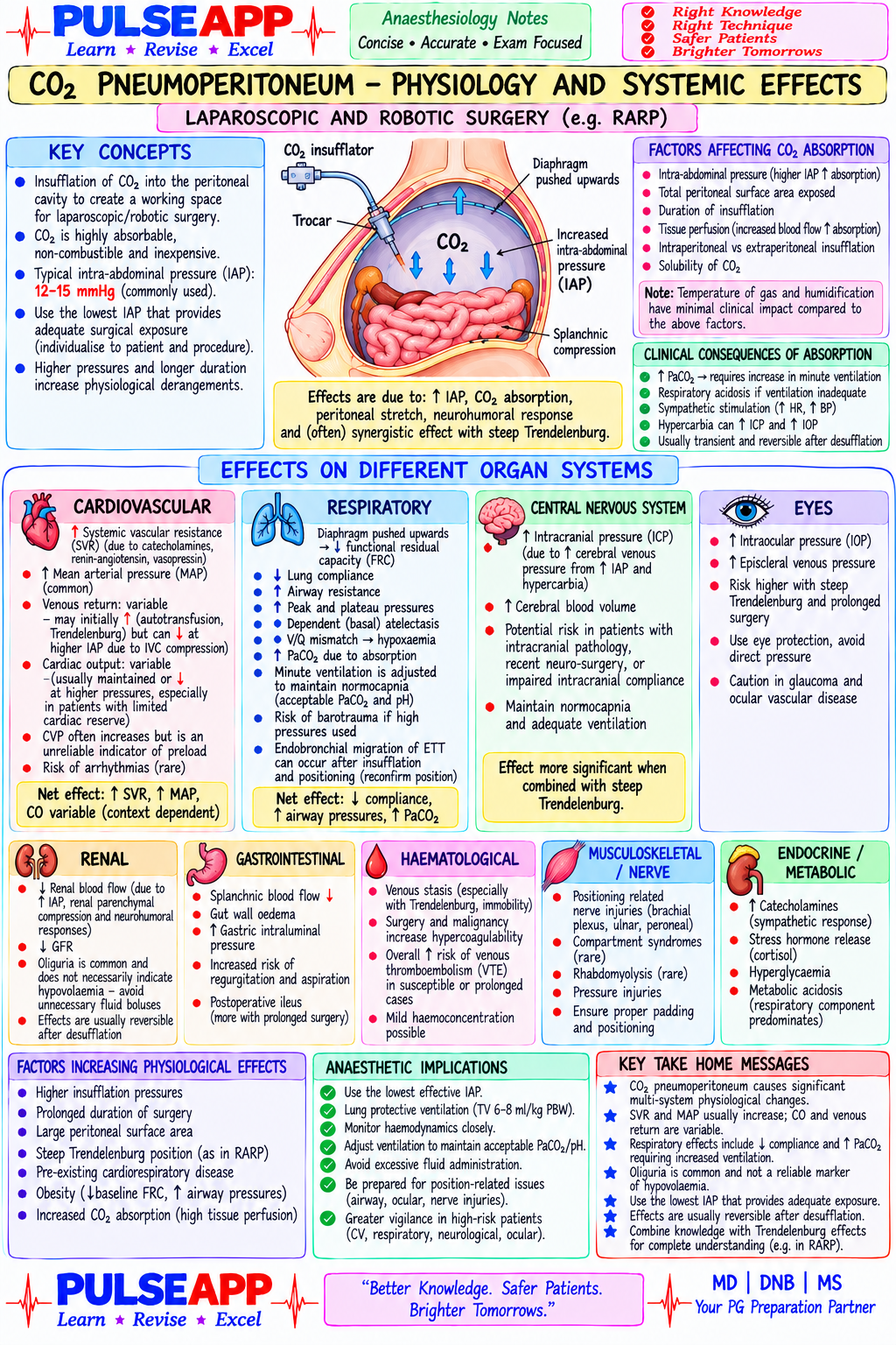
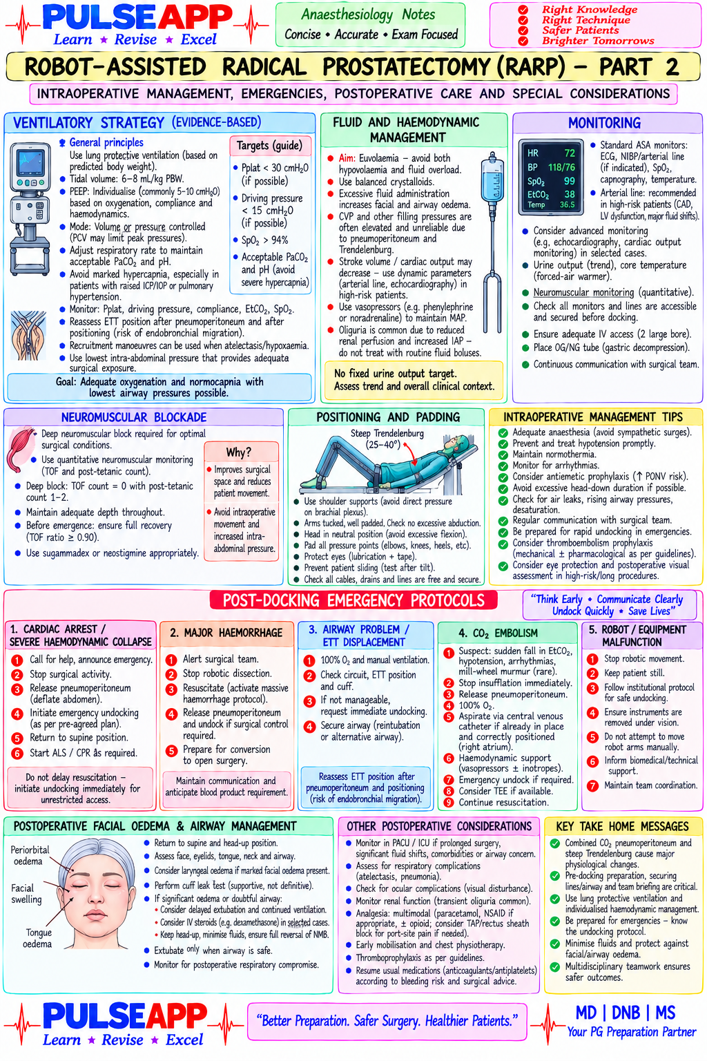
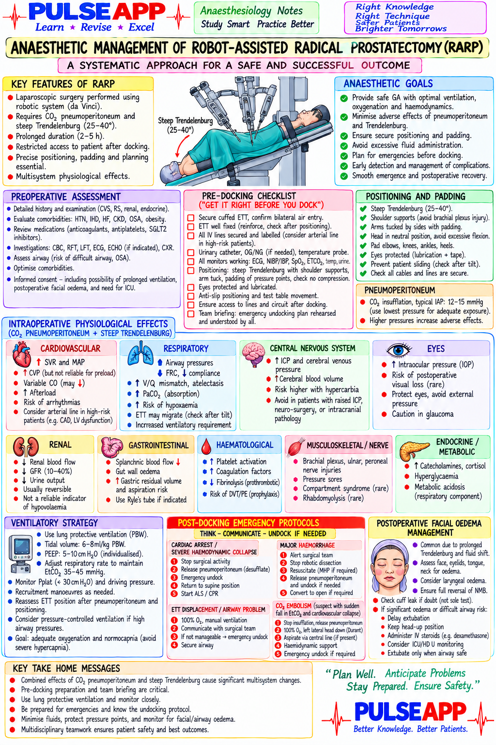
Expand on the anaesthetic considerations for Robot-Assisted Radical Prostatectomy (RARP) specifically addressing: the intraoperative physiological effects of the combined steep Trendelenburg &amp; CO&#8322; pneumoperitoneum on each organ system, pre-docking checklist, ventilatory strategy, post-docking emergency protocols, and postoperative facial oedema management.

description Clinical Response
⚙ Core Concept
RARP combines steep Trendelenburg (30–45°), prolonged pneumoperitoneum, and robotic docking, creating physiological derangement in every major organ system while severely restricting patient access. The pre-docking checklist and emergency protocols must be established before the case begins.
A. Combined Physiological Effects — Organ by Organ4 marks
SystemEffectMagnitude
RespiratoryFRC ↓30–50%, peak pressures ↑40–50%, PaCO₂ ↑10–20mmHgMajor
CardiovascularInitial ↑CO from venous return, then ↓CO as IAP≥15mmHg compresses IVCMajor, vasopressors often needed
ICPImpaired cerebral venous drainage + hypercapnia → ↑ICP 8–15mmHgSignificant if pre-existing pathology
IOPVenous congestion → IOP may double (15→30+mmHg)Important for case duration
RenalIAP compresses renal vein → ↓RBF → transient oliguriaExpected, resolves after desufflation
Hepatic/splanchnicIAP compresses portal vein → ↓hepatic blood flowModerate, relevant for prolonged cases
B. Pre-Docking Checklist2 marks
⚠ Once Docked, Access Is Severely Restricted
ETT secure/taped (armoured preferred); ≥1 large-bore IV; arterial line right radial; urinary catheter; NGT decompression; eyes taped; padding at all pressure points; confirm ventilator settings/emergency drugs accessible.
C. Ventilatory Strategy2 marks

TV 6–7mL/kg IBW; RR ↑15–25% for CO₂ absorption; PEEP 6–10cmH₂O; I:E 1:2 (extend to 1:2.5 if pressures high); permissive hypercapnia (PaCO₂ 50–55) if plateau >30cmH₂O; continuous capnography, ABG every 60–90min.

D. Postoperative Facial Oedema — Airway Safety2 marks

After prolonged Trendelenburg (3–5h): facial/conjunctival/laryngeal oedema. Perform cuff leak test before extubation; IV dexamethasone 8mg; head elevated 30–60 min pre-extubation; have Airway Exchange Catheter ready.

🎤 Viva Corner
Q. During a 4-hour RARP, peak airway pressure rises 22→42cmH₂O and SpO₂ falls to 88% despite FiO₂ 1.0. Differential and management?
Differentials: ETT obstruction/migration (suction, check bilateral breath sounds), progressive atelectasis (recruitment manoeuvre + PEEP), pneumothorax (CO₂ tracking through diaphragmatic defect or trocar injury — needle decompression if tension), endobronchial intubation (withdraw ETT 1–2cm), bronchospasm (salbutamol/deepen volatile/IV magnesium). Simultaneously: FiO₂ 1.0, inform surgeon to consider reducing insufflation pressure/desufflation, have vasopressors ready.
★ Examiner's Pearl
The pre-docking checklist is the clinical safety application examiners require. Cuff-leak test after prolonged Trendelenburg is the single most important RARP-specific extubation safety measure. IOP/ICP effects demonstrate comprehensive knowledge.
Gainsburg DM (Minerva Anestesiol 2011). Awad H et al (J Robotic Surg 2009). POVL Study Group (Anesthesiology 2012).
lock Protected Clinical Document • Pulse App • Screenshots Strictly Prohibited
QUESTION 13 person Asked by .
bookmark_add

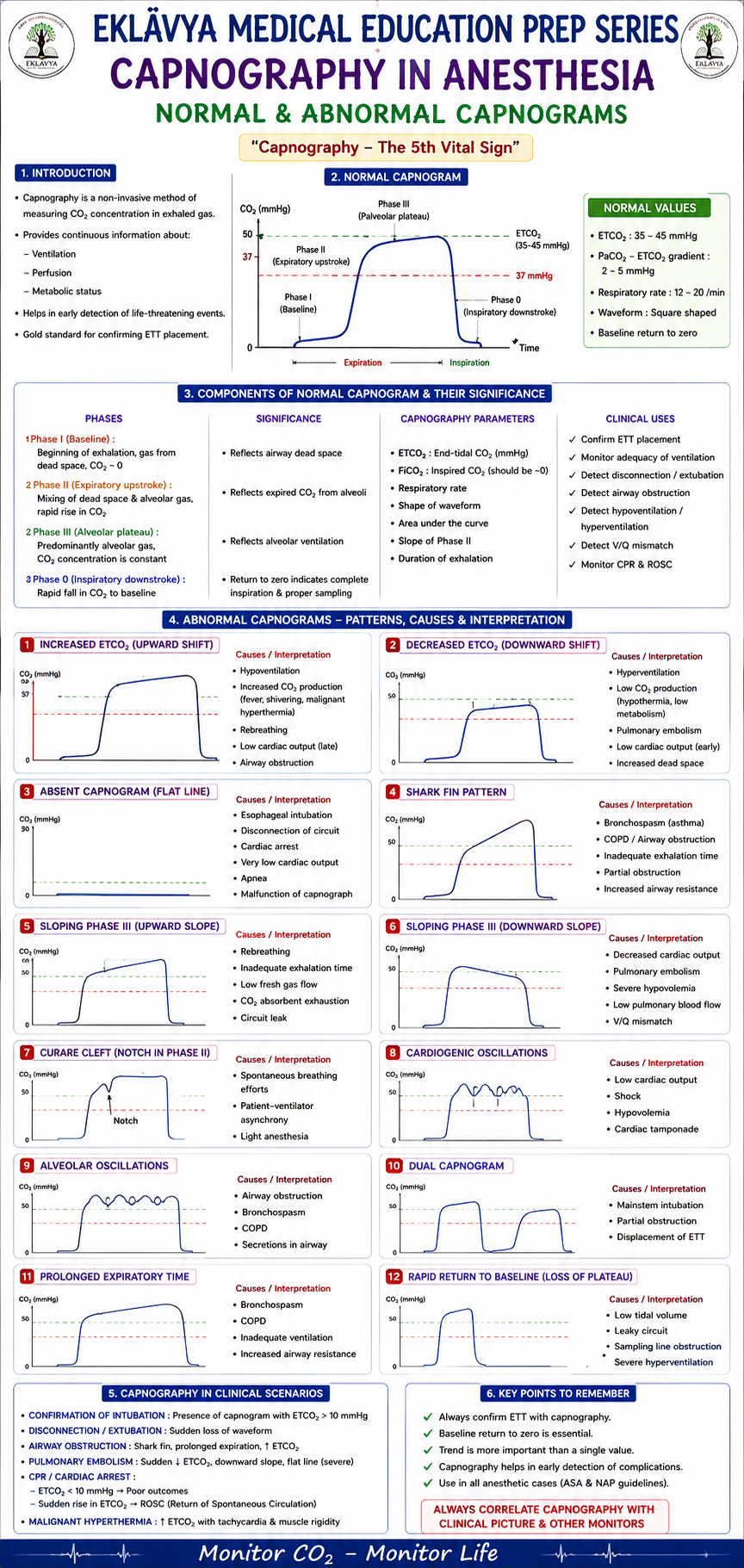
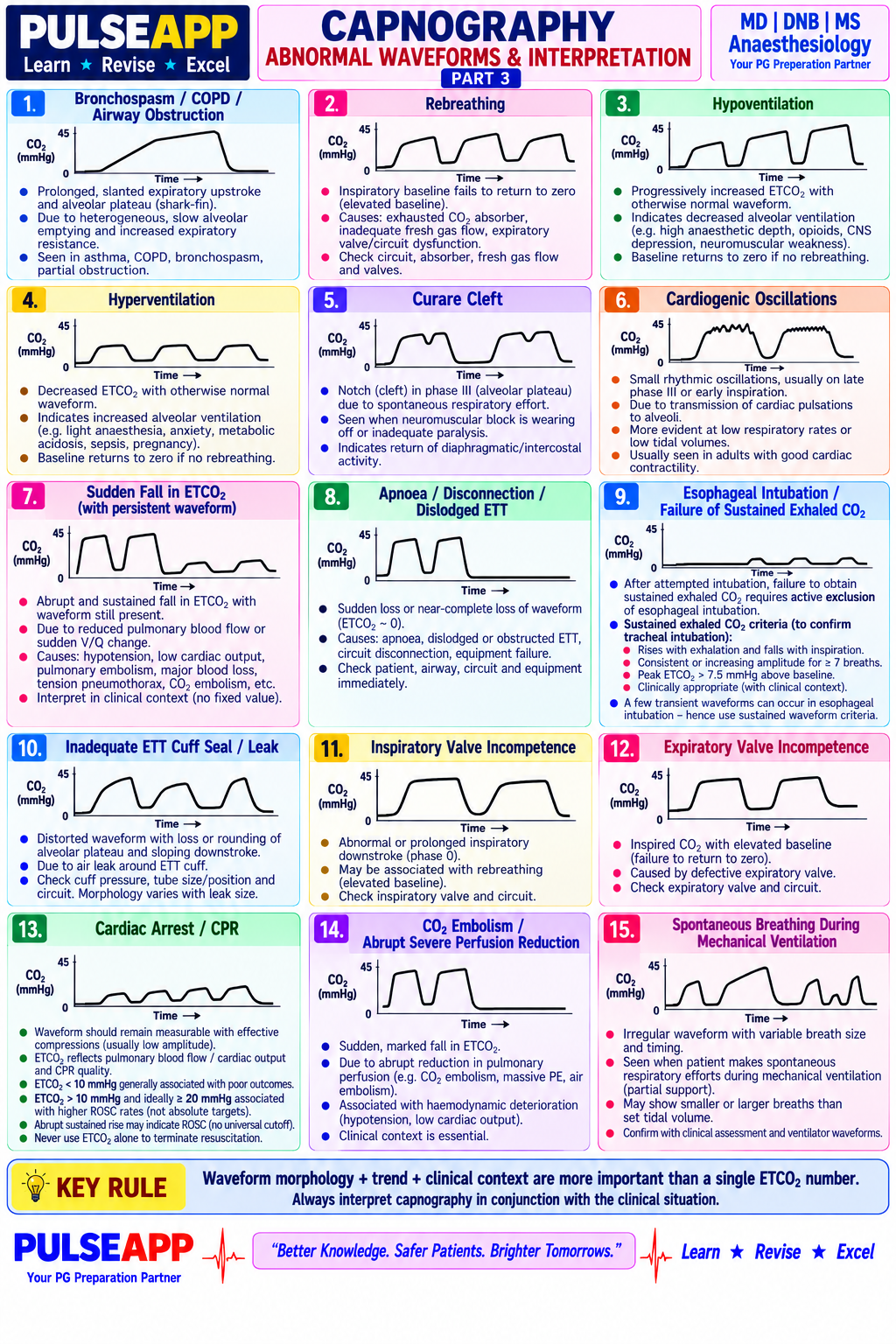
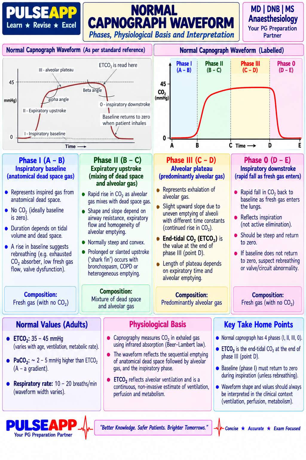
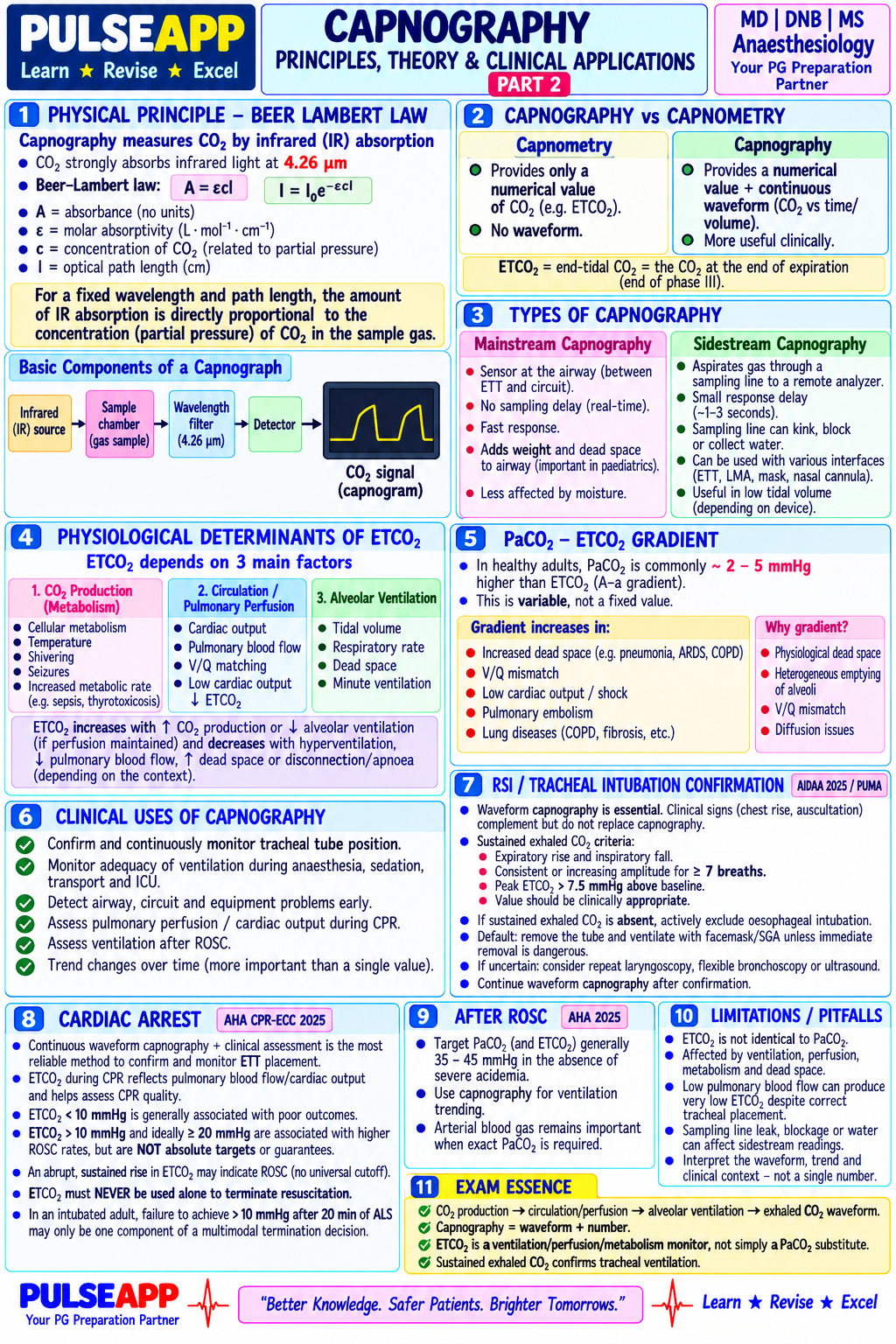
Explain the physical principle of capnography using Beer-Lambert law. Describe the four phases of the normal capnograph waveform and their physiological basis. Discuss the interpretation of abnormal waveform patterns and the specific role of ETCO&#8322; monitoring in RSI confirmation and cardiac arrest.

collections Question Diagrams & Reference Images (1)
Question Reference Diagram
zoom_in View Image
description Clinical Response
⚙ Core Concept
Capnography is arguably the most information-dense monitor in anaesthesia — simultaneously confirming ETT placement, ventilation adequacy, airway patency, pulmonary perfusion, and metabolic state. A sudden rise in ETCO₂ to ≥35mmHg during CPR is the earliest, most specific indicator of ROSC, often preceding detectable pulse by 30–60 seconds.
A. Physical Principle — Beer-Lambert Law2 marks

CO₂ absorbs infrared light at 4.26μm (mid-IR). Absorbance = ε × C × L — absorbed light directly proportional to CO₂ concentration.

Sidestream: gas aspirated at 150mL/min, slight delay (1–3s), suitable for non-intubated patients. Mainstream: sensor at airway, no delay, requires ETT.

B. Normal Four-Phase Waveform3 marks
PhaseGas SampledCO₂Abnormal Meaning
I — BaselineAnatomical dead space≈0mmHg↑baseline → rebreathing/exhausted soda lime
II — Ascending limbDead space washing to alveolarRising sharplyProlonged slope → obstructive disease/cuff leak
III — Alveolar plateauPure alveolar gas (=ETCO₂)35–45mmHgSloped 'shark fin' → bronchospasm/COPD
0 — Inspiratory downstrokeFresh gas replaces expired CO₂Falls to zeroSlow return → rebreathing
C. Abnormal Waveform Patterns3 marks
PatternDiagnosis
ETCO₂→0 (flat line)Oesophageal intubation (most critical), disconnection, complete obstruction
Sudden abrupt fallMassive PE, cardiac arrest, massive haemorrhage
Progressive ↑ETCO₂Hypoventilation, malignant hyperthermia (FIRST SIGN), fever, laparoscopic CO₂ absorption
'Shark fin' sloped Phase IIIBronchospasm/COPD
Cardiogenic oscillationsBenign; small rhythmic oscillations synchronous with heart rate
D. ETCO₂ in RSI and Cardiac Arrest2 marks

RSI: persistent consistent waveform over ≥6 breaths confirms tracheal placement; flat line/2 waves then zero suggests oesophageal intubation.

Cardiac arrest: ETCO₂<10mmHg = poor CPR quality/poor prognosis; sudden rise to ≥35–40mmHg = ROSC indicator, precedes palpable pulse.

🎤 Viva Corner
Q. After RSI, the first two breaths show small CO₂ waveforms but then flat-line from breath three onward. Is the ETT in the trachea?
No — this is the characteristic pattern of oesophageal intubation. Brief initial waveforms come from expelled gastric CO₂, which is then exhausted; tracheal intubation produces a consistent waveform every breath. Immediate action: remove ETT, mask ventilate with 100% O₂, re-intubate with video laryngoscopy, confirm with sustained waveform over ≥6 breaths.
★ Examiner's Pearl
State CO₂'s specific IR absorption wavelength (4.26μm). All four phases with correct CO₂ content must be reproduced. The ROSC indicator (sudden ETCO₂ rise ≥35–40mmHg during CPR) with the action (stop compressions, check pulse) is most tested.
Bhavani-Shankar Kodali (Anesthesiology 2013). Miller's Anaesthesia 9th Ed, Ch 44. AHA/ACC 2020 Guidelines (Circulation 2020).
lock Protected Clinical Document • Pulse App • Screenshots Strictly Prohibited
QUESTION 14 person Asked by .
bookmark_add

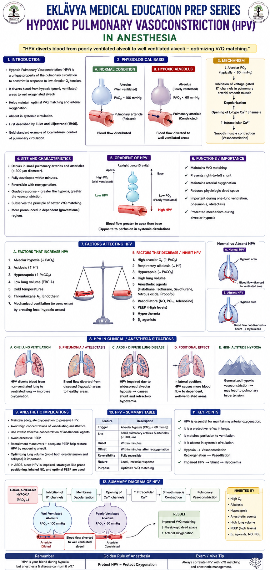
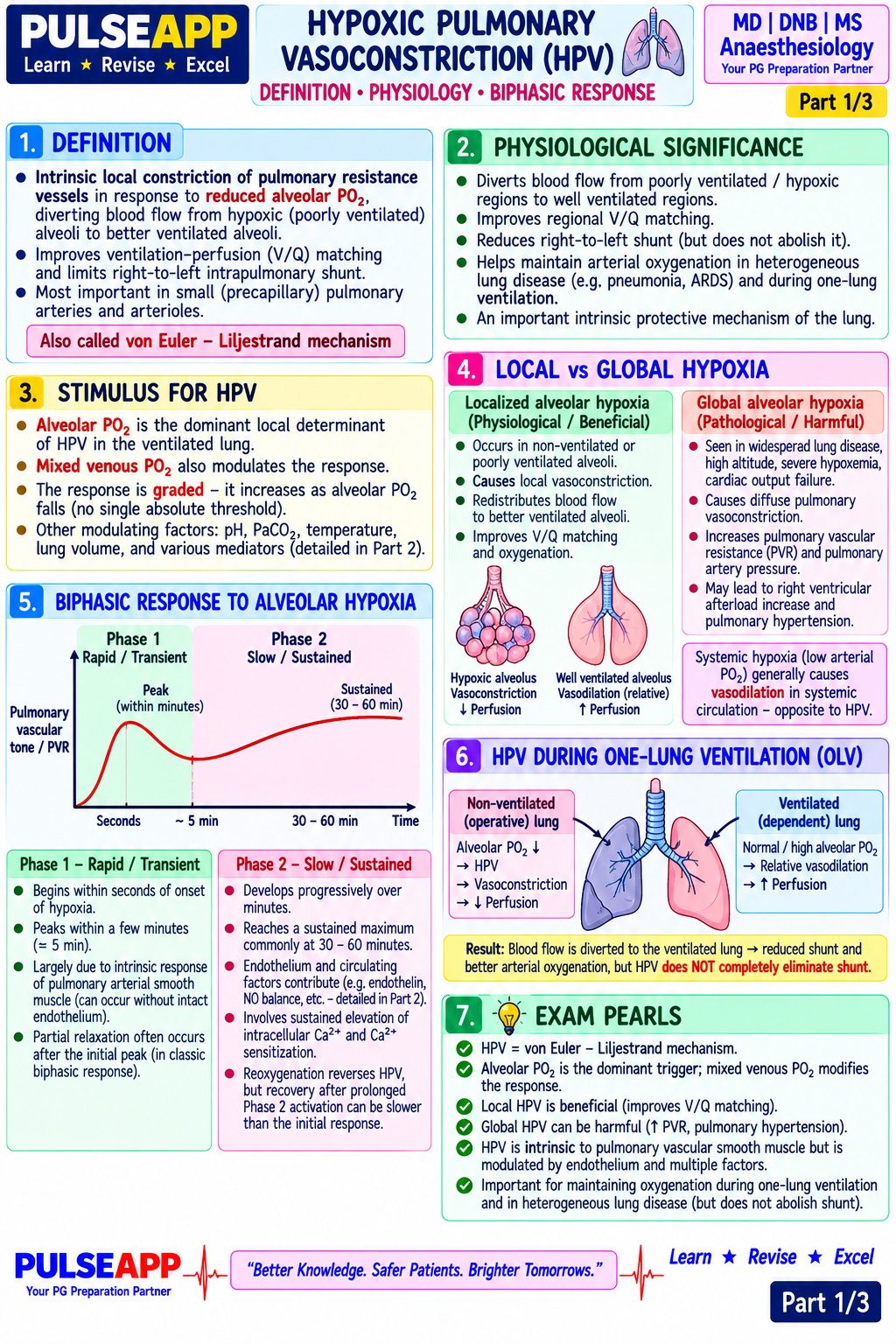
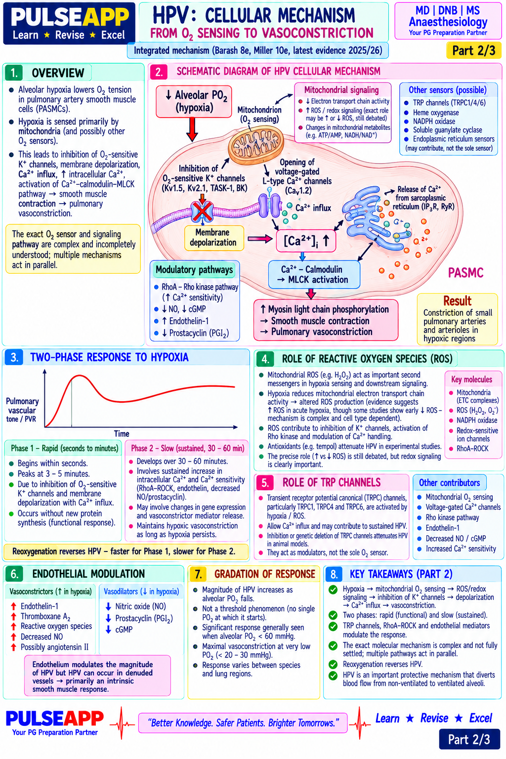
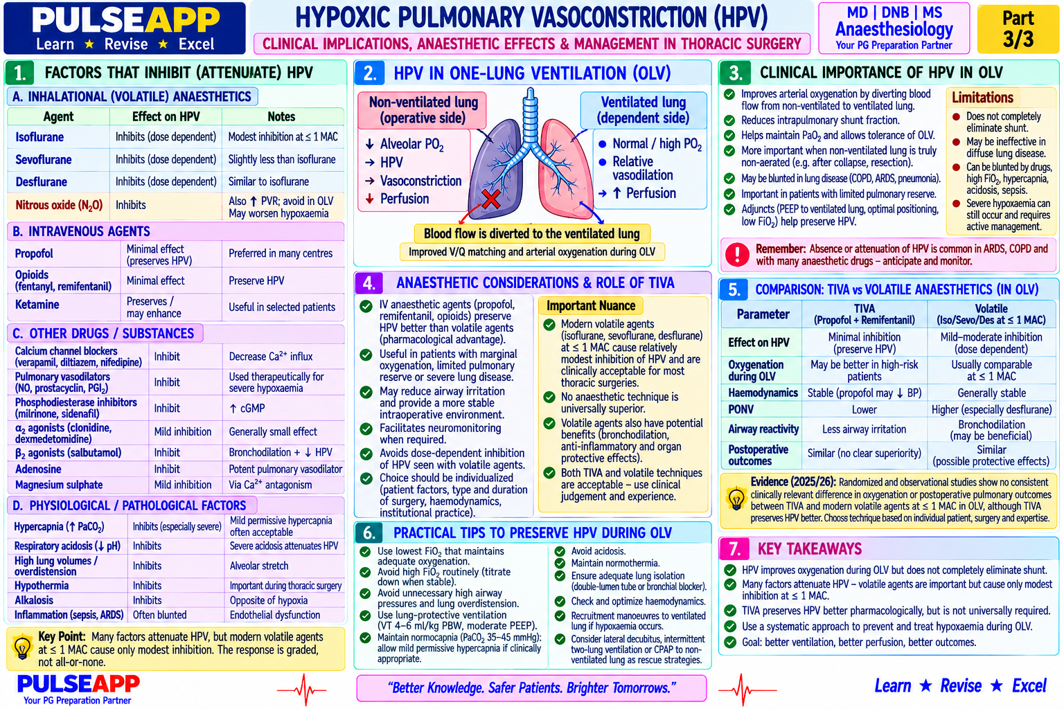
Define HPV. Describe the two-phase cellular mechanism (TRPV channels, ROS, mitochondrial O&#8322; sensing). List factors that inhibit HPV including volatile anaesthetic agents. Explain its clinical importance during one-lung ventilation and why TIVA is preferred for thoracic surgery.

collections Question Diagrams & Reference Images (1)
Question Reference Diagram
zoom_in View Image
description Clinical Response
⚙ Core Concept
HPV is the lung's built-in mechanism for optimising V/Q matching — poorly ventilated (hypoxic) alveoli trigger arteriolar constriction, diverting blood to better-ventilated units. Unlike systemic vasculature (which dilates in hypoxia), pulmonary vasculature uniquely constricts. Anaesthetic agents, particularly volatiles, inhibit this reflex dose-dependently.
A. Definition1 mark

Intrinsic response of pulmonary arteriolar smooth muscle to regional alveolar hypoxia (PAO₂<70mmHg) — can reduce blood flow to a collapsed segment by ~50%.

B. Cellular Mechanism — Two-Phase Response4 marks

Phase 1 (seconds–minutes): ↓PAO₂ → altered mitochondrial ROS signalling → inhibits voltage-gated K⁺ channels (Kv1.5/Kv2.1) → depolarisation → L-type Ca²⁺ channel activation → Ca²⁺ influx → MLCK activation → smooth muscle contraction. Simultaneously ↓NO/prostacyclin removes tonic vasodilation.

Phase 2 (hours–days): Prolonged hypoxia → HIF-1α stabilisation → ↑endothelin-1, ↑VEGF, ↓eNOS — sustained structural component underlying pulmonary hypertension of chronic hypoxaemia.

C. Factors Modulating HPV3 marks
FactorEffectMechanism
Volatile agents (dose-dependent)INHIBIT HPV (most important)Activate K⁺ channels + ↑NO/PGI₂; halothane>isoflurane≈sevoflurane≈desflurane
Propofol (TIVA)Does NOT inhibit HPVNo effect on PVSMC tone at clinical doses
VasodilatorsInhibit HPVNon-selective pulmonary vasodilation
HypocapniaInhibits HPVCO₂ has vasoconstrictor pulmonary effects; avoid hyperventilation
AcidosisAugments HPVPotentiates K⁺ channel inhibition
Infection/inflammationInhibits HPV locallyCytokines override HPV precisely where most needed
D. Clinical Importance During OLV2 marks

During OLV, maximal HPV stimulus in the collapsed lung reduces its flow by ~50%, but ~35–50% shunt persists. TIVA preserves HPV → 15–25mmHg higher PaO₂ vs equiMAC volatile.

Stepwise hypoxia management: FiO₂ 1.0 → PEEP to ventilated lung → recruitment → CPAP to operative lung → brief two-lung ventilation as last resort.

🎤 Viva Corner
Q. Why does pneumonia cause hypoxaemia relatively resistant to oxygen supplementation, via the HPV mechanism?
Consolidated, non-ventilated pneumonic segments produce massive local cytokines/NO/prostaglandins that specifically INHIBIT HPV precisely where it is most needed — blood continues flowing through non-ventilated alveoli at near-normal rates, creating true shunt physiology. Supplemental O₂ cannot reach completely consolidated alveoli, so hypoxaemia is relatively refractory to FiO₂.
★ Examiner's Pearl
The Kv channel→depolarisation→L-type Ca²⁺ channel→contraction mechanism distinguishes a comprehensive answer. Volatile agents inhibit HPV; propofol does not — this is the key clinical application fact for TIVA preference in OLV.
Lumb AB, Slinger P (Anesthesiology 2015). Archer SL, Michelakis ED (N Engl J Med 2009). Della Rocca G et al (Anesth Analg 2001). Miller's Anaesthesia 9th Ed, Ch 68.
lock Protected Clinical Document • Pulse App • Screenshots Strictly Prohibited
QUESTION 15 person Asked by .
bookmark_add

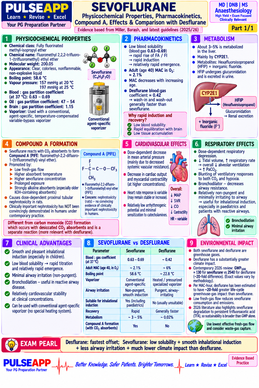
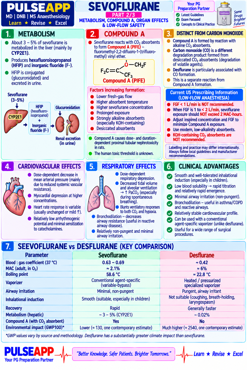
Describe the physicochemical properties of sevoflurane. Explain its pharmacokinetics (blood-gas partition coefficient, MAC, onset and offset). Discuss Compound A formation, cardiovascular and respiratory effects, clinical advantages, and environmental impact compared to desflurane.

collections Question Diagrams & Reference Images (1)
Question Reference Diagram
zoom_in View Image
description Clinical Response
⚙ Core Concept
Sevoflurane is the dominant halogenated volatile globally — its low pungency (smooth inhalational induction), low blood-gas partition coefficient (rapid onset/offset), cardiovascular stability, and favourable environmental profile (GWP 130 vs desflurane 2540) make it broadly preferred.
A. Physicochemical Properties2 marks
PropertySevofluraneComparison
Molecular weight200 g/molHeavier than desflurane (168), lighter than isoflurane (184)
Boiling point58.5°CHigher than desflurane (23.5°C, needs heated vaporiser)
Blood-gas coefficient (λ)0.652nd lowest after desflurane (0.42); rapid equilibration
MAC (40yr adult, O₂)2.0%With 65% N₂O: ~0.66%
B. Pharmacokinetics2 marks

Low λ → poor blood sink → rapid rise in alveolar concentration → fast induction/emergence. Metabolism: CYP2E1 → hexafluoroisopropanol (non-toxic, glucuronidated) + inorganic fluoride (peaks 15–30μmol/L, below the 50μmol/L nephrotoxic threshold; no demonstrated clinical nephrotoxicity).

C. Compound A2 marks
⚠ Formation and Significance
Base-catalysed (NaOH/KOH in soda lime) beta-elimination, worse at high temperature/low flow/desiccated absorbent. Nephrotoxic in rats (high renal beta-lyase activity); human beta-lyase activity 10–30× lower — NO clinically significant renal injury demonstrated in humans. FDA recommends minimum FGF 1L/min; many societies consider this conservative.
D. Cardiovascular Effects2 marks
ParameterEffectComparison
ContractilityDose-dependent ↓~20% at 1MACSimilar to isoflurane
Heart rateMinimal changeUnlike desflurane (tachycardia) and halothane (bradycardia)
Catecholamine sensitisationMinimalSafe with adrenaline infiltration, unlike halothane
Ischaemic preconditioningProtective via KATP/PKCReduces perioperative MI in cardiac surgery
E. Respiratory Effects & Advantages over Desflurane2 marks

Non-pungent — ONLY volatile suitable for inhalational induction in adults/children. Bronchodilator; useful in asthma.

vs desflurane: GWP 130 vs 2540 (19× lower); atmospheric lifetime 1.1yr vs 14yr; no sympathetic activation tachycardia; standard room-temperature vaporiser vs heated pressurised TEC-6.

🎤 Viva Corner
Q. A 5-year-old needs gas induction for tonsillectomy. Why sevoflurane specifically, and what concentration?
Non-irritant (no coughing/breath-holding/laryngospasm), low blood-gas coefficient gives rapid loss of consciousness (30–60s). Prime circuit with 8% sevoflurane in 8L/min O₂; once unconscious reduce to 3–4% and establish IV access, then 2–2.5% maintenance. Caveat: >1.5MAC in children can provoke epileptiform EEG activity — keep ≤2MAC during induction.
Q. What is anaesthetic preconditioning and how does sevoflurane achieve it?
Brief myocardial exposure to a volatile agent reduces ischaemia-reperfusion injury. Mechanism: sevoflurane activates mitochondrial K-ATP channels → attenuates mitochondrial permeability transition pore opening during reperfusion → less apoptosis; also activates PKC-ε. Clinical evidence links sevoflurane maintenance in cardiac surgery to lower troponin release and AF incidence vs propofol TIVA.
★ Examiner's Pearl
State λ=0.65 and MAC=2.0% as specific numbers. Compound A: name mechanism, state rat vs human toxicity difference, and FDA FGF recommendation (1L/min) — all separately marked. Anaesthetic preconditioning (mitoKATP, mPTP) distinguishes an advanced answer.
Stabernack CR et al (Anesth Analg 2000). Lerman J et al (Anesthesiology 1994). De Hert SG et al (BJA 2005). Ryan SM, Nielsen CJ (BJA 2010). Miller's Anaesthesia 9th Ed, Ch 26.
lock Protected Clinical Document • Pulse App • Screenshots Strictly Prohibited
QUESTION 16 person Asked by .
bookmark_add

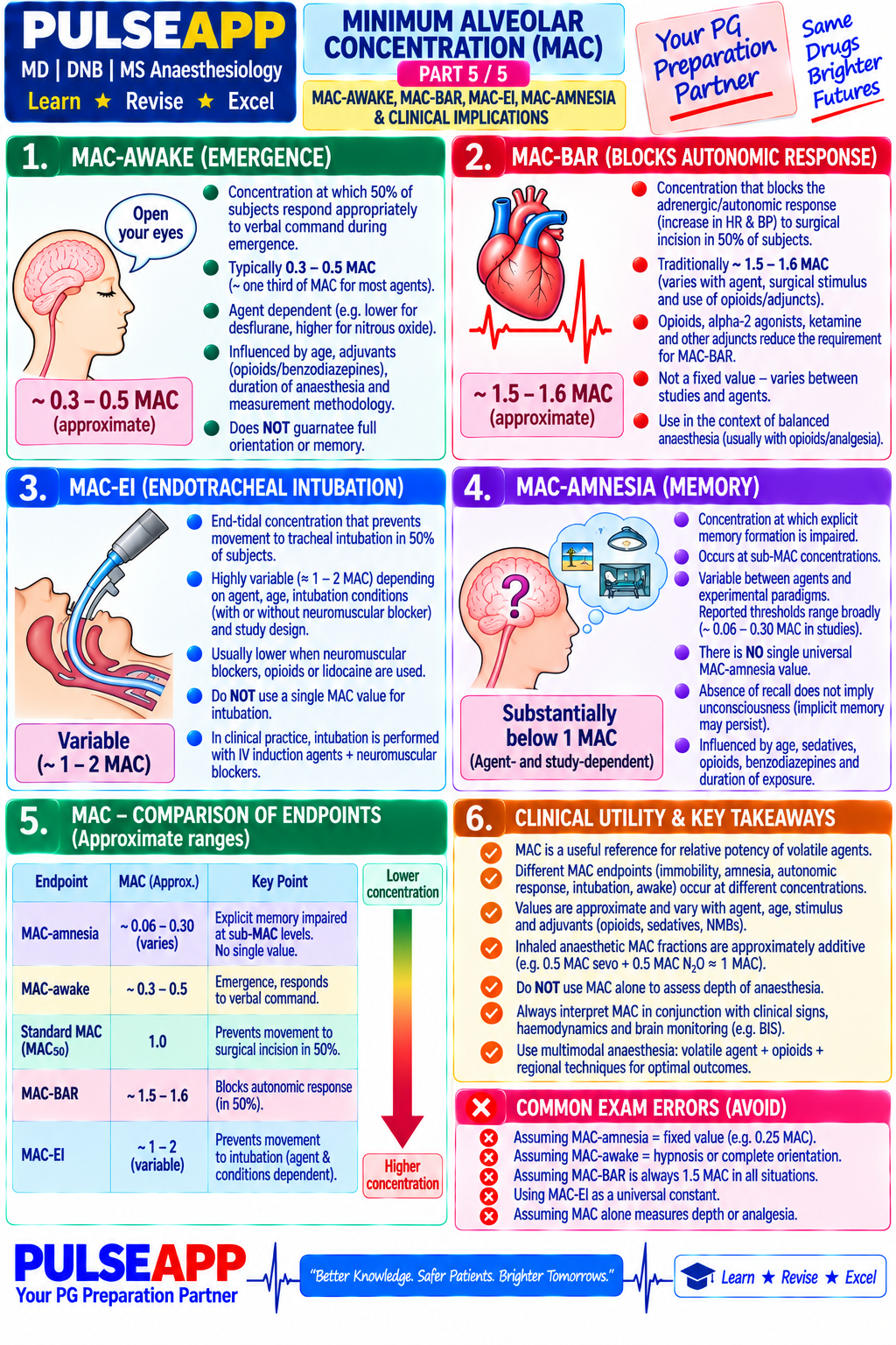
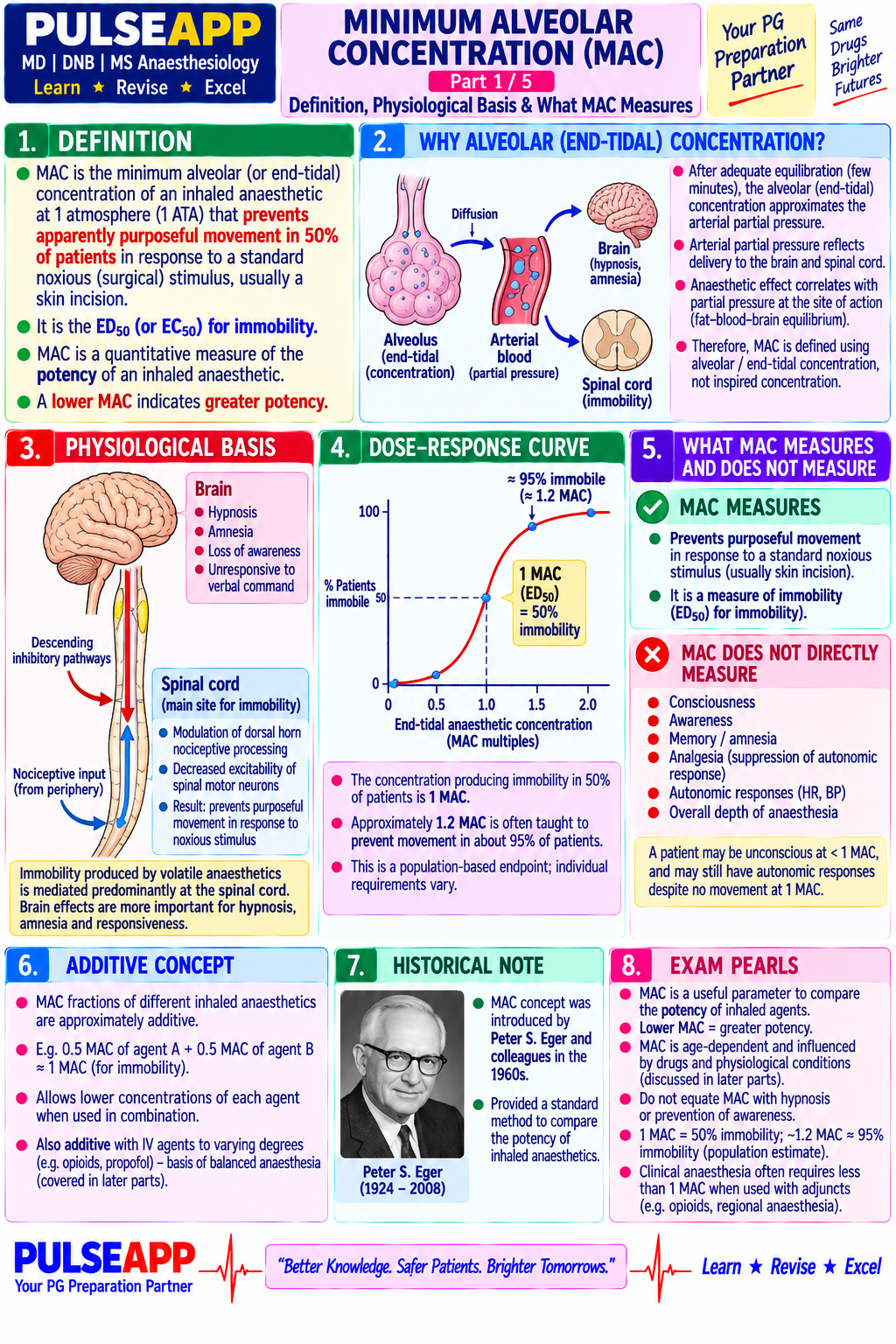
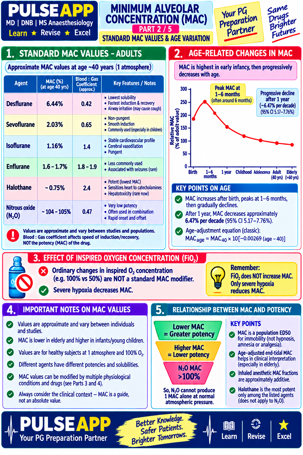
Define MAC. Explain its physiological basis and what it measures. List the standard MAC values for common volatile agents. Describe factors that increase and decrease MAC. Explain the concepts of MAC-awake, MAC-BAR, and MAC-intubation and their clinical utility.

collections Question Diagrams & Reference Images (5)
Question Reference Diagram
zoom_in View Image
Question Reference Diagram
zoom_in View Image
Question Reference Diagram
zoom_in View Image
Question Reference Diagram
zoom_in View Image
Question Reference Diagram
zoom_in View Image
description Clinical Response
⚙ Core Concept
MAC is the single most important pharmacodynamic parameter in volatile anaesthetic pharmacology — the inhalational equivalent of ED50 for immobility. It reflects spinal cord-mediated IMMOBILITY, not unconsciousness (which occurs at lower concentrations, MAC-awake).
A. Definition and Physiological Basis2 marks

MAC = alveolar concentration at 1 atm preventing purposeful movement to skin incision in 50% of unpremedicated subjects at steady state (age 40, reference standard).

Why alveolar? At steady state alveolar partial pressure = brain partial pressure (Henry's law); alveolar concentration is measurable via ETCO₂-like monitoring.

MAC measures spinal cord-mediated immobility, NOT unconsciousness (which occurs at ~0.3–0.4 MAC = MAC-awake). At 1MAC, 50% move; 1.3MAC≈ED95.

B. Standard MAC Values1 mark
AgentMAC in O₂ (40yr adult)MAC with 65% N₂O
Halothane0.75%0.29%
Isoflurane1.15%0.50%
Sevoflurane2.0%0.66%
Desflurane6.0%2.8%
Nitrous oxide105%N/A
C. Factors Modifying MAC4 marks
FactorDirectionMagnitude
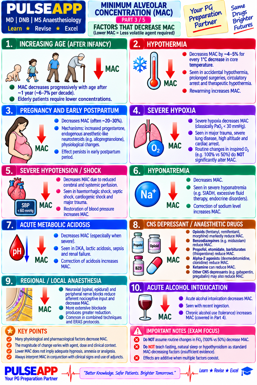
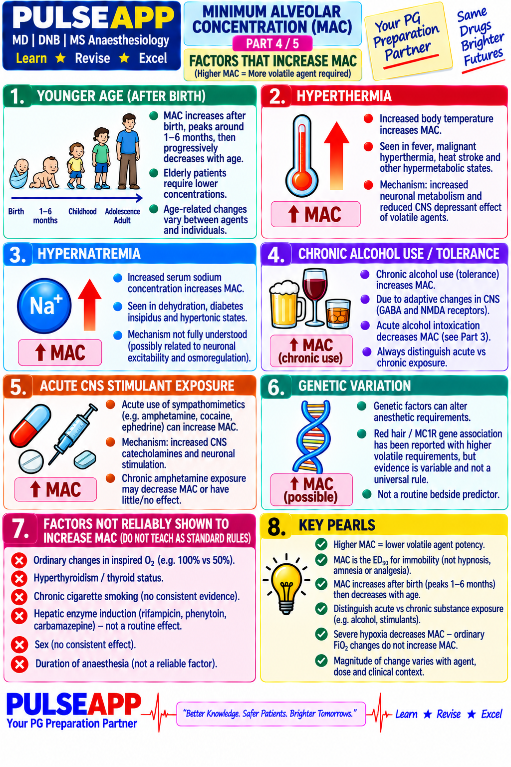
Age~6% per decade above 40
Hypothermia~5% per 1°C reduction
Nitrous oxide65% N₂O contributes ~0.62 MAC-equivalents (additive)
Opioids/alpha-2 agonists/benzodiazepinesRemifentanil can reduce MAC ~50%; dexmedetomidine 35–50%
Pregnancy~25–40% lower (progesterone effect)
Hyperthyroidism/fever~5% per °C
Chronic alcohol useCross-tolerance

Not significantly affected: sex, height, duration of anaesthesia, PaCO₂ (20–90mmHg), PaO₂>40mmHg, mild acid-base changes.

D. MAC Variants3 marks
VariantDefinitionApprox ValueClinical Use
MAC-awakeAlveolar conc. at which 50% respond to verbal command~0.3–0.4 MACLower limit for safe anaesthesia; correlates to BIS 60
MAC-intubationPrevents laryngeal/respiratory reflex response to intubation~1.3 MACExplains why induction alone insufficient without NMB/opioids
MAC-BARBlocks adrenergic (autonomic) response to incision in 50%~1.4–1.7 MACConcentration for haemodynamic stability without opioids
🎤 Viva Corner
Q. A 75-year-old on remifentanil 4ng/mL TCI and N₂O 65% requires sevoflurane. Target concentration and calculation?
Age adjustment: 2.0% × (1−0.21) = 1.58% (35yr above ref, 6%/decade). N₂O contributes 0.62 MAC-equiv: 1.58% × (1−0.62) = 0.60%. Remifentanil reduces MAC ~50%: 0.60% × 0.5 = ~0.3% sevoflurane target. This is close to MAC-awake, so BIS monitoring (target 40–60) must guide final titration rather than relying solely on calculation.
★ Examiner's Pearl
MAC values for sevoflurane (2.0%), desflurane (6.0%), isoflurane (1.15%), N₂O (105%) are tested numerically. Factor magnitudes (age 6%/decade, hypothermia 5%/°C, pregnancy 25–40%) need direction AND magnitude. MAC-awake/MAC-BAR definitions with clinical use are most tested.
Eger EI et al (Anesthesiology 1965). Merkel G, Eger EI (Anesthesiology 1963). Aranake A et al (Anaesthesia 2013). Miller's Anaesthesia 9th Ed, Ch 26.
lock Protected Clinical Document • Pulse App • Screenshots Strictly Prohibited
QUESTION 17 person Asked by .
bookmark_add

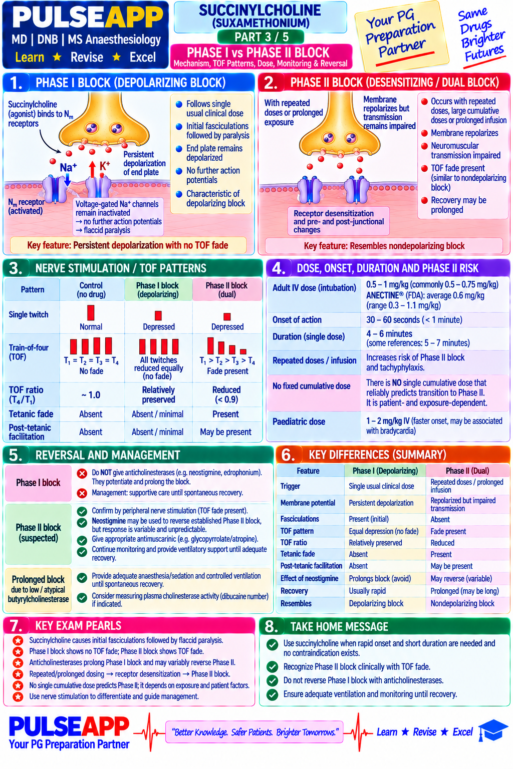
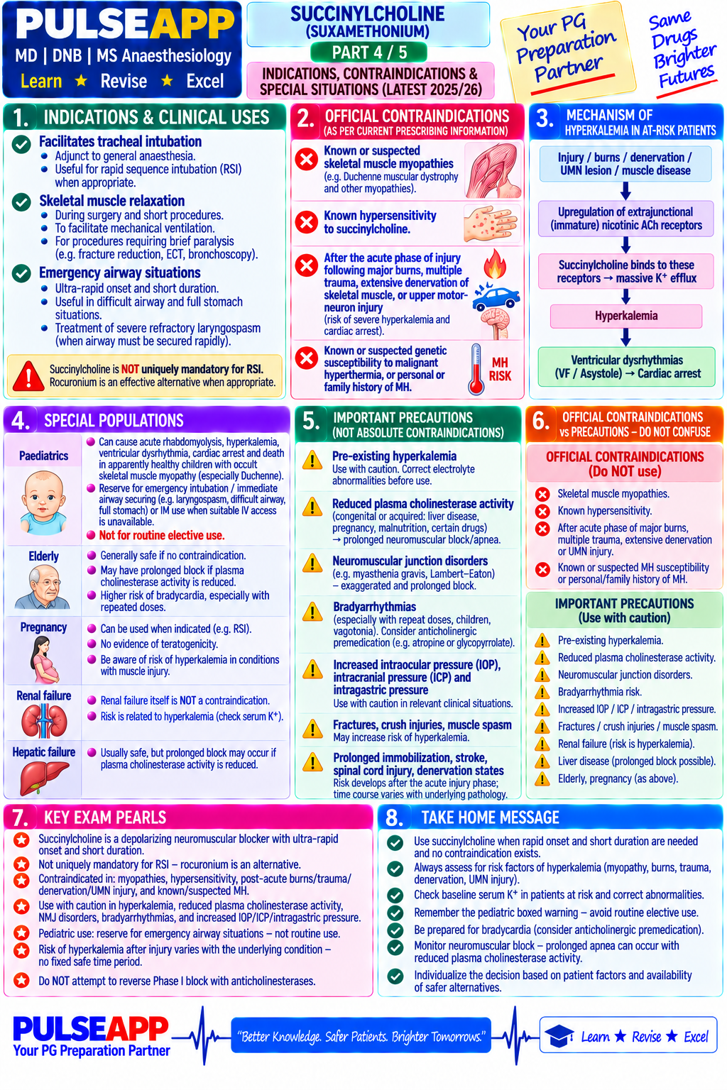
Describe the chemical structure and mechanism of action of succinylcholine. Distinguish Phase I (depolarising) from Phase II (dual block). State its unique indications. Comprehensively list its contraindications and complications with mechanisms.
collections Question Diagrams & Reference Images (1)
Question Reference Diagram
zoom_in View Image
description Clinical Response
⚙ Core Concept
Succinylcholine is the only depolarising NMB in clinical use — its unmatched onset (60–90s) and short duration (~10min) make it uniquely suitable for RSI, but it carries an extensive list of potentially fatal contraindications, meaning it should be reserved for specific indications.
A. Structure and Mechanism2 marks

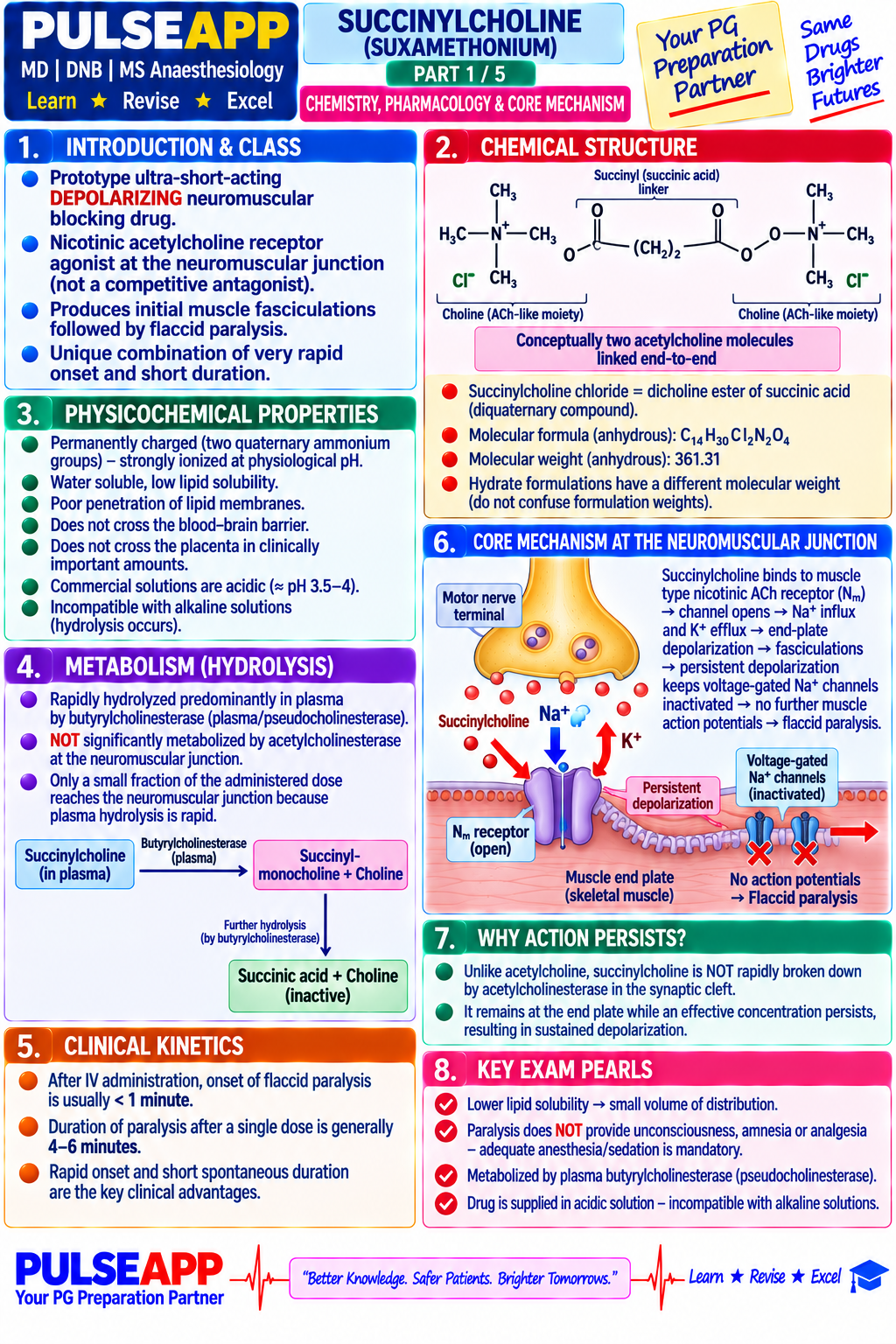
Two ACh molecules joined via acetyl groups (bisquaternary ammonium).
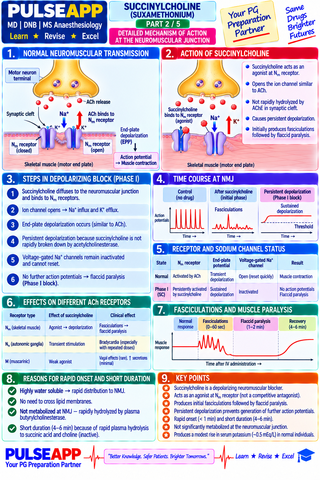
Phase I block: binds and ACTIVATES nicotinic AChR (fasciculations) → NOT hydrolysed by AChE (requires plasma pseudocholinesterase) → sustained depolarisation → Na⁺ channel inactivation → flaccid paralysis. NO fade on TOF; augmented (not antagonised) by anticholinesterases.
Hydrolysis by plasma pseudocholinesterase → succinylmonocholine → choline+succinic acid; duration 10–15 min; onset 60–90s at 1–1.5mg/kg.

B. Phase II (Dual) Block1 mark

With repeated/prolonged dosing (>3–5mg/kg), block develops non-depolarising characteristics: FADE on TOF, post-tetanic potentiation, PARTIALLY reversible by neostigmine — from receptor desensitisation.

C. Indications1 mark
  • RSI — fastest onset of all NMBs with short duration; rocuronium+sugammadex now an equivalent alternative
  • Laryngospasm treatment: 0.5–1mg/kg IV (or 3–4mg/kg IM)
  • Brief procedures (ECT) where rapid offset is advantageous
D. Contraindications and Complications6 marks
ComplicationMechanismContext
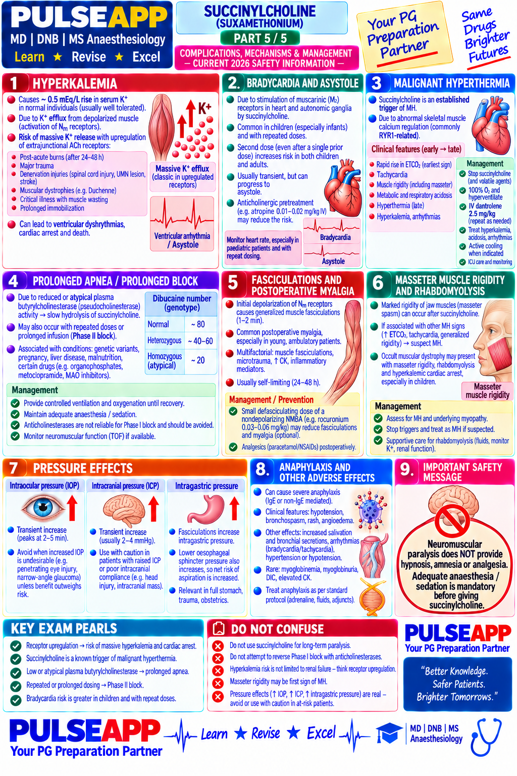
Hyperkalaemia → cardiac arrest (most important)Extrajunctional AChR upregulation → K⁺ rise 5–10mEq/LDenervation >48h, burns >10% BSA, immobility, myopathies
Malignant hyperthermia triggeringPotent MH trigger, especially combined with volatileKnown/suspected MH susceptibility, family history
Pseudocholinesterase deficiencyNot hydrolysed → paralysis hours–daysAtypical PChE (DN<30), liver disease, pregnancy
Masseter muscle rigidityMay herald MH or be isolated responseCancel surgery, monitor for MH signs
↑Intraocular pressureFasciculations contract extraocular musclesRelative contraindication in open globe injury
↑Intracranial pressureTransient, modest riseRelative contraindication in raised ICP (debated)
MyotoniaSustained contraction instead of relaxation → jaw lockAbsolute contraindication in myotonic conditions
Bradycardia/asystoleMuscarinic (M2) SA node stimulationAlways give atropine pretreatment in paediatric RSI
🎤 Viva Corner
Q. List five absolute contraindications with one-line mechanisms.
1) MH history/susceptibility — RyR1-mediated Ca²⁺ release crisis. 2) Denervation >48h (paraplegia) — extrajunctional AChR upregulation → K⁺ efflux 5–10mEq/L. 3) Burns >10% BSA (48h–2yr) — same mechanism. 4) Myotonic conditions — sustained contraction, jaw lock. 5) Homozygous pseudocholinesterase deficiency (DN<30) — paralysis for hours–days.
★ Examiner's Pearl
Hyperkalaemia mechanism (extrajunctional AChR upregulation → K⁺ rise 5–10mEq/L → cardiac arrest) is the single most important safety fact. Phase I vs Phase II distinction (fade, anticholinesterase response) is the most tested pharmacological distinction. Dibucaine number values (80/60/20) are specific tested numbers.
Naguib M et al (Anaesth Intensive Care 2001). Gronert GA (Anesthesiology 2001). Rosenberg H et al (Orphanet J Rare Dis 2007). Miller's Anaesthesia 9th Ed, Ch 34.
lock Protected Clinical Document • Pulse App • Screenshots Strictly Prohibited
QUESTION 18 person Asked by .
bookmark_add

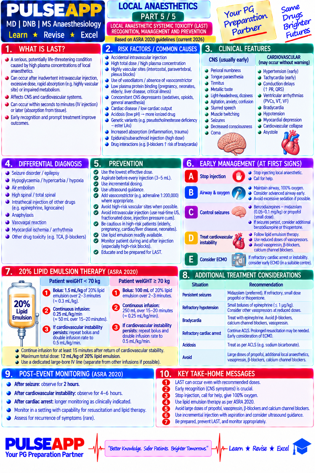
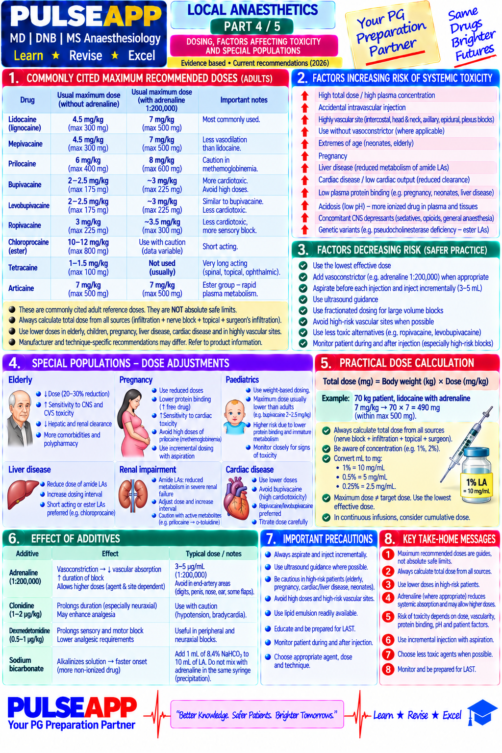
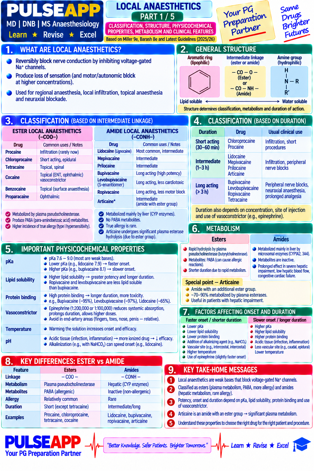
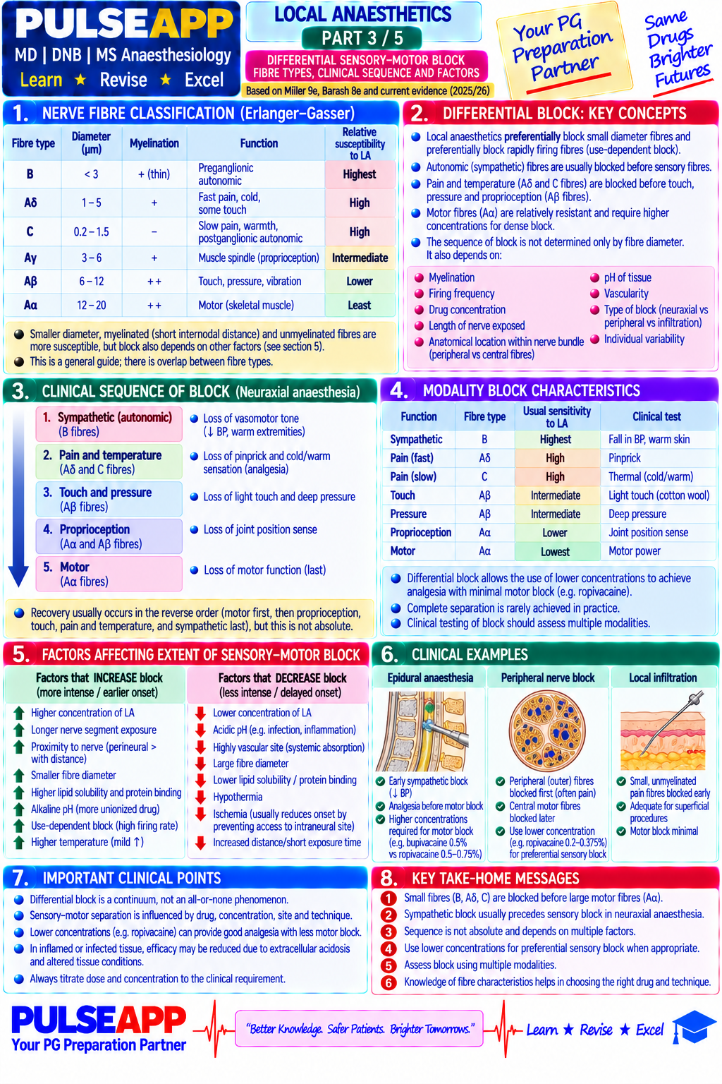
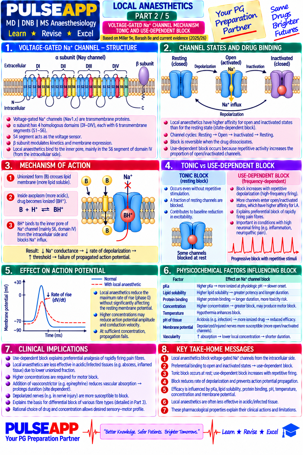
Classify local anaesthetics. Describe the voltage-gated Na&#8314; channel mechanism and tonic/use-dependent block. Explain differential sensory-motor block with fibre types. State maximum safe doses. Outline LAST recognition and lipid emulsion resuscitation.

collections Question Diagrams & Reference Images (2)
Question Reference Diagram
zoom_in View Image
Question Reference Diagram
zoom_in View Image
description Clinical Response
⚙ Core Concept
Local anaesthetics share a single mechanism — reversible blockade of voltage-gated Na⁺ channels — producing a range from 30-minute cocaine topical to 72-hour liposomal bupivacaine. Onset, duration, and cardiotoxicity all trace to pKa, protein binding, and lipid solubility. LAST from inadvertent intravascular injection is a dangerous complication; 20% lipid emulsion is the specific antidote.
A. Classification — Ester vs Amide2 marks
ClassLinkageMetabolismExamples
Esters–COO–Plasma pseudocholinesterase, t½ minutesCocaine, tetracaine, benzocaine, chloroprocaine, procaine
Amides–NH–CO–Hepatic CYP450, t½ 1–3hLidocaine, bupivacaine, ropivacaine, levobupivacaine, prilocaine
B. Ion Channel Mechanism2 marks

Unionised LA crosses axonal membrane, re-ionises intracellularly, and binds the Na⁺ channel from the cytoplasmic face (domain IV-S6) → blocks Na⁺ influx → conduction fails.

Use-dependent block: the receptor site is accessible only when channels are OPEN/INACTIVATED. High-frequency-firing C fibres (pain) accumulate more block at a given concentration — the basis of differential block.

C. Structure-Activity Relationships2 marks
PropertyEffectCorrelate
pKaLower pKa → more unionised → faster onsetLidocaine pKa7.9 faster than bupivacaine pKa8.1
Protein bindingHigher → longer durationBupivacaine 95% bound → 6–8h; lidocaine 65% → 1–2h
Lipid solubilityHigher → greater potencyBupivacaine ~4× more potent (and toxic) than lidocaine
D. Differential Block2 marks

Order of block: autonomic B fibres → C fibres (pain/temp) → A-delta → A-beta (touch) → A-alpha (motor). C fibres block first (small, high-frequency firing); motor blocked only at higher concentrations.

Walking epidural application: 0.0625–0.1% bupivacaine gives sensory block while preserving motor function.

E. Maximum Safe Doses & LAST Management2 marks
AgentMax dose (plain)Max dose (+adrenaline)
Lidocaine3–4 mg/kg7 mg/kg
Bupivacaine2–2.5 mg/kg3 mg/kg
Ropivacaine3 mg/kg
Prilocaine5–6 mg/kg8 mg/kg
⚠ LAST — ASRA 2023 Management
CNS: circumoral tingling, tinnitus → seizures → coma. CVS (later, worse with bupivacaine): widened QRS, VT/VF. Management: STOP injection, 100% O₂, benzodiazepines for seizures (avoid propofol if CVS compromise); 20% lipid emulsion 1.5mL/kg bolus then 0.25mL/kg/min ×30–60min (max 12mL/kg); reduced-dose adrenaline (≤1mcg/kg) if arrest; ECMO if refractory.
🎤 Viva Corner
Q. Why does bupivacaine have dramatically greater cardiotoxicity than lidocaine?
'Fast in, slow out' — bupivacaine binds cardiac Na⁺ channels rapidly during systole but dissociates extremely slowly during diastole, accumulating with each heartbeat (use-dependent trapping), causing progressive conduction slowing and refractory VF. Lidocaine dissociates rapidly during diastole ('fast in, fast out'), preventing cumulative block. This kinetic difference is the molecular basis for bupivacaine's poor-prognosis cardiac arrest, requiring lipid emulsion.
★ Examiner's Pearl
Maximum dose table with specific numbers is the most tested table. LAST lipid emulsion dose (1.5mL/kg bolus then 0.25mL/kg/min, 20% concentration) must be stated exactly. The 'fast in, slow out' bupivacaine cardiotoxicity kinetics vs lidocaine is the key mechanistic comparison.
Neal JM et al, ASRA LAST Advisory 2023. Butterworth JF, Strichartz GR (Anesthesiology 1990). Weinberg GL (Anesthesiology 2012). Miller's Anaesthesia 9th Ed, Ch 30.
lock Protected Clinical Document • Pulse App • Screenshots Strictly Prohibited
QUESTION 19 person Asked by .
bookmark_add

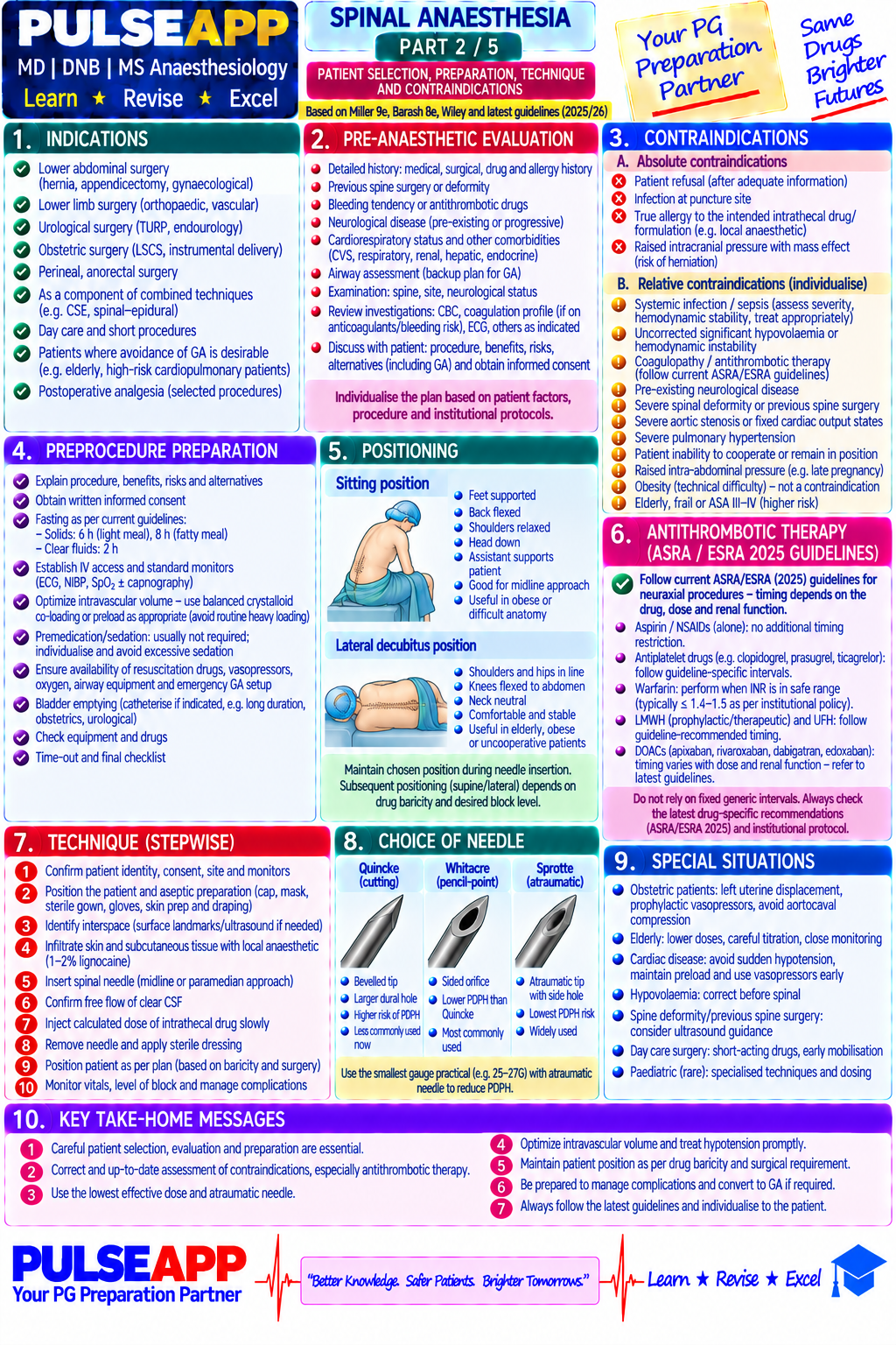
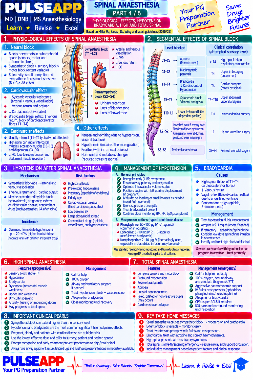
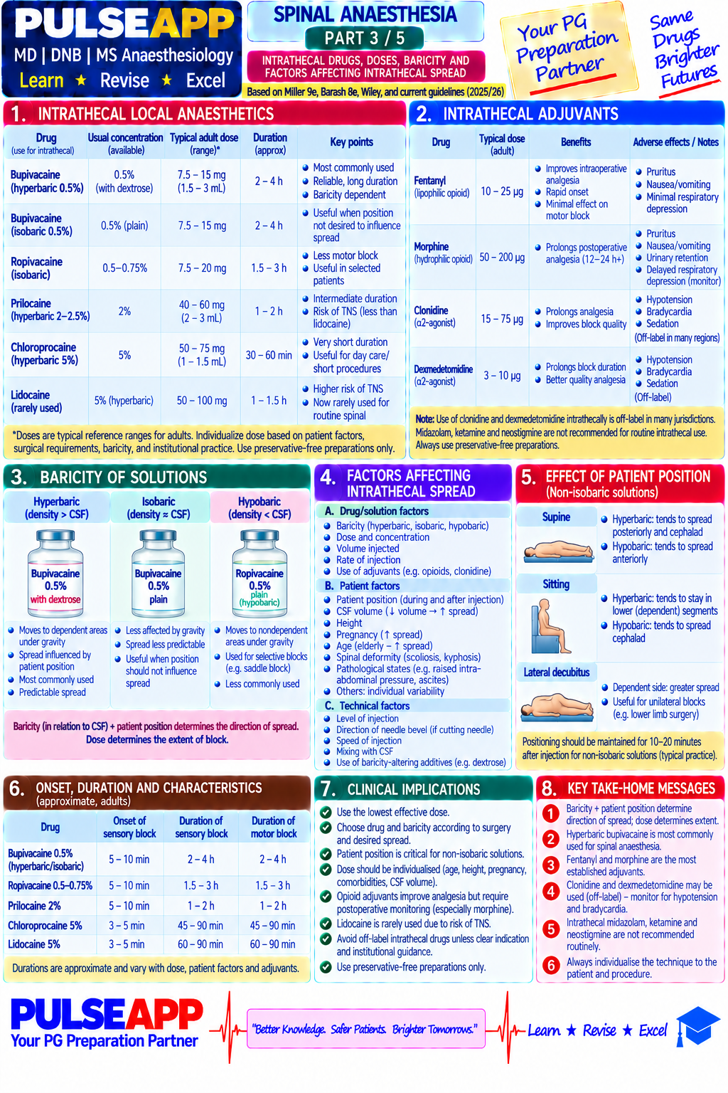
Describe the relevant anatomy for spinal anaesthesia. Outline patient selection, technique, choice of drugs and doses, factors affecting intrathecal spread, and the management of complications including total spinal, post-dural puncture headache, and hypotension.

collections Question Diagrams & Reference Images (1)
Question Reference Diagram
zoom_in View Image
description Clinical Response
⚙ Core Concept
Spinal anaesthesia is the most commonly performed regional technique globally — rapid onset, dense block, low drug dose, and extensive evidence base make it preferred for lower abdominal/pelvic/perineal/lower limb surgery. Complications are predictable and manageable with proper technique.
A. Anatomy2 marks

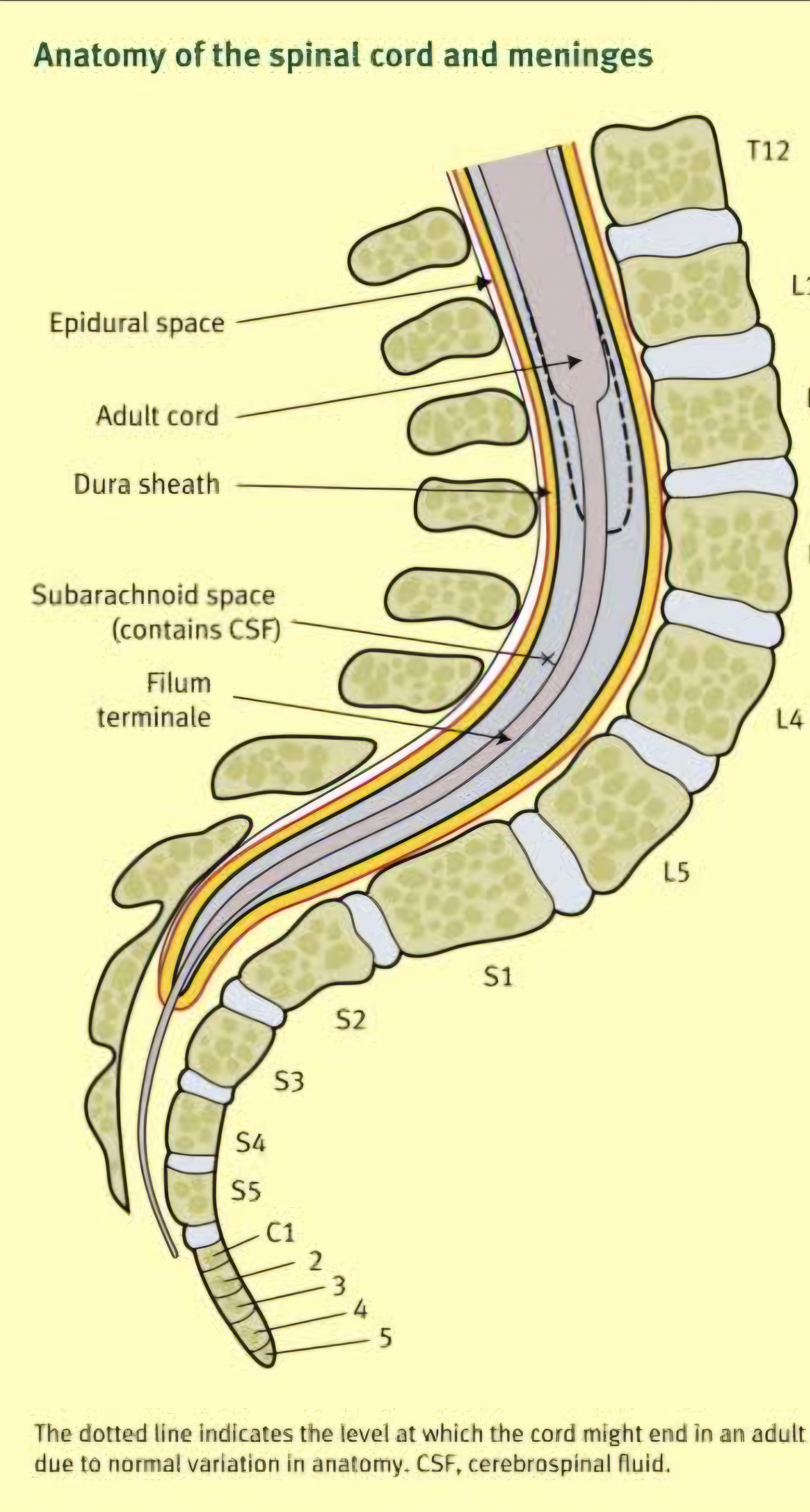
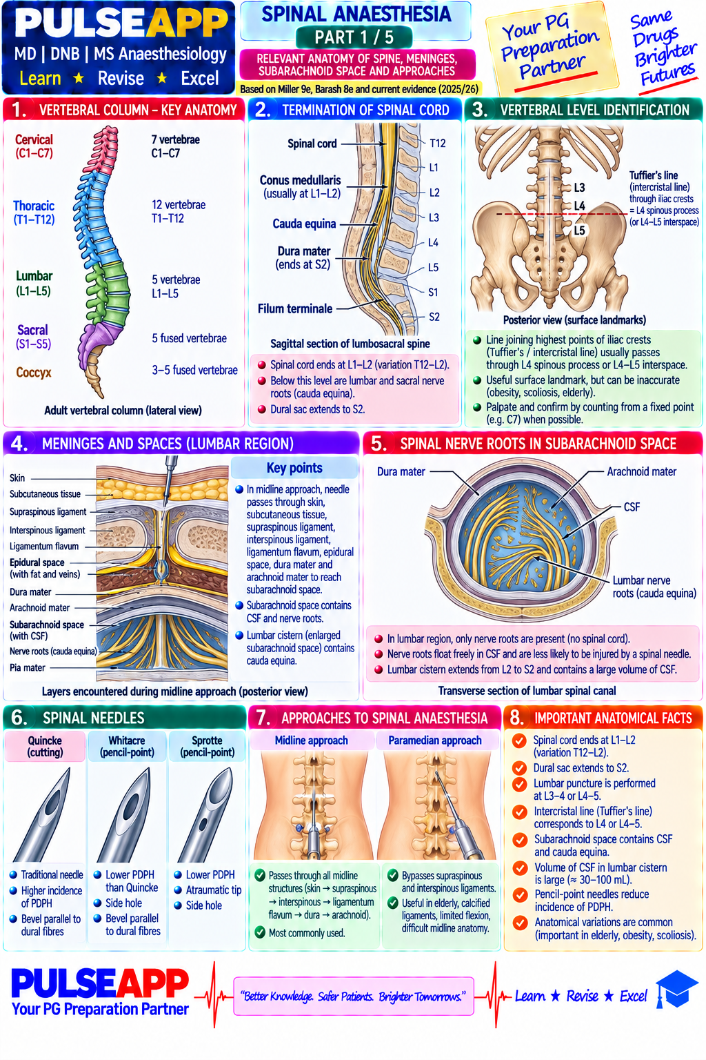
Layers penetrated: skin → subcutaneous fat → supraspinous ligament → interspinous ligament → ligamentum flavum (LOR) → epidural space → dura mater → arachnoid mater (true puncture) → subarachnoid space (CSF).

Safe insertion: L3–L4 or L4–L5 (conus ends at L1 in adults, L3 in neonates). CSF volume ~35–60mL, lower in elderly/obese/pregnant → wider spread per dose.

B. Technique2 marks

Position: sitting or lateral decubitus, full lumbar flexion. Needle: 25–26G pencil-point (Whitacre/Sprotte) — lower PDPH (1–2%) vs cutting bevel (10–15%).

Identification: free-flowing clear CSF confirms subarachnoid position; rotate 90° if no CSF; withdraw if blood-stained or paraesthesia.

C. Drugs and Doses2 marks
DrugBaricityDoseDuration
Heavy bupivacaine 0.5%Hyperbaric — gold standard2–4mL (most); 1.5–2mL for CS2–4h motor, 3–5h sensory
Isobaric bupivacaine 0.5%Position-independent2–3mL lower limb/pelvicSimilar
Intrathecal fentanylAdjuvant10–25mcgEnhances quality, no resp depression
Intrathecal morphineAdjuvant100–300mcg (preservative-free only)12–24h postop analgesia; delayed resp depression risk 6–24h
D. Determinants of Spread2 marks

Baricity is most important: hyperbaric sinks to dependent areas; isobaric is position-independent. Position during/after injection determines settling in hyperbaric solutions. Dose/volume matters most for isobaric; age/height affect spread per dose.

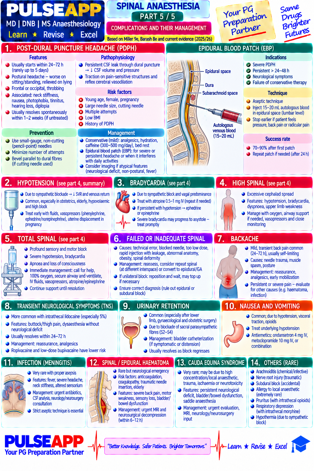
E. Complications2 marks
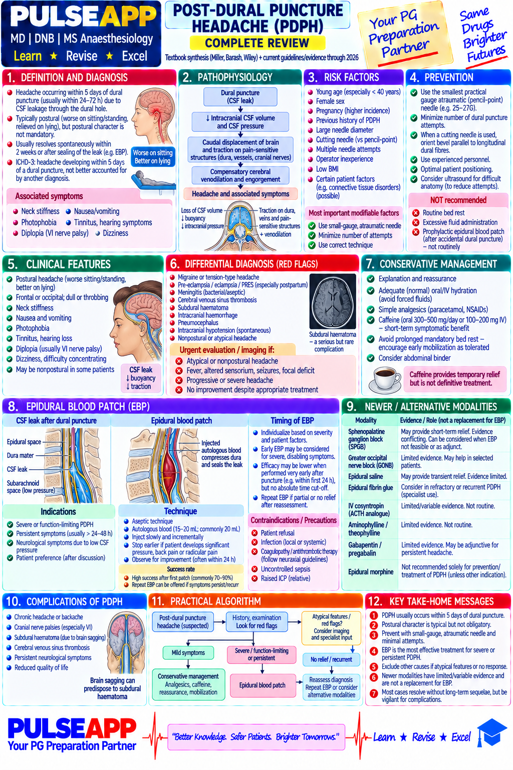
ComplicationMechanismManagement
Hypotension (up to 30%)Sympathetic block, vasodilationFluid co-load, phenylephrine/ephedrine, atropine if bradycardic
High/total spinalBlock ascends to C3–C5, phrenic paralysis100% O₂, secure airway, vasopressors, CPR if arrest
PDPHCSF leak, low pressure tractionConservative; epidural blood patch 15–20mL if persists >24h
TNSBilateral buttock/leg pain 6–24h, no deficitNSAIDs; avoid hyperbaric 5% lidocaine
Urinary retentionSacral parasympathetic blockCatheterise until resolved
🎤 Viva Corner
Q. During spinal for CS, the patient becomes unconscious and apnoeic 3 minutes after injection. What has happened and what do you do?
Total spinal — cephalad spread to C3–C5 blocking the phrenic nerve, plus cardiac sympathetic/cervical sympathetic block causing bradycardia/hypotension and cerebral hypoperfusion. Obstetric emergency: call for help, 100% O₂, RSI (thiopentone+suxamethonium) and intubate/ventilate, ephedrine+phenylephrine, atropine if bradycardic, left lateral tilt, IV fluid bolus; if arrest — CPR with perimortem CS within 5 min if no ROSC. Full recovery expected once block recedes (3–5h) if oxygenation/haemodynamics maintained.
★ Examiner's Pearl
State all layers penetrated in order — specifically tested. Baricity concept with clinical application (hyperbaric for CS in supine → T4 block) is most tested. Intrathecal morphine dose with delayed respiratory depression monitoring requirement is a specific tested protocol.
Cousins MJ, Bridenbaugh PO, Neural Blockade, 4th Ed. Hadzic A, Textbook of Regional Anesthesia, 2nd Ed. Dyer RA et al (Anesthesiology 2008). Sng BL et al (Cochrane 2018).
lock Protected Clinical Document • Pulse App • Screenshots Strictly Prohibited
QUESTION 20 person Asked by .
bookmark_add

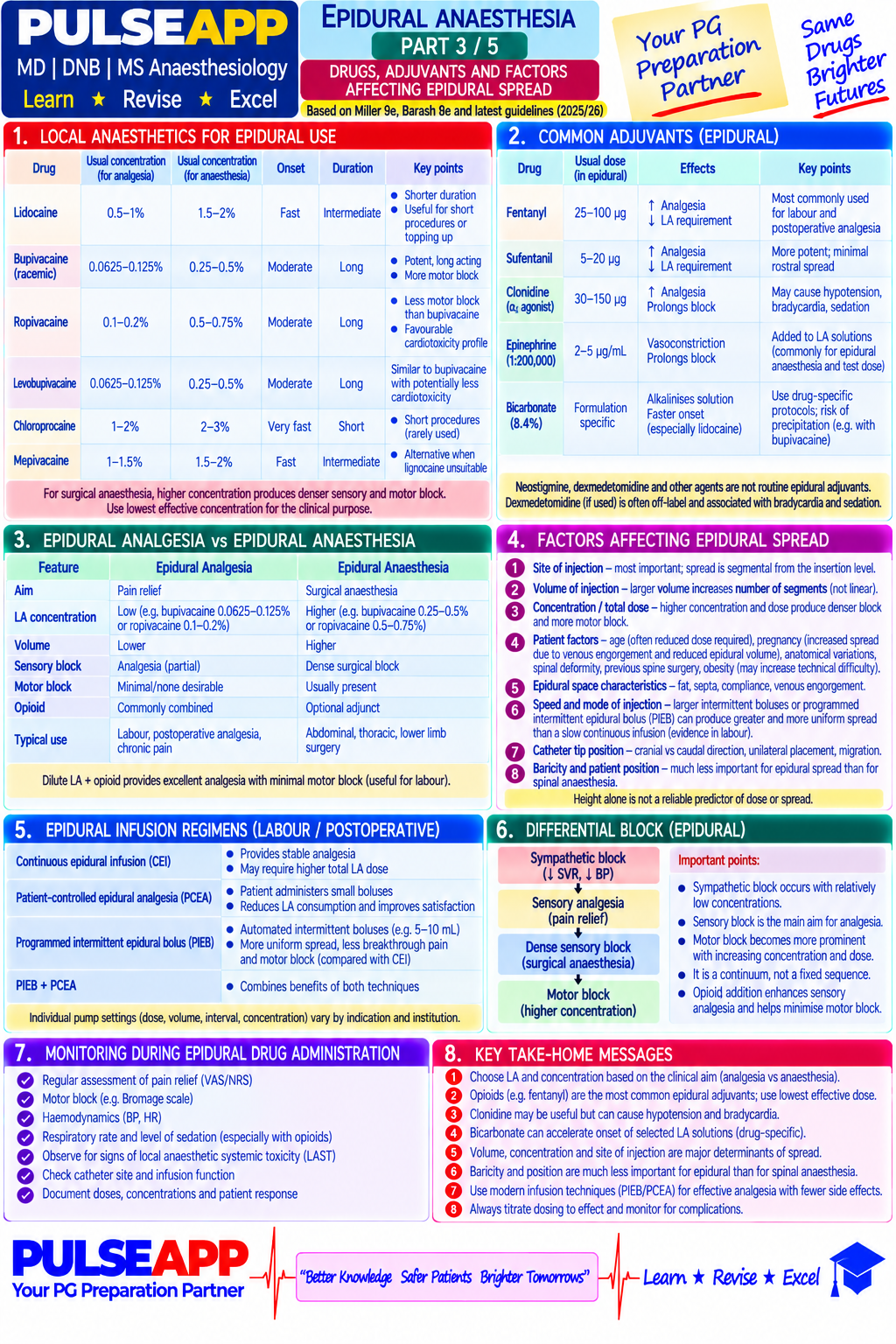
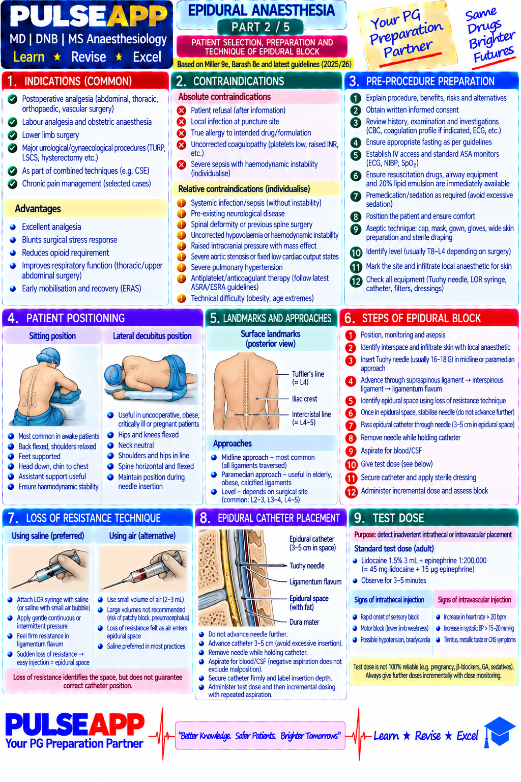
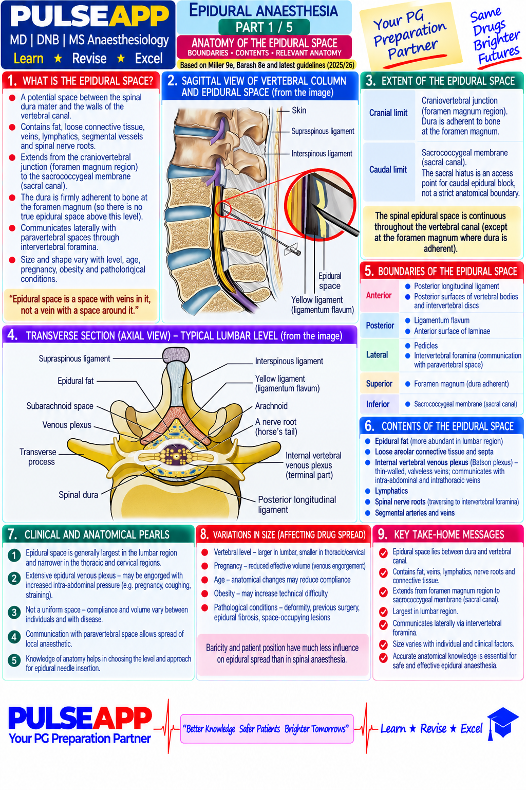
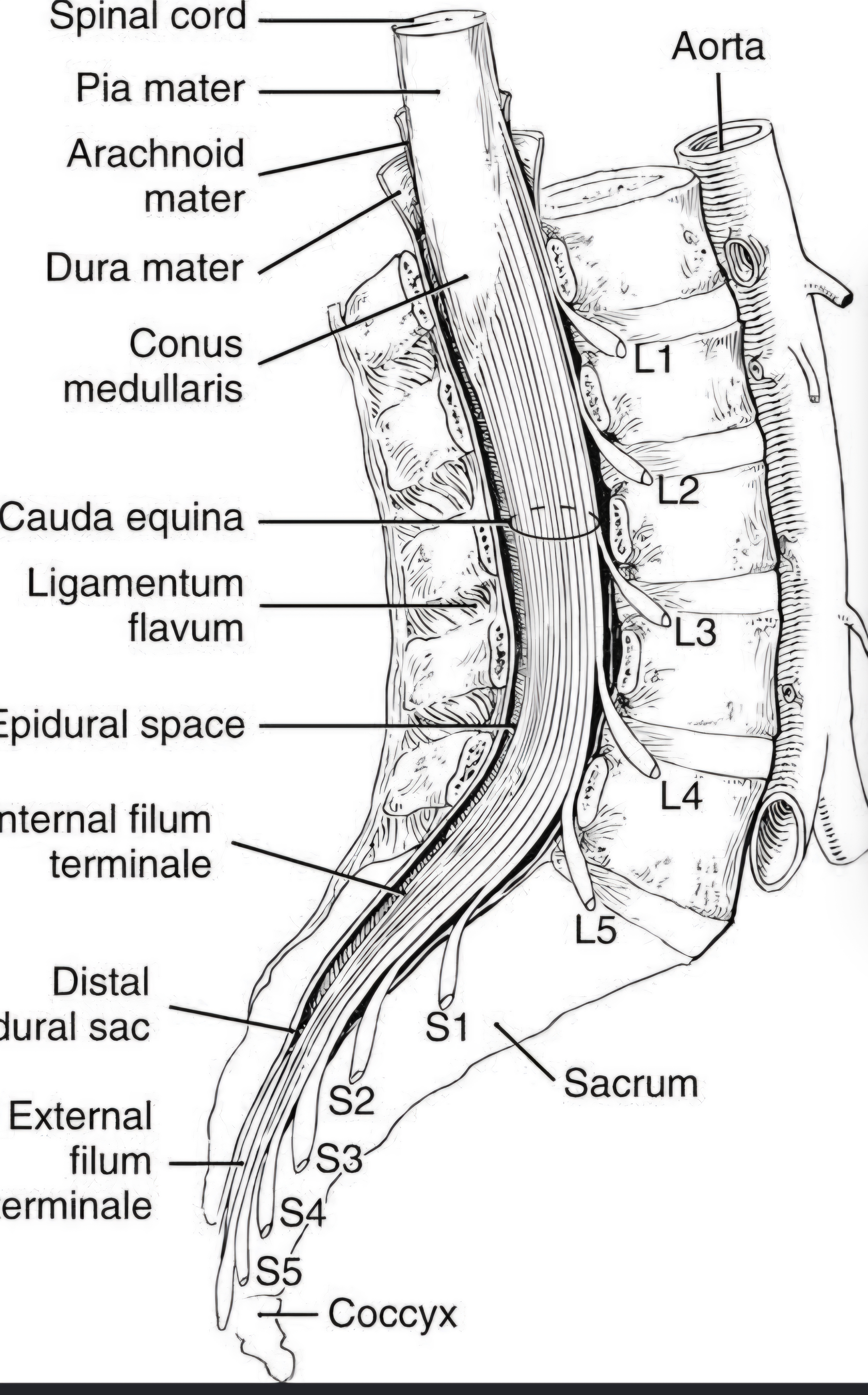
Describe the boundaries and contents of the epidural space. Outline the technique of epidural block including loss of resistance, test dose, and catheter placement. Discuss factors affecting LA spread, drug choices for epidural analgesia vs anaesthesia, and management of complications.

collections Question Diagrams & Reference Images (1)
Question Reference Diagram
zoom_in View Image
description Clinical Response
⚙ Core Concept
The epidural space is a potential space filled with fat, blood vessels, and nerve roots, not empty air. Its anatomy explains why volume (not concentration) primarily determines spread, and why elderly/pregnant patients need less drug. Epidural catheter analgesia is the gold standard for major abdominal/thoracic postop pain.
A. Epidural Space — Boundaries & Contents2 marks
BoundaryStructure
SuperiorFusion of dura/periosteum at foramen magnum
InferiorSacrococcygeal membrane
AnteriorPosterior longitudinal ligament
PosteriorLigamentum flavum (characteristic loss of resistance)
LateralPedicles/intervertebral foramina

Contents: epidural fat (LA depot), Batson's venous plexus (engorged in pregnancy → reduced volume), spinal nerve roots. LF thickest at L3–L4 (5–6mm).

B. Technique2 marks

Loss of Resistance (LOR): 16–18G Tuohy needle through supraspinous→interspinous→ligamentum flavum with continuous pressure on saline-filled syringe; sudden easy injection confirms entry.

Catheter threaded 3–5cm into space. Test dose: 3mL 2% lidocaine + 1:200,000 adrenaline — IV catheter → tachycardia ≥20bpm within 60s; intrathecal catheter → dense bilateral motor block within 3–5min.

C. Factors Affecting Spread2 marks
FactorEffect
VolumeMost important — ~1–1.5mL per spinal segment
ConcentrationDetermines intensity (motor vs sensory), NOT spread
AgeElderly: greater spread per volume (less epidural fat)
PregnancyReduce dose 25–30% (engorged Batson's plexus)
D. Drug Choices2 marks
Clinical GoalDrug/Concentration
Labour (walking epidural)Bupivacaine 0.0625–0.1% + fentanyl 2mcg/mL
Postop analgesia (major abdo)Bupivacaine 0.125% or ropivacaine 0.2% + fentanyl
Surgical anaesthesia (CS top-up)2% lidocaine + 1:200,000 adrenaline, incremental to T4
Thoracic epidural (thoracotomy)Ropivacaine 0.2% + fentanyl/sufentanil, inserted T4–T8
E. Complications2 marks

Accidental dural puncture (1–2%): re-site adjacent level, or thread intrathecal catheter for continuous spinal, or blood patch. Epidural haematoma (<1:150,000): urgent MRI, decompression within 8h. Epidural abscess: MRI, IV antibiotics ± drainage. High/total epidural: manage as total spinal.

🎤 Viva Corner
Q. After the test dose, heart rate suddenly rises 75→115bpm. What has happened and what do you do?
Positive intravascular test dose — catheter tip is in an epidural vein; the adrenaline component caused β1 stimulation. Do NOT inject further; withdraw catheter, wait for tachycardia to resolve, re-site at same/adjacent level, aspirate before re-testing. The test dose protocol exists specifically to detect intravascular placement before a full therapeutic dose (which would cause LAST) is given.
★ Examiner's Pearl
All five epidural space boundaries with structures must be reproduced exactly. Test dose content and positive responses (tachycardia ≥20bpm=IV; dense motor block=intrathecal) with timing are most tested. Epidural haematoma decompression within 8h is a specific tested emergency threshold.
Miller's Anaesthesia 9th Ed, Ch 56. Cousins MJ, Bridenbaugh PO, Neural Blockade, 4th Ed. Hogan QH (Reg Anesth 1996). ASRA Anticoagulation Guidelines 2018.

Showing 1120 of 337 questions

account_tree

Subcategory Tree

Explore Anesthesia subcategories

folder_special Anesthesia
Main
lock

Category Subscription

Subscribe to Anesthesia to unlock this module and all nested subcategories.

  • check_circle Access Anesthesia & all subcategories
  • check_circle Detailed, Peer-Reviewed Answers
  • check_circle High-yield visual aids & imaging
Get Category Subscription arrow_forward
Secure 256-bit SSL Connection

Anatomical Models

Explore high-fidelity 3D visualizations included in premium modules.

Case Reviews

Real-world clinical scenarios narrated by senior consultants.

Pulse App