Anesthesia | Eklavya Medical
vaccines
medical_services Main Specialty Domain lock Subscription Required

Anesthesia

Clinical anesthesiology, local and general anesthesia delivery systems, monitoring, and perioperative care.

verified Verified Medical Faculty menu_book 337 Q&A Modules
lock Protected Clinical Document • Pulse App • Screenshots Strictly Prohibited
QUESTION 21 person Asked by .
bookmark_add

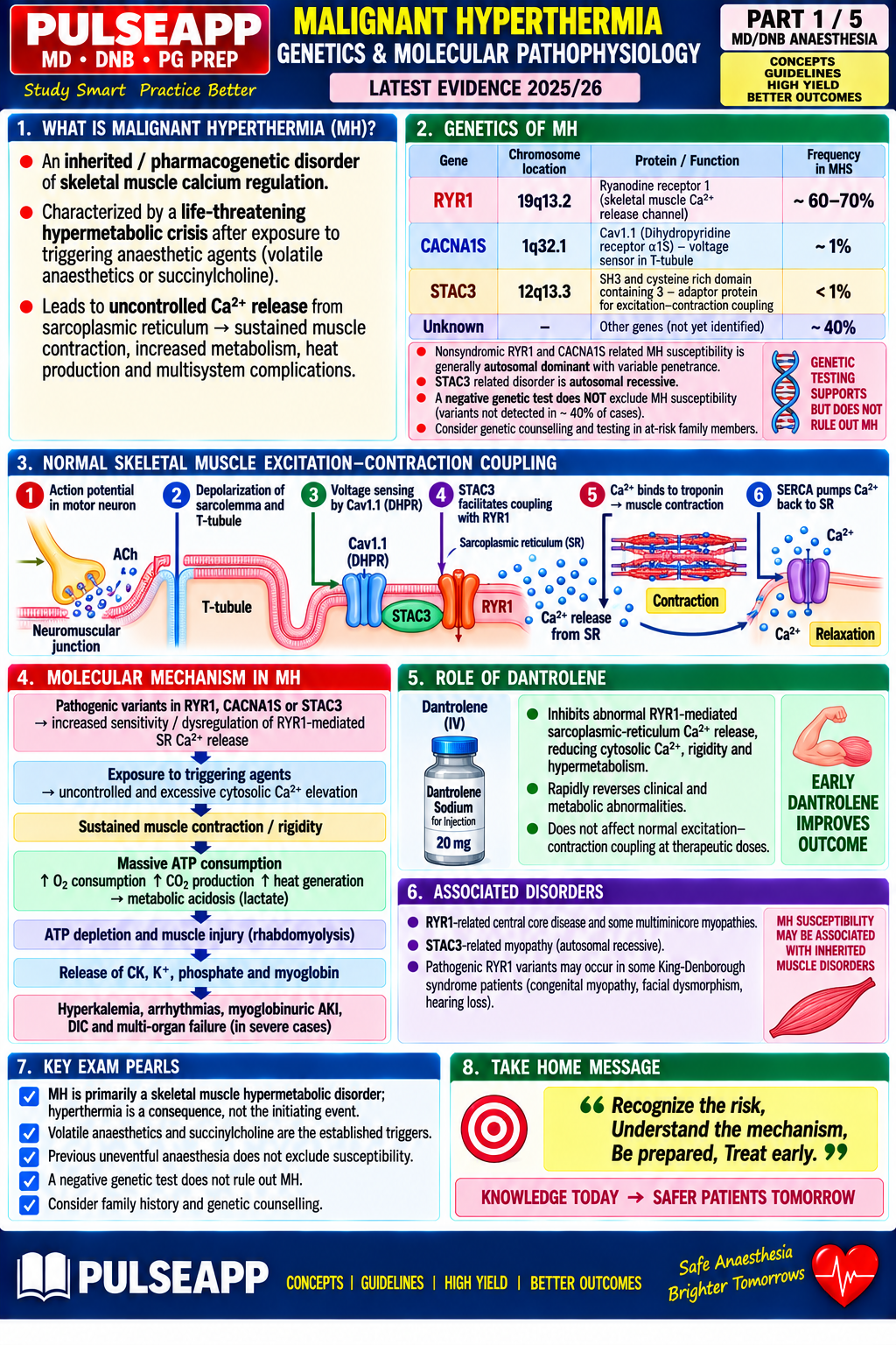
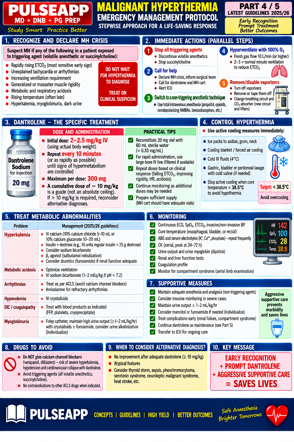
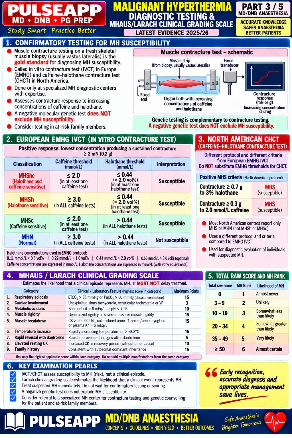
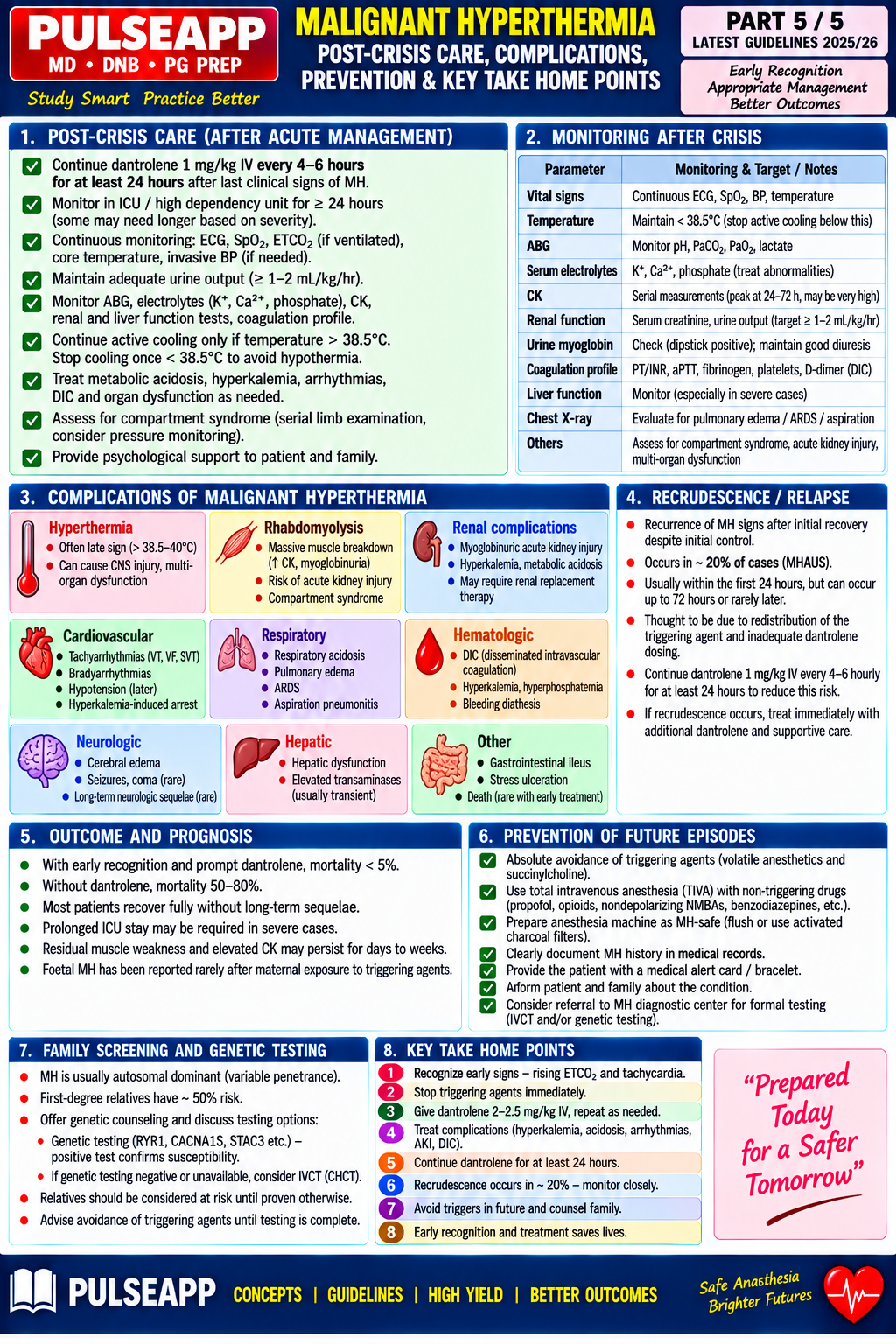
Describe the genetic and molecular pathophysiology of malignant hyperthermia. List triggering agents. Outline the clinical features (CHCT/IVCT grading, MHAUS clinical grading scale). Describe the emergency management protocol including dantrolene dosing and post-crisis care.
collections Question Diagrams & Reference Images (1)
Question Reference Diagram
zoom_in View Image
description Clinical Response
⚙ Core Concept
MH is a pharmacogenetic disorder of skeletal muscle calcium regulation that, when triggered by specific anaesthetic agents, produces an uncontrolled hypermetabolic state. Without dantrolene, cardiac arrest and multi-organ failure are nearly inevitable; with early treatment, survival exceeds 90%.
A. Genetics and Molecular Pathophysiology3 marks

Autosomal dominant; RYR1 mutations (~70%), CACNA1S (~1%). Normal contraction: T-tubule depolarisation → DHPR conformational change → RyR1 opens → controlled SR Ca²⁺ release → contraction → SERCA reuptake.

MH event: mutant RyR1 is abnormally sensitive/slow to close; trigger contact → massive uncontrolled Ca²⁺ release → sustained contraction (rigidity) → ATP consumed at enormous rate → anaerobic glycolysis → lactic acidosis → heat → hyperthermia → rhabdomyolysis → AKI, hyperkalaemia, arrhythmia.

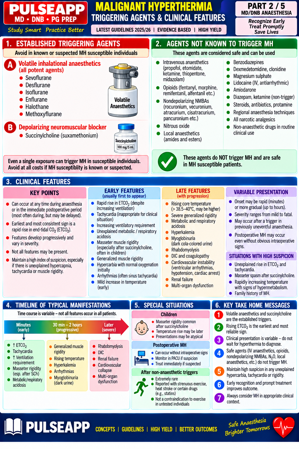
B. Triggering Agents1 mark
⚠ ALL Volatile Agents + Succinylcholine
Every halogenated volatile (no 'safer' one) and succinylcholine (combined trigger fastest/most severe). SAFE: propofol, all IV agents, opioids, non-depolarising NMBs, benzodiazepines, LAs, N₂O — TIVA is the standard for MH-susceptible patients.
C. Clinical Features (MHAUS Grading)2 marks
SignDetailsSpecificity
↑ETCO₂ (EARLIEST sign)Rapid rise despite adequate ventilationMost sensitive early warning
Masseter rigidityJaw stiffness 30–60s after succinylcholinePathognomonic concern, cancel elective surgery
TachycardiaHR 140–180Non-specific but common
Hyperthermia (NOT first sign)May exceed 40°C, rises 1°C/3–5minLate presentation indicates diagnostic delay
Muscle rigidityGeneralised, 'stiff as a board'Highly specific under volatile anaesthesia
Metabolic acidosisMixed, lactate>10mmol/LCombined with ↑ETCO₂ = strong indicator
RhabdomyolysisCK peaks 12–24h laterConfirms diagnosis retrospectively
D. Emergency Management4 marks
⚠ MH Emergency Protocol
1) STOP all triggers immediately. 2) Call for help, activate MH protocol. 3) Hyperventilate 100% O₂ at max FGF. 4) DANTROLENE immediately — do not wait for confirmation. 5) Active cooling. 6) Treat metabolic derangements. 7) Convert to TIVA + non-depolarising NMB if surgery must continue.
DantroleneDetail
MechanismBinds FKBP12 → stabilises RyR1 in CLOSED state → stops uncontrolled Ca²⁺ release; only agent targeting the molecular defect
Initial dose2.5mg/kg IV bolus, repeat q5–10min to max 10mg/kg
Maintenance1mg/kg IV q4–6h for 24–48h — prevents recrudescence (can recur 24–36h later)
Preparation20mg/vial + 60mL sterile water/vial; 70kg pt at 2.5mg/kg needs ~9 vials

Cooling: ice packs axillae/groin/neck, cold IV saline 4°C 15mL/kg, target <38.5°C. Hyperkalaemia: calcium gluconate, insulin+dextrose, bicarbonate. Rhabdomyolysis: fluids, urine alkalinisation to prevent AKI. Arrhythmia: procainamide/lidocaine, avoid CCBs.
🎤 Viva Corner
Q. Twenty minutes into a lap chole under sevoflurane, ETCO₂ rises 35→62mmHg, temp 38.9°C rising, HR 145, patient rigid. Diagnosis and immediate management?
Fulminant Malignant Hyperthermia. STOP sevoflurane immediately; call for help and assign roles; hyperventilate 100% O₂ at 15L/min; convert to TIVA if NMB needed for closure; administer dantrolene 2.5mg/kg IV ASAP, repeat q5min to max 10mg/kg; active cooling with cold IV saline and ice packs; send ABG, CK, U&Es, urinalysis; postop: dantrolene 1mg/kg q4–6h ×24–48h, ICU admission, renal protection, report to MHAUS, arrange CHCT/IVCT and genetic testing.
★ Examiner's Pearl
RYR1 gene mechanism (uncontrolled SR Ca²⁺ release) is the most tested molecular fact. Dantrolene dose (2.5mg/kg initial, max 10mg/kg, then 1mg/kg q4–6h ×24–48h) must be stated fully. ETCO₂ rise as the EARLIEST sign (before hyperthermia) is a commonly missed sequence fact.
Rosenberg H et al (Orphanet J Rare Dis 2007). Hopkins PM (BJA 2000). MHAUS Guidelines 2023. Miller's Anaesthesia 9th Ed, Ch 32.
lock Protected Clinical Document • Pulse App • Screenshots Strictly Prohibited
QUESTION 22 person Asked by .
bookmark_add

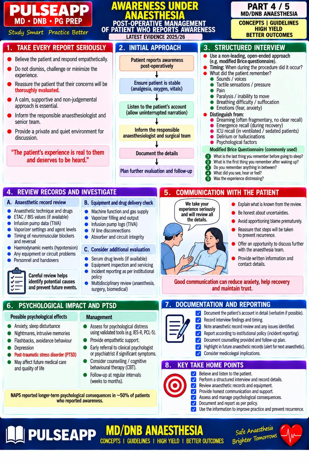
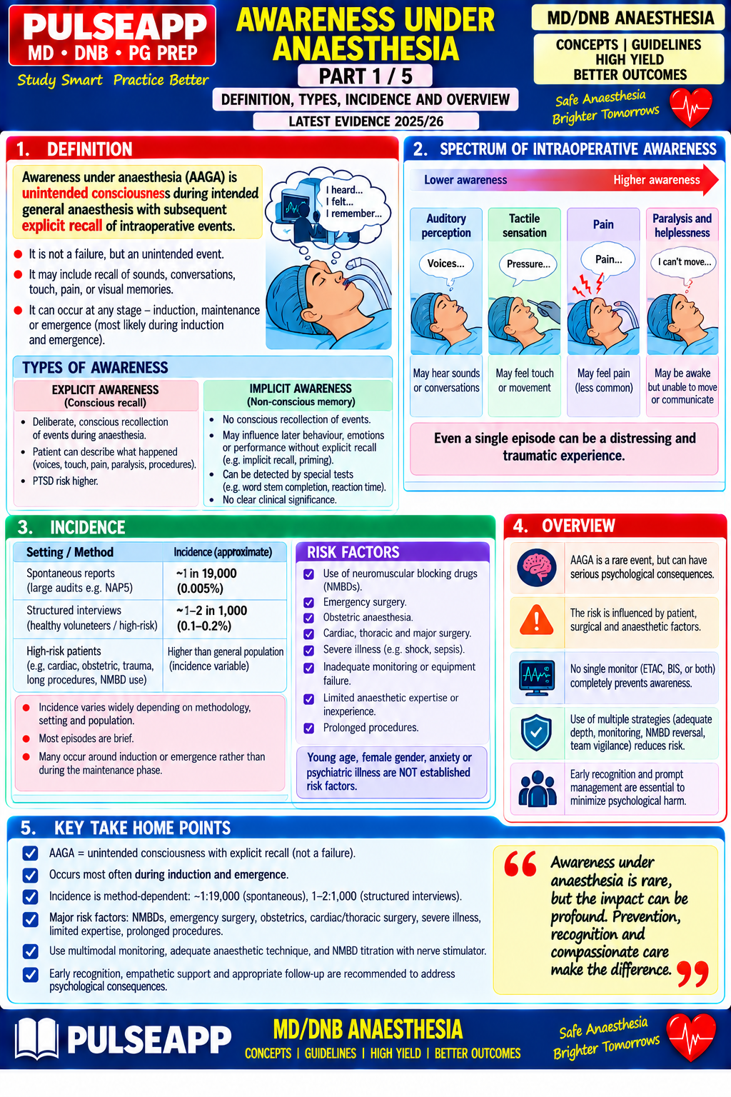
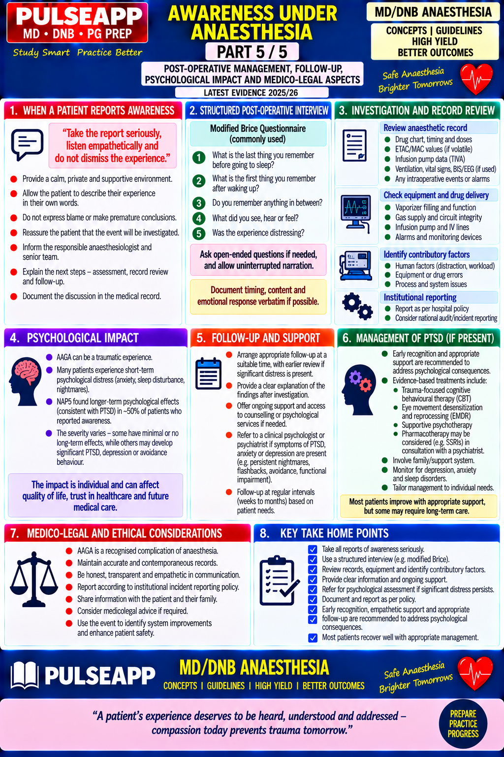
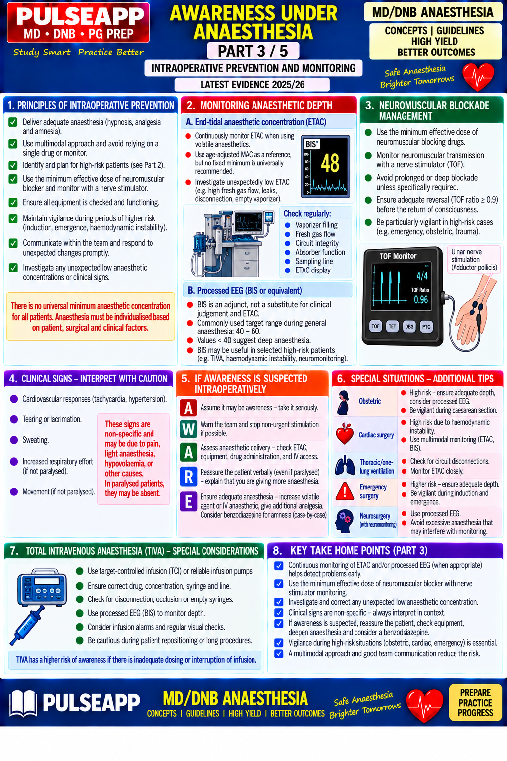
Define awareness under anaesthesia. Classify types (explicit vs implicit). State the incidence and identify high-risk patient groups. Describe preventive strategies including BIS monitoring. Outline the management of the patient who reports awareness post-operatively including PTSD considerations.

collections Question Diagrams & Reference Images (2)
Question Reference Diagram
zoom_in View Image
Question Reference Diagram
zoom_in View Image
description Clinical Response
⚙ Core Concept
Awareness under anaesthesia affects ~1–2 patients per 1000 GAs (0.1–0.2%), causing psychological harm ranging from mild discomfort to PTSD in a significant proportion. It represents a major medicolegal risk and ethical failure of the fundamental duty to ensure unconsciousness.
A. Definition and Classification2 marks
TypeDefinitionIncidence
Explicit awareness with recallConscious during surgery AND recalls events postop~0.1–0.2% (1–2/1000)
Explicit awareness without recallShows signs of consciousness but no postop recallMore common, difficult to quantify
Implicit awarenessSubcortical processing without conscious recallPoorly defined, controversial
Dreaming during anaesthesiaHypnagogic dreams during light anaesthesia/emergence, NOT true awareness~20% of patients, usually benign
B. Incidence and High-Risk Populations2 marks

General incidence: Sandin (Lancet 2000) 0.18%; NAP5 UK (2014) 1 in 19,600 (0.005%) with potential harm.

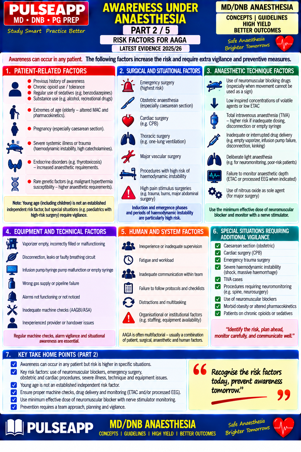
  • Obstetric emergency CS under GA — highest risk, 1 in 250 (0.4%)
  • Cardiac surgery — 1 in 500 (opioid-based technique, bypass dilutes volatile)
  • Trauma RSI — 1 in 500 (haemodynamic instability limits depth)
  • Difficult airway/failed intubation, chronic alcohol/opioid use, equipment failure (vaporizer/pump/disconnection)
C. Prevention Strategies3 marks

Maintain volatile ≥0.7 MAC-equivalent (above MAC-awake 0.3–0.4); benzodiazepine premedication in high-risk groups; TIVA requires reliable propofol delivery with anti-free-flow pumps and pressure-monitoring lines.

✅ BIS Evidence — B-Aware vs B-Unaware
B-Aware (2004): BIS reduced awareness in high-risk patients. B-Unaware/BAG-RECALL (2008/2011): ETAC≥0.7MAC was NON-INFERIOR to BIS for volatile anaesthesia. Conclusion: ETAC is primary monitor for volatiles; BIS is essential specifically for TIVA (no ETAC available) and equipment-failure detection.
D. Post-awareness Management3 marks

If detected intraoperatively: immediately deepen anaesthesia, reassure verbally, give midazolam 2mg IV for amnesia.

Postop: structured interview (Modified Brice Interview) in all high-risk/reporting patients. Transparent disclosure is ethically mandatory. PTSD develops in ~30% of those with awareness — early psychological referral (CBT/EMDR) can prevent full PTSD. Document, report via incident system/national database, investigate the anaesthetic record for the likely cause.

🎤 Viva Corner
Q. Why are paralysed patients who become aware more likely to develop PTSD than non-paralysed aware patients?
A non-paralysed aware patient can move/signal, prompting prompt deepening of anaesthesia. A paralysed aware patient experiences complete helplessness — fully conscious, possibly in pain, but totally unable to signal distress despite maximal effort. This total loss of agency/control while experiencing a terrifying event is precisely the psychological profile most strongly predictive of PTSD, and may also prolong the duration of the awareness episode since the team cannot be alerted.
Q. The B-Unaware trial showed BIS was not superior to ETAC for awareness prevention. Does this mean BIS has no value?
No — for volatile-based anaesthesia, ETAC≥0.7MAC gives equivalent protection and is simpler. BIS retains critical value for: TIVA (no ETAC exists for propofol — BIS is the primary depth monitor), detecting equipment failure (vaporizer/circuit failure shown by rising BIS with falling ETAC), and patients with unusual pharmacological requirements (tolerance, genetic variation).
★ Examiner's Pearl
Incidence (0.1–0.2% overall, 0.4% obstetric emergency CS) with study citations (Sandin 2000, NAP5 2014) shows evidence-based knowledge. B-Aware vs B-Unaware conclusions (BIS=ETAC for volatile; BIS essential for TIVA) is the most tested controversy. PTSD link (30%, worsened by paralysis) distinguishes comprehensive answers.
Sandin RH et al (Lancet 2000). Avidan MS et al (NEJM 2008; Lancet 2011). Myles PS et al (Lancet 2004). Royal College of Anaesthetists NAP5 2014.
lock Protected Clinical Document • Pulse App • Screenshots Strictly Prohibited
QUESTION 23 person Asked by .
bookmark_add

Define PONV and describe its pathophysiology including the neurotransmitter pathways and the chemoreceptor trigger zone (CTZ). Describe the Apfel simplified risk score. Outline a risk-stratified prophylaxis and treatment protocol including the drugs, mechanisms, and doses for each antiemetic class.

collections Question Diagrams & Reference Images (1)
Question Reference Diagram
zoom_in View Image
description Clinical Response
⚙ Core Concept
PONV affects ~20–30% of surgical patients and up to 70–80% of high-risk patients. Despite being preventable through risk-stratified multimodal prophylaxis, it remains underreported and undertreated. The Apfel score targets prophylaxis at high-risk patients using combination antiemetics on different receptor pathways.
A. Pathophysiology — Pathways and CTZ3 marks

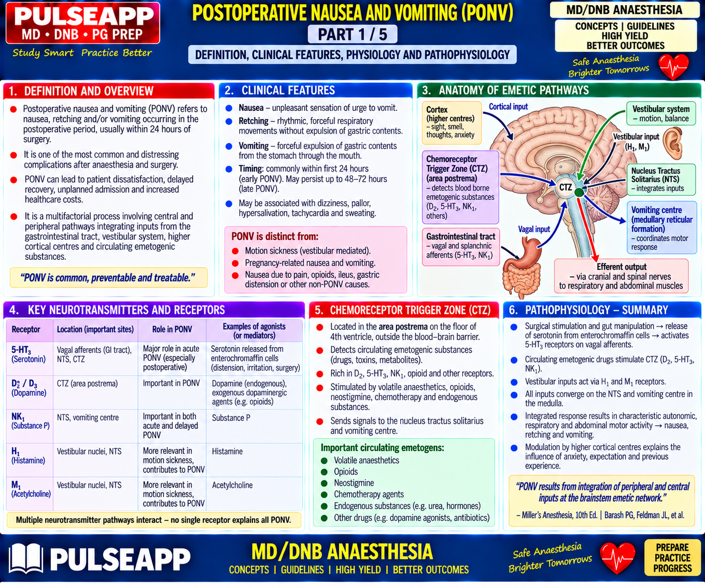
The vomiting centre (medullary reticular formation) receives afferent input from four sources, each with specific neurotransmitters — the targets of antiemetic drugs.

Afferent SourceNeurotransmittersAntiemetic Target
CTZ (area postrema, outside BBB)Dopamine D2, Serotonin 5-HT3, Substance P (NK1)D2 antagonists, 5-HT3 antagonists, NK1 antagonists, steroids
Vestibular systemHistamine H1, Acetylcholine M1H1 antagonists (cyclizine), anticholinergics (scopolamine)
GI tract afferentsSerotonin (from enterochromaffin cells), Substance P5-HT3 antagonists, metoclopramide, NK1 antagonists
Cerebral cortexMultipleAnxiolytics, TIVA (propofol has direct antiemetic action)
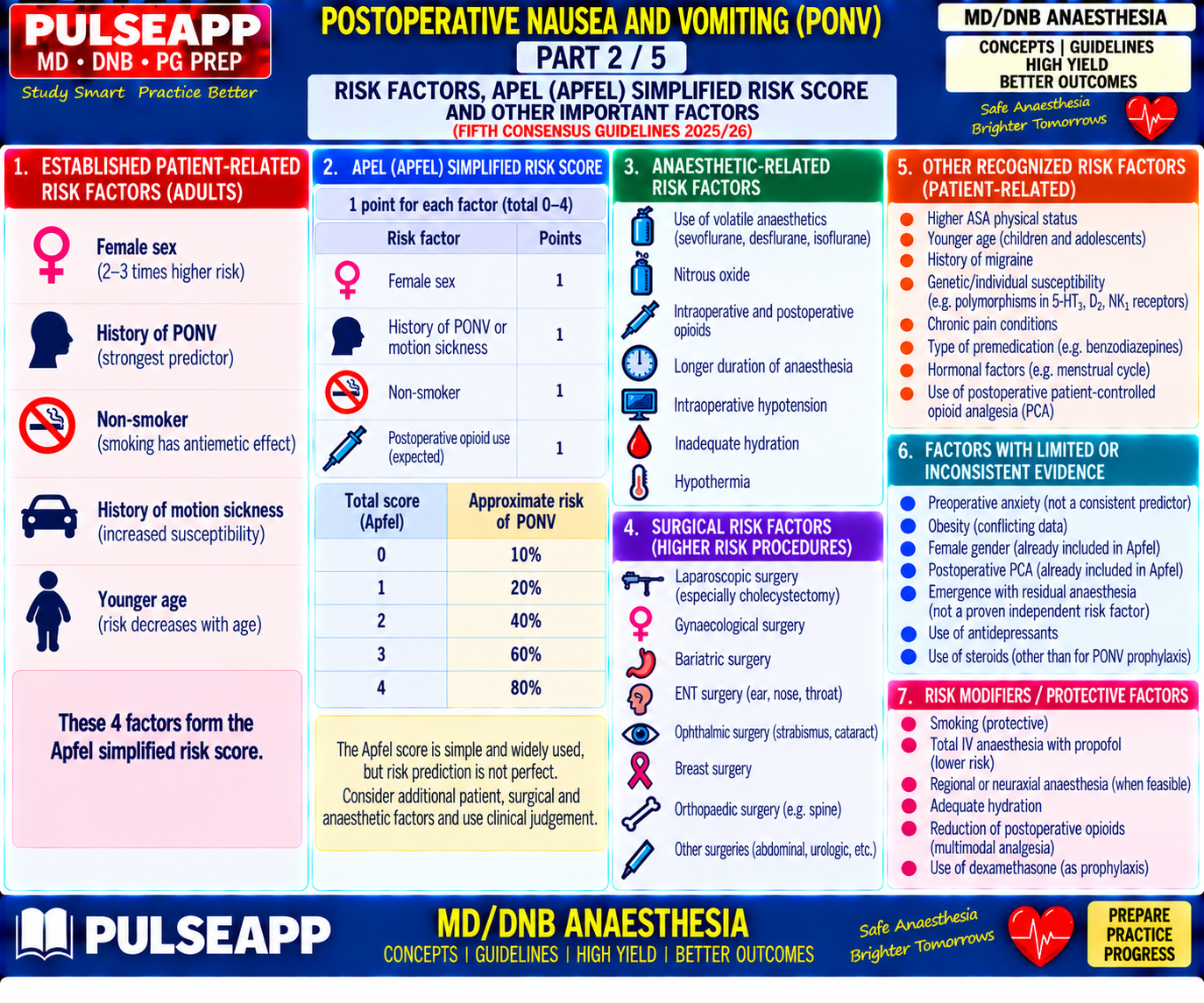
B. Apfel Simplified Risk Score2 marks
✅ Four Risk Factors (Each = 1 Point)
1) Female sex. 2) Non-smoker. 3) History of PONV/motion sickness. 4) Postoperative opioid use expected.
Score→Risk: 0=10%, 1=20%, 2=40%, 3=60%, 4=80%.
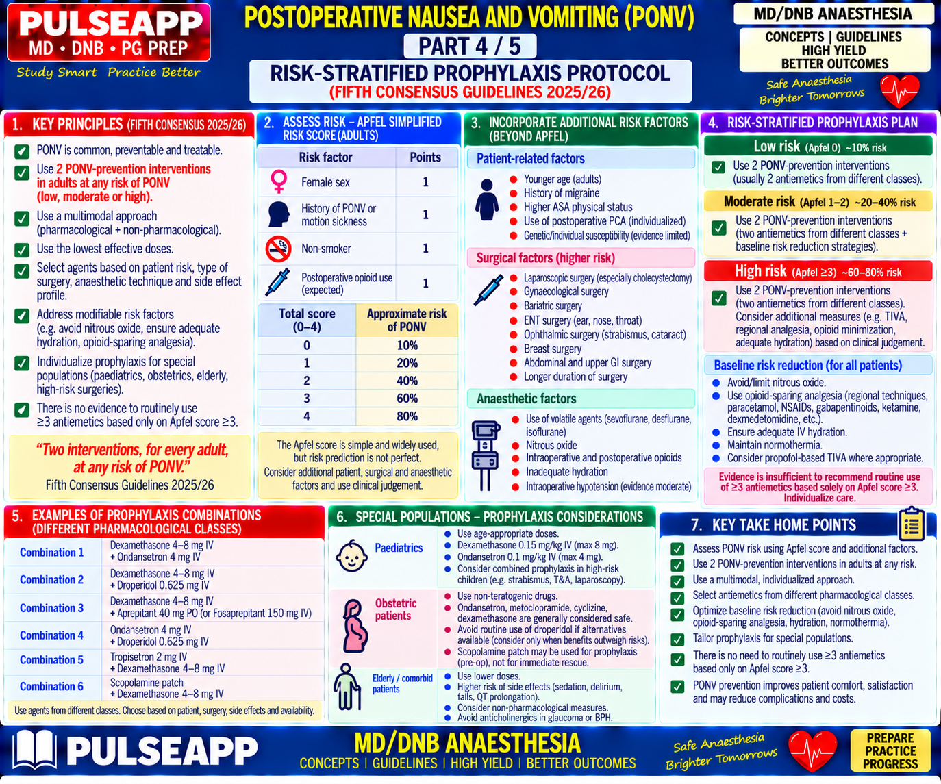
C. Baseline Risk Reduction1 mark
  • TIVA with propofol instead of volatile (reduces PONV 25–30%)
  • Minimise opioids via multimodal analgesia
  • Adequate IV hydration
  • Avoid N₂O and neostigmine where possible (use sugammadex)
  • Adequate preoperative anxiolysis
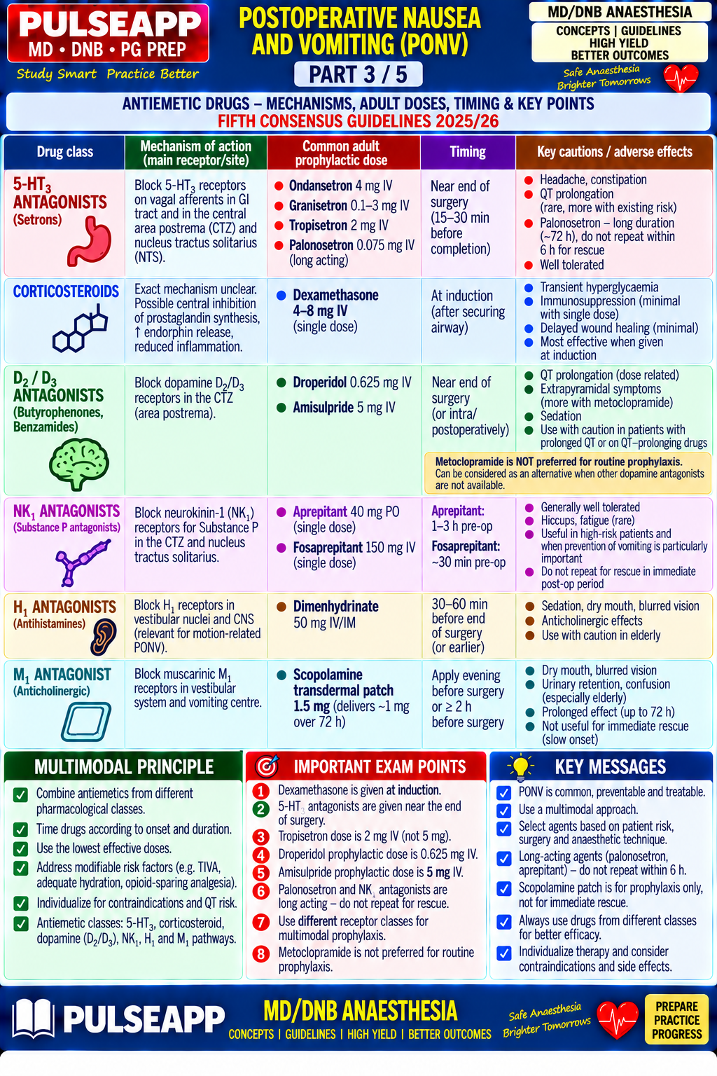
D. Antiemetic Drug Classes4 marks
ClassMechanismDoseNotes
5-HT3 antagonists (ondansetron)Blocks 5-HT3 in CTZ/vagal afferents4mg IV at END of surgeryFirst-line; QTc prolongation risk
Corticosteroids (dexamethasone)Reduces prostaglandin synthesis/serotonin release4–8mg IV at INDUCTION (slow onset)Give early — NOT at end of surgery
D2 antagonists (droperidol)Blocks D2 in CTZ0.625–1.25mg IV at endQTc prolongation dose-related
NK1 antagonists (aprepitant)Blocks substance P at NK140–80mg oral 1–2h pre-opMost effective single agent, especially delayed PONV
Antihistamines (cyclizine)H1/M1 blockade50mg IV/IM at endBest for vestibular-mediated nausea
Propofol (TIVA)Central 5-HT3/D2 modulationRescue: 20mg IV bolusIntrinsic antiemetic effect
E. Risk-Stratified Protocol0 marks

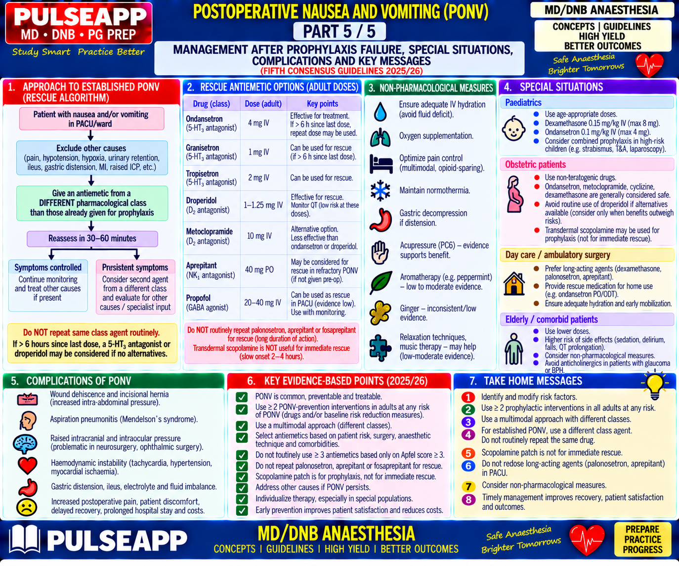
Apfel 0–1: minimal/no prophylaxis. Apfel 2: two antiemetics, different classes. Apfel 3: three antiemetics + consider TIVA. Apfel 4: maximum multimodal (TIVA + 3–4 antiemetic classes + scopolamine patch). Treatment of established PONV: use a DIFFERENT class from any prophylaxis already given; do not repeat the same agent within 6 hours.

🎤 Viva Corner
Q. A 35yo female non-smoker with motion sickness history undergoes lap chole and is expected to need postop morphine. Apfel score and full strategy?
Apfel score = 4/4 (~80% risk). Strategy: scopolamine patch the night before; TIVA (propofol-remifentanil) instead of volatile; multimodal analgesia (paracetamol, ketorolac, port-site LA infiltration) to minimise opioid; dexamethasone 8mg IV at INDUCTION; ondansetron 4mg IV at END of surgery; droperidol 0.625mg IV at end (third class); adequate hydration; if PONV occurs postop, treat with a different class (e.g. metoclopramide) from those already given.
Q. Why is dexamethasone given at induction rather than at the end of surgery, unlike other antiemetics?
Dexamethasone's antiemetic effect is genomic/anti-inflammatory (reduced prostaglandin synthesis, reduced serotonin release), requiring gene transcription and new protein synthesis — onset takes 60–90 minutes to peak. Given at induction, its peak effect coincides with emergence and the highest-risk early recovery period; given at the end of surgery, its peak effect would arrive after PONV has already had time to occur.
★ Examiner's Pearl
Apfel four factors with exact risk percentages (0=10% to 4=80%) must be reproduced numerically. Dexamethasone timing (INDUCTION, not end) with the reason is the most tested timing fact. The antiemetic class table covering all four receptor pathways (5-HT3/D2/NK1/H1) must be complete.
Apfel CC et al (Anesthesiology 1999). Gan TJ et al, Fourth Consensus Guidelines (Anesth Analg 2020). Miller's Anaesthesia 9th Ed, Ch 96.
lock Protected Clinical Document • Pulse App • Screenshots Strictly Prohibited
QUESTION 24 person Asked by .
bookmark_add

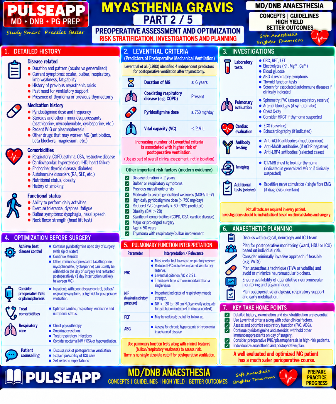
Discuss the pathophysiology of Myasthenia Gravis. Outline the preoperative assessment including Leventhal's criteria, intraoperative anaesthetic considerations, choice of neuromuscular blockers, and postoperative ventilation planning for a patient undergoing thymectomy.

collections Question Diagrams & Reference Images (1)
Question Reference Diagram
zoom_in View Image
description Clinical Response
⚙ Core Concept
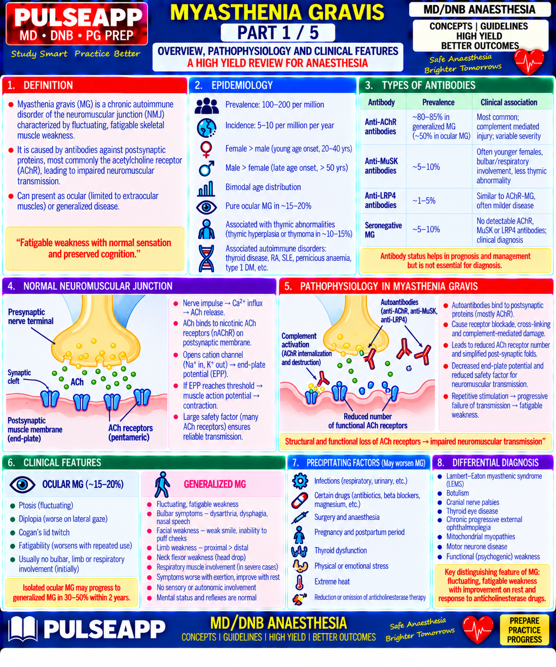
Myasthenia Gravis is the prototype autoimmune neuromuscular junction disease — antibody-mediated destruction of postsynaptic nicotinic acetylcholine receptors produces fatigable weakness that affects bulbar, respiratory, and limb muscles in a characteristic pattern. For the anaesthesiologist, MG represents perhaps the highest-stakes perioperative challenge in neuromuscular pharmacology: these patients have dramatically altered sensitivity to ALL neuromuscular blocking agents, require meticulous respiratory assessment and planning, and have a defined risk of postoperative myasthenic crisis that must be anticipated and prepared for before the patient enters the operating theatre. (Miller's Anaesthesia 9th Ed; Baraka A — MG and anaesthesia; Oh TE — Intensive Care Manual; Leventhal SR et al. — Predicting the need for post-thymectomy ventilation)
A. Pathophysiology of Myasthenia Gravis2 marks

Autoimmune mechanism: MG results from autoantibody-mediated destruction and functional impairment of the postsynaptic nicotinic acetylcholine receptor (nAChR) at the neuromuscular junction; Anti-AChR antibodies are present in 85% of patients (complement-activating IgG1 and IgG3); Anti-MuSK (musclespecific kinase) antibodies in 10%; seronegative MG (~5%) — antibodies against LRP4 or other targets Thymic abnormality in ~80%: thymoma in 10–15% (often aggressive, paraneoplastic); thymic hyperplasia in 60–70%; the thymus drives autoreactive T-cell sensitization against AChR — thymectomy produces remission in 30–40%, improvement in 70–80%

NMJ consequence: Anti-AChR Ab → complement activation → receptor degradation, cross-linking and endocytosis → fewer functional AChRs → smaller end-plate potential for each ACh quantum released → more quanta needed to reach action potential threshold → with repetitive stimulation, the ACh store depletes faster than it is replenished → progressive failure of neuromuscular transmission = FATIGABLE WEAKNESS — the hallmark of MG

Osserman Classification Features Anaesthetic Risk

Class I — Ocular Ptosis and diplopia only; no bulbar or limb weakness Lowest risk; rarely requires post-op ventilation

Class IIa — Mild generalised Generalised weakness, predominantly limb; mild bulbar Moderate risk; monitor closely post-op

Class IIb — Moderate generalised Moderate generalised weakness with significant bulbar involvement High risk; plan for post-op ventilatory support

Class III — Acute severe Rapid onset of severe generalised weakness; respiratory muscles ± Very high risk; plan ICU post-op

Class IV — Late severe Severe generalised from Class I/II after ≥2 years Very high risk

Class V — Intubated Requiring intubation for respiratory failure (myasthenic crisis) In crisis; stabilise before any elective surgery

B. Preoperative Assessment3 marks
Respiratory Assessment

Spirometry: FVC and FEV1 — FVC <2.9 L = high post-operative ventilation risk; FVC <40 mL/kg = very high risk

NIF (Negative Inspiratory Force): worse than −25 cmH₂O = likely ventilator-dependent post-operatively

ABG: CO₂ retention (elevated PaCO₂) indicates severe disease with impaired ventilatory reserve

CT chest: thymoma size, tracheal deviation, substernal extension, mediastinal involvement

⚠ Leventhal's Criteria — Predicting Need for Post-thymectomy Ventilation
1. Duration of MG >6 years 2. Chronic respiratory disease (COPD, bronchiectasis) 3. Pyridostigmine dose >750 mg/day 4. Vital capacity <2.9 L ≥3 criteria present → plan for postoperative mechanical ventilation. Counsel patient preoperatively. Book ICU bed. Grading score 0–12: score <12 = likely extubation; ≥12 = anticipate prolonged ventilation.
Drug Management Pre-operatively

Drug Perioperative Management Rationale Pyridostigmine (neostigmine, Continue on morning of surgery with sip of water; do NOT Abrupt withdrawal → myasthenic crisis; helps maintain NMJ function mestinon) abruptly withdraw through induction Steroids (prednisolone) Continue; give stress dose hydrocortisone 100 mg IV at Adrenal suppression from long-term steroids; surgical stress dose induction required Azathioprine, mycophenolate Continue; note immunosuppression increases infection risk Abrupt cessation can worsen disease Plasmapheresis (3–5 sessions) If poorly controlled — start 2–4 weeks before elective Removes circulating anti-AChR antibodies; temporary improvement Plasmapheresis (3–5 sessions) If poorly controlled — start 2–4 weeks before elective Removes circulating anti-AChR antibodies; temporary improvement thymectomy (4–8 weeks) IVIG 2 g/kg over 5 days Alternative to plasmapheresis for preoperative optimisation Modulates immune response; effect lasts 4–8 weeks

Drugs to AVOID in MG
⚠ Drugs That Worsen Neuromuscular Transmission in MG
Antibiotics: aminoglycosides (gentamicin, tobramycin — block presynaptic Ca²⁺ channels reducing ACh release), fluoroquinolones, polymyxins Cardiovascular: beta-blockers, quinidine, procainamide, calcium channel blockers Other: magnesium sulphate (reduces ACh release), D-penicillamine (induces anti-AChR antibodies), phenytoin, chloroquine, statins (rare but documented) ALL neuromuscular blocking agents: exquisitely sensitive — even "non-depolarising" drugs at fractions of normal doses produce profound, prolonged block
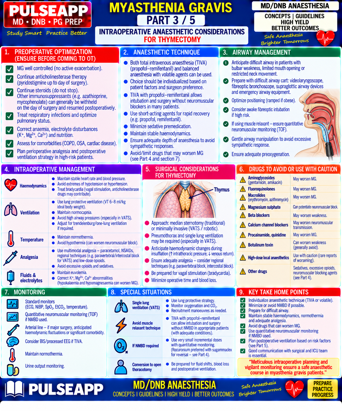
C. Intraoperative Anaesthetic Management3 marks
Induction

Standard IV induction: propofol or thiopentone; ketamine relatively contraindicated (increases sympathetic tone and may worsen laryngospasm in bulbar MG)

Airway: careful assessment for bulbar weakness and aspiration risk; rapid sequence induction if bulbar MG present (aspiration risk from impaired swallowing) Topical airway anaesthesia for awake fiberoptic intubation if severely compromised

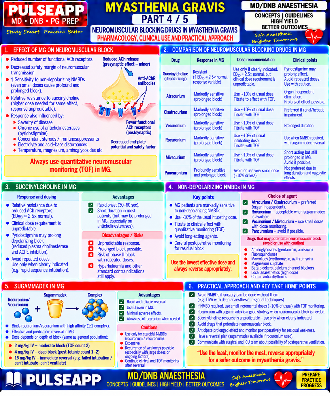
Neuromuscular Blocking Agents — The Critical Decision
✅ NMB Strategy in MG
Ideal approach: AVOID neuromuscular blockers entirely — most thymectomies can be performed without NMBs using deep volatile anaesthesia for relaxation (sevoflurane MAC 1.5–2.0 reduces NMJ transmission and provides adequate surgical conditions for thoracic approach). If NMBs required: Succinylcholine: REDUCED sensitivity (AChRs are already reduced in number → need relatively MORE succinylcholine for a given block); doses 1.5– 2 mg/kg may be needed; however, pyridostigmine inhibits plasma cholinesterase → prolongs succinylcholine action → unpredictable; use with caution Non-depolarising NMBs: MARKEDLY INCREASED sensitivity — use 10–20% of normal intubating dose; rocuronium 0.1–0.2 mg/kg (vs normal 0.6 mg/kg); vecuronium 0.01–0.02 mg/kg; atracurium 0.1–0.15 mg/kg; ALWAYS monitor with TOF — do not give further doses without TOF guidance Mivacurium: shortest-acting NDMR; metabolised by plasma cholinesterase (similarly prolonged by pyridostigmine); avoid Sugammadex: drug of choice for reversal if rocuronium used — provides reliable, complete reversal regardless of depth of block; 2–4 mg/kg depending on TOF count; particularly valuable in MG where residual block is catastrophic
Intraoperative Monitoring

Continuous quantitative NMJ monitoring (acceleromyography/TOF-Watch) at the ulnar nerve throughout — mandatory; set a TOF ratio threshold of ≥0.9 for extubation, same as in non-MG patients but even more critically enforced

Standard monitoring: SpO₂, ETCO₂, invasive arterial BP (for blood gas assessment and beat-to-beat BP during sternotomy/VATS approach), CVP if mediastinal surgery

Temperature monitoring — hypothermia worsens NMJ transmission impairment in MG BIS or entropy monitoring for depth of anaesthesia — important if volatile agents are used as the primary "muscle relaxant" at high doses

Anaesthetic Maintenance

TIVA preferred by many centres: propofol + remifentanil infusion; remifentanil provides excellent intraoperative analgesia with very brief post-infusion effects; propofol does not significantly affect NMJ function; no volatile agent effects on respiratory function at emergence

If volatile used: sevoflurane preferred (less airway irritation, suitable for potential bronchoscopy); desflurane is acceptable; halothane AVOIDED (exacerbates NMJ block)

D. Postoperative Management & Myasthenic Crisis2 marks

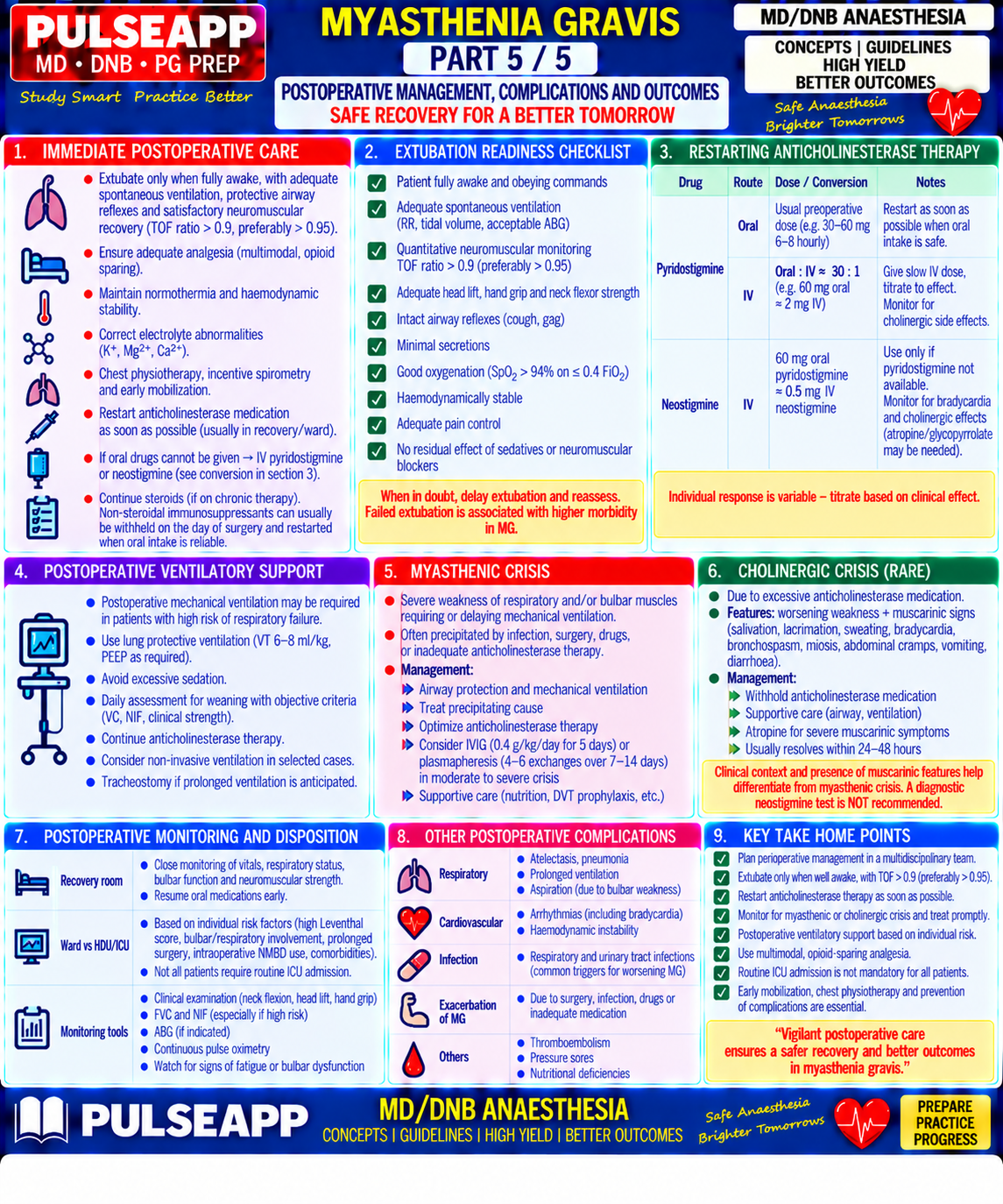
Parameter Detail Extubation TOF ratio ≥0.9 (quantitative monitoring); FVC ≥15 mL/kg (ideally ≥20 mL/kg); NIF better than −25 cmH₂O; alert, following commands; adequate criteria (MG) swallow/gag reflex; normothermia; pain controlled; no ongoing residual NMB Post-op Plan for ICU admission; ventilate overnight; gradually wean using pressure support; daily extubation readiness assessment; restart pyridostigmine via ventilation (if NGT when bowel sounds return Leventhal ≥3) Myasthenic Acute respiratory failure from worsening MG — triggers: inadequate immunosuppression, infection, stress, drugs, surgery itself; treatment: intubate crisis and ventilate, plasmapheresis (most rapid effect), IVIG, increase steroids, eliminate trigger; withhold anticholinesterases if excessive secretions (cholinergic crisis possible)

Cholinergic Both cause weakness — differentiate by edrophonium (Tensilon) test: 2 mg IV → if myasthenic crisis → temporary improvement; if cholinergic crisis crisis vs → worsens (excessive ACh); or by clinical signs: cholinergic = SLUDGE (salivation, lacrimation, urination, defecation, GI cramps, emesis) + myasthenic bradycardia + miosis crisis Analgesic Thoracic epidural or paravertebral block (VATS/sternotomy); minimise systemic opioids (respiratory depression particularly hazardous); NSAIDs + strategy paracetamol as baseline multimodal; avoid morphine PCA without close monitoring

🎤 Viva Corner
Q. A 45-year-old MG patient (Class IIb, pyridostigmine 600 mg/day, FVC 2.2 L) is scheduled for thymectomy. Leventhal score? Will you extubate at end of surgery?
Leventhal criteria assessment: Duration of MG — not stated, assume <6 years = 0; Chronic respiratory disease — not mentioned = 0; Pyridostigmine dose >750 mg/day — no, 600 mg = 0; Vital capacity <2.9 L — YES, FVC 2.2 L = 1. Score = 1/4 criteria. However, the FVC of 2.2 L is critically important individually — it falls below the 2.9 L threshold, which alone is a significant respiratory risk factor. Combined with Class IIb (moderate bulbar involvement), this patient has significant postoperative respiratory risk. My plan: I would NOT plan routine extubation in theatre. Instead, I would plan for post-operative ICU admission with the patient sedated and ventilated overnight, with daily extubation readiness assessment using FVC ≥15 mL/kg, NIF <−25 cmH₂O, and TOF ≥0.9. I would preoperatively arrange plasmapheresis (3–5 sessions) or IVIG to optimise NMJ function before surgery. For the anaesthetic: avoid NMBs entirely — use propofol/remifentanil TIVA with sevoflurane 1.5 MAC for muscle relaxation if needed; thoracic epidural for analgesia. If NMBs used, only rocuronium at 10–20% of normal dose with TOF guidance and reverse with sugammadex.
Q. Why are MG patients paradoxically RESISTANT to succinylcholine but SENSITIVE to non-depolarising NMBs?
The paradoxical pharmacological sensitivities in MG arise directly from the same pathology — reduced numbers of functional postsynaptic nAChRs — but affect the two drug classes through opposite mechanisms. Non-depolarising NMBs (rocuronium, vecuronium, atracurium) work by competitive antagonism at the nAChR — they occupy the receptor without activating it. In MG, where only a fraction of normal nAChRs are functional, a much smaller absolute number of receptors needs to be blocked to prevent neuromuscular transmission. Even a small dose of a non-depolarising NMB occupies the critically limited remaining functional receptors, producing profound block — hence MG patients are exquisitely SENSITIVE to non-depolarising NMBs. Succinylcholine, by contrast, works by ACTIVATING the nAChR (depolarisation block) — it needs to bind and activate sufficient receptors to produce sustained depolarisation and block. With far fewer functional receptors available in MG (the rest having been destroyed by anti-AChR antibodies), more succinylcholine drug per kg is needed to occupy enough of the reduced receptor pool to produce adequate depolarisation for block — hence relative RESISTANCE to succinylcholine. The other complicating factor is that pyridostigmine (which MG patients take) inhibits plasma pseudocholinesterase, prolonging succinylcholine's duration unpredictably. This combination — unpredictable dose requirement AND unpredictable duration — makes succinylcholine pharmacologically hazardous in MG patients and explains why avoiding NMBs entirely or using only small-dose non-depolarising agents with rigorous TOF monitoring is the preferred strategy.
Q. Your MG patient is in the ICU on post-op day 2 after thymectomy, on pressure support ventilation. They suddenly become increasingly weak with excessive secretions, bradycardia and miosis. How do you distinguish myasthenic crisis from cholinergic crisis, and what is the immediate management?
The clinical picture — excessive secretions (bronchorrhoea), bradycardia, and miosis — strongly suggests a CHOLINERGIC CRISIS (excess acetylcholine from overstimulation with anticholinesterase drugs) rather than a myasthenic crisis. Both produce weakness and potential respiratory failure, but the SLUDGE syndrome (Salivation, Lacrimation, Urination, Defecation, GI cramps, Emesis) along with bradycardia (muscarinic effects) and miosis are characteristic of cholinergic excess. The Tensilon (edrophonium) test historically differentiated them: 2 mg IV — if myasthenic crisis, the patient transiently improves (more ACh available is beneficial); if cholinergic crisis, worsening occurs (additional ACh on top of already-excess ACh is harmful). However, this test is risky in a patient who may deteriorate further and should only be performed with full resuscitation equipment immediately available. The safer approach given the clear cholinergic signs: WITHHOLD all anticholinesterases immediately (stop pyridostigmine); give atropine for the bradycardia and excessive secretions (0.6–1.2 mg IV titrated); ensure airway patency with suctioning and adequate ventilatory support (already intubated in ICU); monitor for improvement over next hours. In myasthenic crisis, management would be the opposite: maintain or increase anticholinesterases, consider plasmapheresis or IVIG. Once stable, the neurologist should review dosing and timing of anticholinesterase resumption with careful clinical reassessment.
★ Examiner's Pearl
State Leventhal's four criteria with exact thresholds (duration >6 years; chronic respiratory disease; pyridostigmine >750 mg/day; VC <2.9 L) — ≥3 criteria = plan for post-op ventilation. This table is tested verbatim. The NMB paradox (RESISTANT to succinylcholine, SENSITIVE to non-depolarisers) with the mechanistic explanation is the single most examined pharmacological concept in MG anaesthesia. State both the myasthenic crisis and cholinergic crisis features clearly as examiners specifically test the clinical distinction — SLUDGE + bradycardia + miosis = cholinergic; pure worsening weakness without autonomic features = myasthenic.
Miller RD et al. Miller's Anaesthesia, 9th Ed, Chapter 29 (Neuromuscular Diseases and Anaesthesia). Baraka A. Anaesthesia and myasthenia gravis (BJA 1992;69:227-231). Leventhal SR et al. Predicting the need for postoperative mechanical ventilation in myasthenia gravis (Anesthesiology 1980;53:26-30). Eisenkraft JB et al. Resistance to succinylcholine in myasthenia gravis (Anesthesiology 1988;69:760-763). Drachman DB. Myasthenia gravis (N Engl J Med 1994;330:1797-1810).
lock Protected Clinical Document • Pulse App • Screenshots Strictly Prohibited
QUESTION 25 person Asked by .
bookmark_add

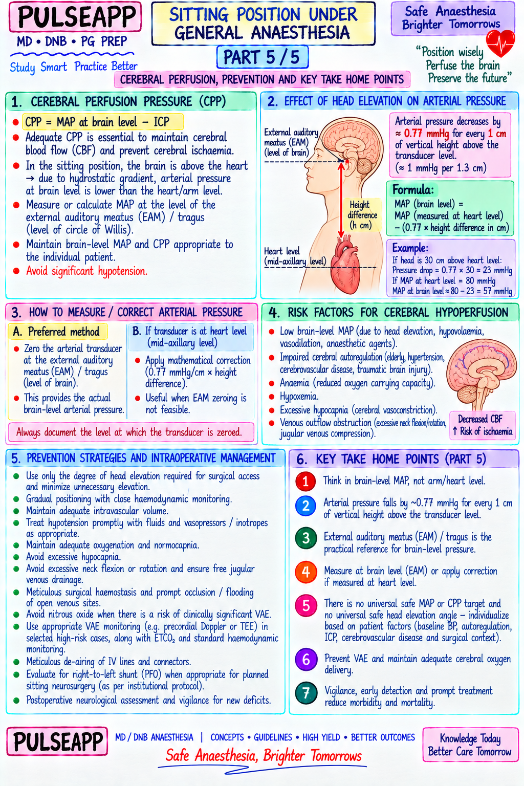
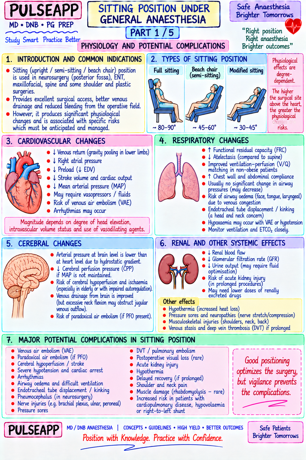
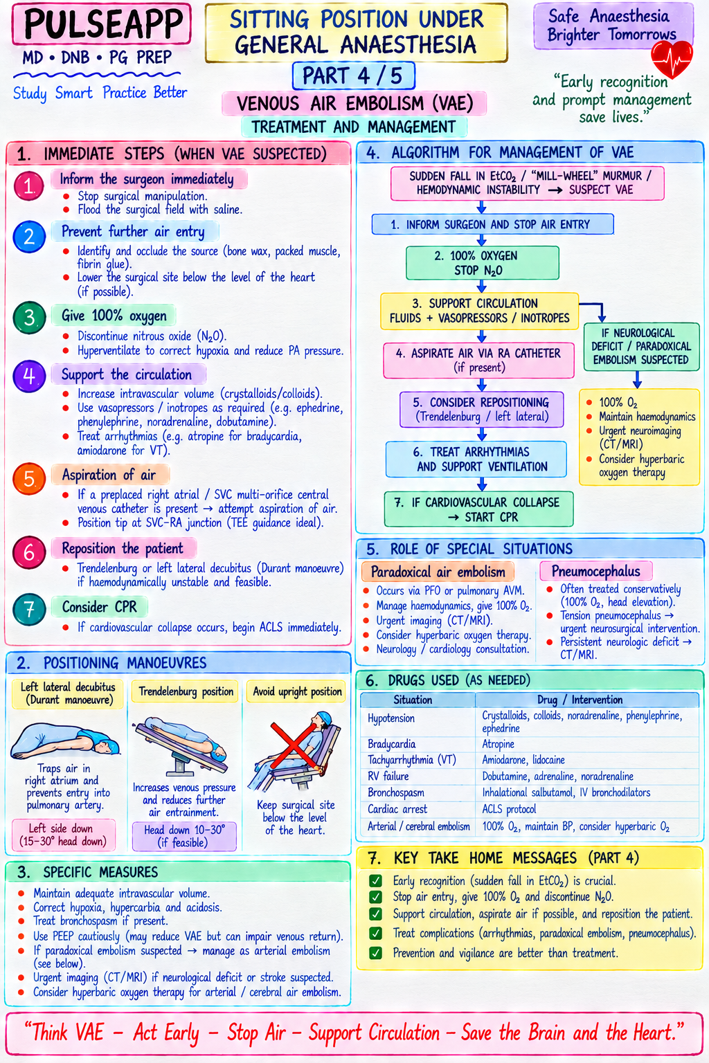
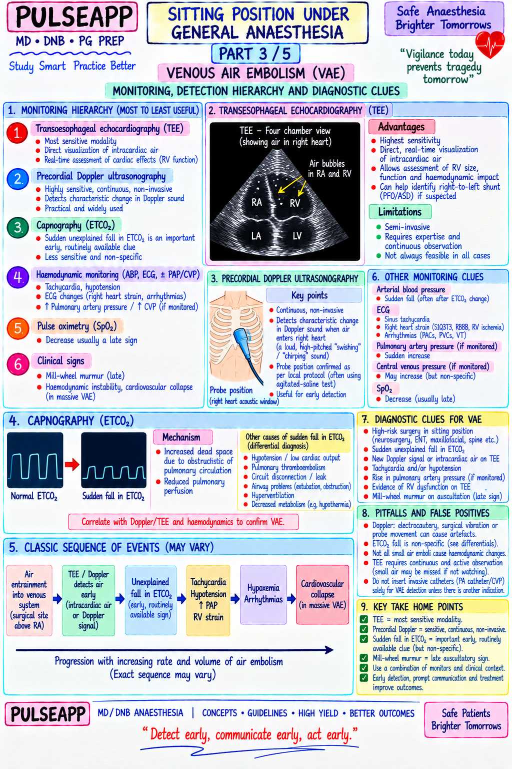
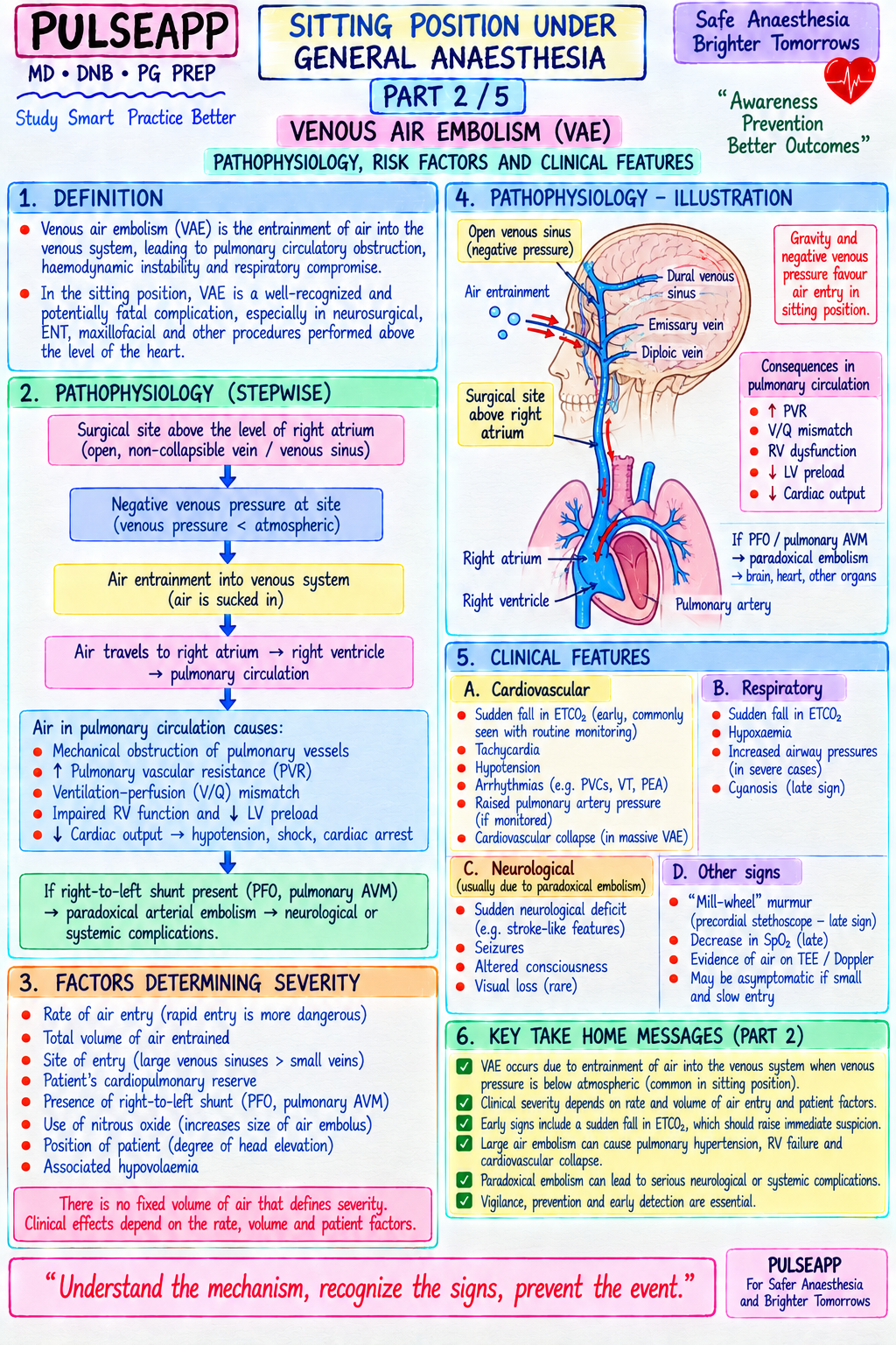
Discuss the physiological complications of the sitting position under general anaesthesia. Detail the pathophysiology, monitoring hierarchy, and treatment of venous air embolism. Explain the cerebral perfusion pressure correction for head height and prevention strategies.

collections Question Diagrams & Reference Images (2)
Question Reference Diagram
zoom_in View Image
Question Reference Diagram
zoom_in View Image
description Clinical Response
⚙ Core Concept
The sitting position creates a unique physiological challenge: the brain is placed at a significantly higher level than the heart, creating both a gravitational threat to cerebral perfusion pressure AND an environment where open venous sinuses at negative pressure relative to atmospheric can entrain room air directly into the right heart — with potentially fatal consequences. The sitting position is used for posterior fossa neurosurgery, shoulder arthroscopy (beach chair), and ENT procedures because it provides optimal surgical access, reduced blood loss, and improved airway access — but at the cost of these specific risks that must be anticipated, monitored, and managed in real time. (Miller's Anaesthesia 9th Ed; Cucchiara RF — Sitting position in neurosurgery; Matjasko J — Incidence of VAE; Porter JM — VAE monitoring)
A. Positions Used and Their Specific Risks1 mark

Beach chair / sitting position (45–90° trunk elevation): posterior fossa neurosurgery (Fowler's position — head in Mayfield pins, trunk upright), shoulder arthroscopy, ENT procedures; creates the risks described below

Key anatomical problem: the brain is above the level of the heart → cerebral venous pressure is subatmospheric (negative) → open dural sinuses and bone marrow sinuses act as open tubes at negative pressure → air can be entrained with each inspiration or surgical manipulation

B. Cardiovascular Complications2 marks

Hypotension — The Most Common Complication Venous pooling in the lower limbs from gravity → ↓ venous return → ↓ cardiac output → ↓ MAP; compounded by general anaesthesia (vasodilation, reduced cardiac reserve)

Critical CPP calculation for sitting position: MAP is measured at arm/heart level, but the brain is above this reference point; for every centimetre of head elevation above the right atrium, subtract 0.77 mmHg from the measured MAP to obtain actual cerebral perfusion pressure at the level of the Circle of Willis: CPP = MAP(measured at heart) − (height of brain above heart in cm × 0.77) − ICP

Target: maintain CPP ≥70 mmHg at brain level; this may require MAP at arm level of 80–100 mmHg if the head is 30–40 cm above the heart (30 cm × 0.77 = 23 mmHg correction)

Management: adequate IV fluid loading before positioning; graduated compression stockings + abdominal binders to reduce venous pooling; vasopressors titrated to maintain corrected CPP ≥70 mmHg; arterial line at the level of the external auditory meatus (EAM) for accurate "brain-level" blood pressure measurement in some centres

C. Venous Air Embolism (VAE) — The Most Feared Complication5 marks
Pathophysiology

Open venous structures in the surgical field (dural sinuses, diploic veins of skull, epidural veins) + subatmospheric venous pressure (brain above the heart) → pressure gradient drives room air into the venous system with each surgeon's cut, dissection, or drill → air enters right atrium and right ventricle → large air bolus can obstruct the right ventricular outflow → pulmonary arterial air lock → acute right heart failure, sudden loss of cardiac output → death. Small boluses: air distributes to the pulmonary vasculature → microvascular obstruction → ventilation-perfusion mismatch → pulmonary hypertension → increased dead space → ETCO₂ falls.

Incidence: 25–40% by precordial Doppler in posterior fossa surgery in the sitting position; clinically significant in 10–15%

Paradoxical Air Embolism (PAE): if a patent foramen ovale (PFO) is present (~25–30% of the general population), air in the right atrium can cross to the left atrium via the PFO → arterial circulation → coronary air embolism (MI), cerebral air embolism (stroke); PAE makes VAE potentially lethal even with small volumes of air

Monitoring for VAE — Sensitivity Hierarchy (Most to Least Sensitive)

Minimum Volume Rank Monitor What It Detects Detectable 1 (Most Precordial Doppler frequency shift from air bubbles in the right heart — characteristic "mill wheel" murmur (loud, As little as 0.05 mL/kg of air sensitive) Doppler churning, machinery-like sound); probe placed over the right cardiac border (4th intercostal space, right sternal edge) 2 Transoesophageal Direct visualisation of air bubbles in right atrium and right ventricle; also detects PFO (bubble test — ~0.05 mL/kg; same echocardiography injection of saline agitated with air through a peripheral vein; bubbles seen crossing from right to left sensitivity as Doppler but (TOE/TEE) atrium = PFO); most specific monitor and can guide aspiration via CVP catheter more specific 3 ETCO₂ Sudden fall in ETCO₂: air emboli create pulmonary dead space (capillaries obstructed → no gas Clinically significant VAE (capnography) exchange → CO₂ not eliminated → ETCO₂ falls despite unchanged ventilation) (~0.5 mL/kg); less sensitive than Doppler for small emboli 4 ETCO₂ − PaCO₂ Pulmonary dead space increase → PaCO₂ rises (CO₂ not eliminated) while ETCO₂ falls or plateaus Significant VAE gradient widens → the arterial-to-end-tidal CO₂ gradient increases (normal <5 mmHg); requires arterial line for simultaneous ABG 5 Expired N₂ monitor Air (78% N₂) entering the pulmonary circulation → N₂ detected in expired gas; requires specialised Small to moderate VAE gas analyser; not routine 6 (Least CVP rise, BP fall, Late signs of haemodynamically significant VAE; CVP rises from right heart outflow obstruction; Large, haemodynamically sensitive) arrhythmia, SpO₂ hypotension from reduced cardiac output; dysrhythmia from right heart distension significant VAE; already a fall crisis

Treatment of VAE — Act Immediately, Act in Sequence
⚠ VAE Emergency — Immediate Sequential Actions
1. NOTIFY THE SURGEON IMMEDIATELY — flood the surgical field with saline (prevents more air entry through open vessels); pack the wound 2. Compress both jugular veins bilaterally (raises venous pressure in the head, creating back-pressure that reduces air entrainment rate) 3. LOWER THE HEAD IMMEDIATELY — reduce the hydrostatic gradient driving air into the venous sinuses; ask surgeon to lower operative table 4. Aspirate air via the multi-orifice CVP catheter positioned in the right atrium (confirmed by fluoroscopy or TOE); aspiration can remove significant quantities of entrained air; most effective with a multi-orifice catheter placed at the junction of SVC and right atrium (guided by ECG P-wave changes) 5. Discontinue N₂O immediately — N₂O diffuses into air-filled spaces (30× faster than N₂ is absorbed), dramatically expanding an air embolus; discontinue N₂O and switch to 100% O₂ 6. Increase venous return — leg compression, IV fluid bolus to raise CVP, Trendelenburg if possible (but may compromise surgical field) 7. Cardiopulmonary resuscitation if cardiac arrest from air lock — CPR may physically dislodge the air lock; left lateral decubitus position (Durant's manoeuvre) — tilts the right ventricular outflow toward the apex, potentially allowing air to move out of the RVOT 8. Hyperbaric oxygen if available post-event — promotes air absorption by creating high pressure differential
D. Neurological Complications of the Sitting Position2 marks

Complication Mechanism Prevention Cerebral Reduced CPP from MAP fall + gravity reduction at brain level; posterior circulation Maintain corrected CPP ≥70 mmHg; arterial line for ischaemia (vertebrobasilar) particularly vulnerable in the sitting position (long route from heart to brain continuous beat-to-beat monitoring; vasopressors; from with gravity against it) avoid excessive head flexion (reduces vertebral artery hypotension blood flow) Quadriplegia Excessive neck flexion → cervical cord ischaemia or direct compression; particularly Lateral skull pins for head fixation; neutral neck position; (catastrophic) dangerous if there is pre-existing cervical stenosis; the "two-finger rule" — maintain at least pre-operative MRI of cervical spine in elderly or patients two finger-breadths between chin and sternum to prevent excessive neck flexion and with known spondylosis; monitor SSEP and MEP vertebral artery compression intraoperatively Paradoxical Air crossing PFO to systemic circulation → cerebral embolism Pre-operative bubble echocardiogram to exclude PFO; air embolism if large PFO present, reconsider sitting position; → stroke intraoperative TOE for detection Peripheral Stretch to brachial plexus from arm position; pressure on ulnar nerve; sciatic nerve stretch Careful arm positioning (neutral, supported, no nerve injuries from extreme hip flexion in beach chair abduction >90°); padding at all bony prominences; avoid extreme hip flexion

🎤 Viva Corner
Q. You are monitoring a posterior fossa craniotomy in the sitting position. The precordial Doppler suddenly produces a loud mill-wheel murmur, ETCO₂ falls from 35 to 18 mmHg, and BP drops from 95 to 65 mmHg. Walk through your immediate management in the correct sequence. This is a significant venous air embolism — confirmed by the Doppler mill-wheel murmur (most sensitive sign), sudden ETCO₂ fall (increased dead space from pulmonary air embolism), and haemodynamic compromise. Immediate sequential actions: First, simultaneously call the surgeon and immediately flood the surgical field with irrigation saline — this is the most important action, stopping further air entrainment at the source. Ask the surgeon to pack and compress the wound and lower the patient's head if possible. Compress both jugular veins bilaterally to raise venous pressure in the head. Aspirate via the CVP catheter positioned in the right atrium — if a multi-orifice right atrial catheter was placed preoperatively (standard practice for posterior fossa sitting surgery), aspirate forcefully with a 20 mL syringe repeatedly. Immediately switch off N₂O and ventilate with 100% O₂ — N₂O will expand any air embolus 30-fold within minutes and must be stopped immediately. Give IV fluid bolus 500 mL to increase CVP and preload. Vasopressor — phenylephrine or noradrenaline — to restore MAP while the source is controlled. If TOE is available, position the probe to visualise the right atrium to guide aspiration catheter placement and assess for PFO air crossing. If cardiac arrest occurs: CPR immediately — compressions may dislodge the RVOT air lock; left lateral decubitus (Durant's manoeuvre); continue aspiration attempts. Post-event: if the patient is resuscitated and stable, consider hyperbaric oxygen for residual air embolism and paradoxical cerebral embolism if there is neurological deterioration. Q. Your patient for posterior fossa surgery in the sitting position has an arterial line at the radial artery showing MAP of 75 mmHg. The head is positioned 35 cm above the right atrium. What is the actual CPP at the level of the Circle of Willis?
Using the hydrostatic correction formula: for every centimetre of head elevation above the right atrium, the effective arterial pressure at brain level is reduced by 0.77 mmHg (this is derived from the density of blood: 1 cmH₂O of fluid column = 0.74 mmHg, converted for blood density). At 35 cm of head elevation: Correction = 35 cm × 0.77 mmHg/cm = 26.95 mmHg ≈ 27 mmHg. Therefore, effective arterial pressure at brain level = MAP measured at arm (right atrium level) − hydrostatic correction = 75 − 27 = 48 mmHg. CPP = MAP at brain level − ICP; if we assume ICP is approximately 10 mmHg (normal), then CPP = 48 − 10 = 38 mmHg. This is critically LOW — the target CPP for neurosurgery is ≥60–70 mmHg. To achieve a CPP of 70 mmHg with ICP of 10 mmHg, we need: MAP at brain level = 70 + 10 = 80 mmHg; MAP at arm level = 80 + 27 = 107 mmHg. Therefore, despite a MAP of 75 mmHg at the arm appearing adequate, the brain is critically underperfused — vasopressors should be titrated to maintain arm-level MAP of at least 100–107 mmHg in this patient. This calculation demonstrates why routine blood pressure monitoring at the arm significantly underestimates the haemodynamic challenge of the sitting position, and why some centres place the arterial transducer at the level of the external auditory meatus (EAM) to measure "brain-level" MAP directly.
Q. Why is N₂O specifically contraindicated once VAE is detected, even though it was safe before the event?
Nitrous oxide is soluble in blood at a rate approximately 34 times faster than nitrogen (the primary component of room air). Once air has entered the systemic venous circulation and the pulmonary vasculature as venous air embolism, N₂O in the inspired gas diffuses from the alveolar capillary blood into the air-filled embolic bubbles down its concentration gradient — the air bubbles contain almost no N₂O initially (they came from room air, which contains no N₂O), whereas the blood passing the embolus contains high N₂O concentrations from the inspired gas. This massive concentration gradient drives N₂O into the bubble much faster than the N₂ within the bubble can be absorbed into the blood (N₂ is 30+ times less soluble), causing the embolus to expand dramatically — an air embolus can expand 3 to 4 times its initial volume within minutes in the presence of high inspired N₂O. This expansion converts a potentially survivable small embolus into a large air lock obstructing the right ventricular outflow — turning a manageable complication into a potentially fatal one. Discontinuing N₂O (and switching to 100% O₂) immediately not only stops this expansion process but also creates a reverse gradient that promotes N₂ absorption from the embolus into the N₂-depleted, O₂-rich blood — gradually shrinking the bubble. This is why the instruction "turn off N₂O" is the third or fourth action in the VAE emergency protocol, immediately after flooding the surgical field and compressing the jugulars.
★ Examiner's Pearl
The CPP correction formula (MAP at arm − 0.77 mmHg per cm of head height − ICP) is a numerically-tested calculation in DNB written papers — practice calculating it with different head heights. The VAE monitoring hierarchy (Precordial Doppler most sensitive → TOE → ETCO₂ fall → CVP rise/BP fall) must be reproduced in order; examiners specifically ask for the "most sensitive monitor for VAE" — state Precordial Doppler with the 0.05 mL/kg threshold. The N₂O expansion mechanism (solubility 34× greater than N₂ → diffuses into air bubble → expansion → fatal air lock) is a mechanistic explanation that distinguishes thorough answers.
Miller RD et al. Miller's Anaesthesia, 9th Ed, Chapter 45 (Positioning) and Chapter 70 (Neurosurgical Anaesthesia). Cucchiara RF et al. Venous air embolism in upright neurosurgical patients (Anesthesiology 1984;60:100-107). Matjasko J et al. Incidence of venous air embolism during craniotomy (Anesthesiology 1985;62:246-250). Porter JM et al. Comparison of arterial and jugular venous oxygen saturation (BJA 2001). Durant TM et al. Pulmonary air embolism (Am Heart J 1947;33:269-281).
lock Protected Clinical Document • Pulse App • Screenshots Strictly Prohibited
QUESTION 26 person Asked by .
bookmark_add

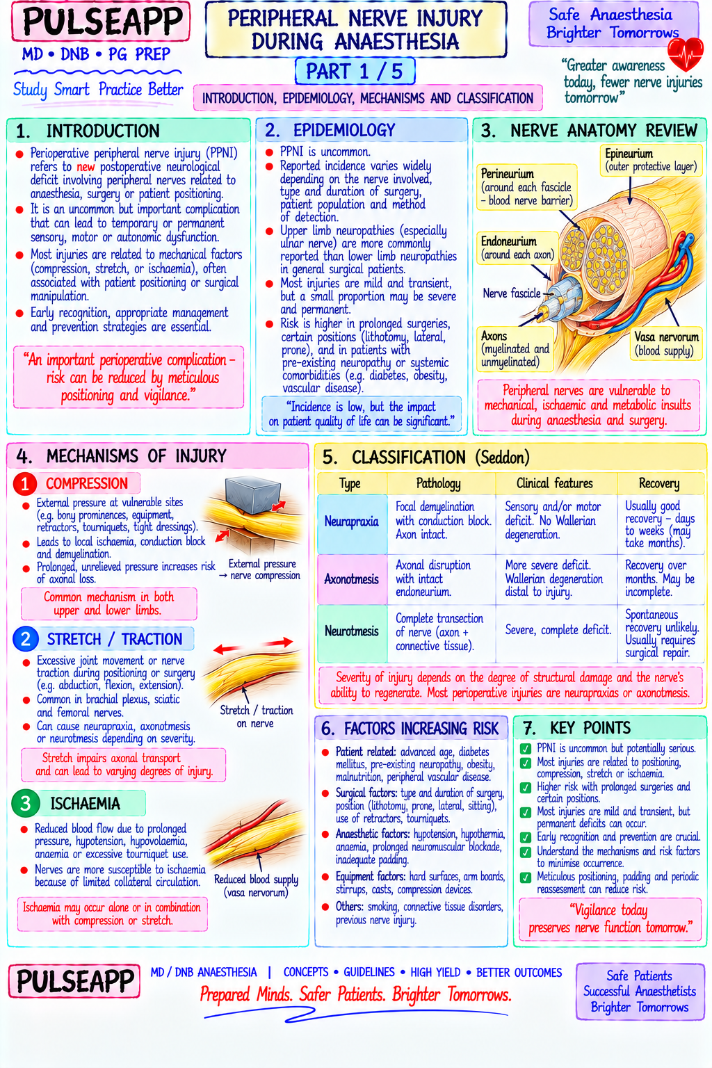
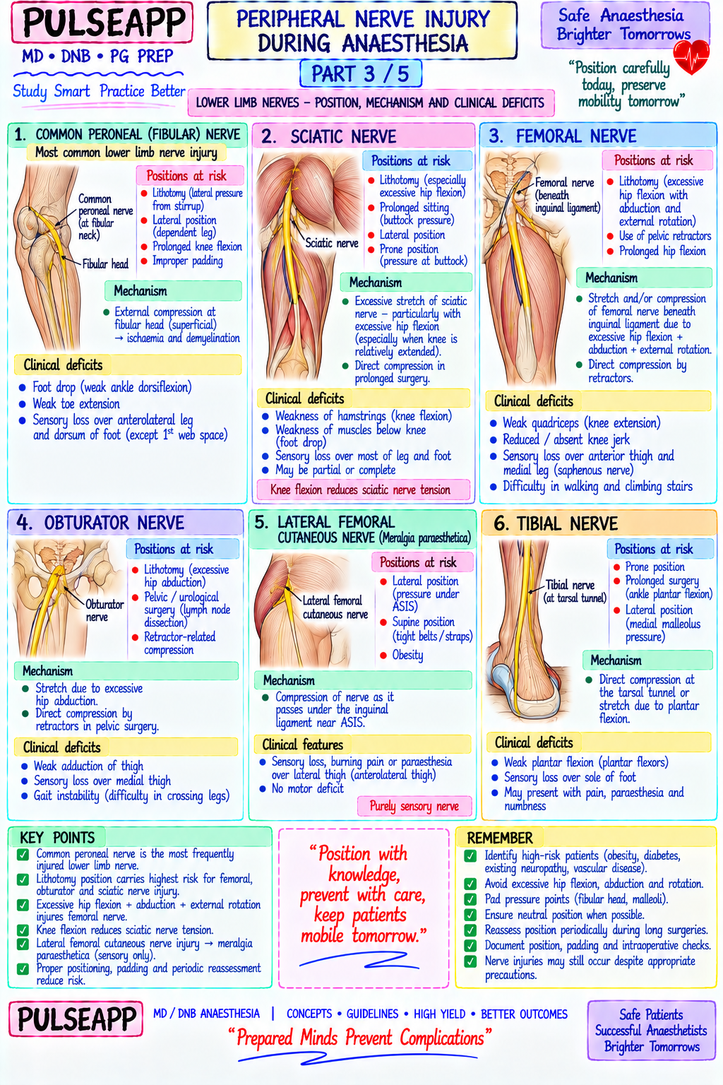
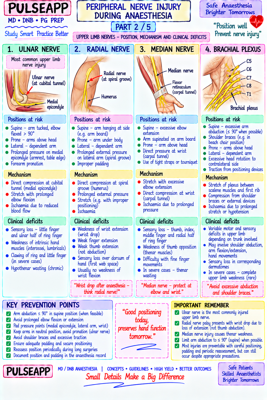
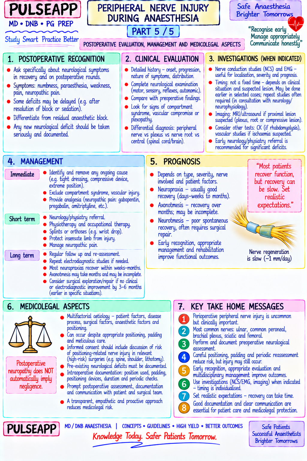
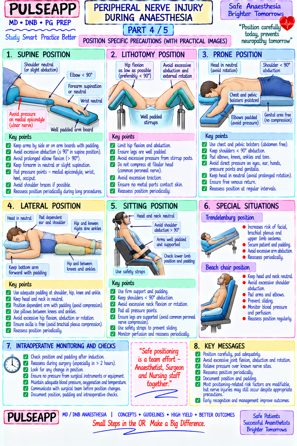
Describe the three mechanisms of peripheral nerve injury during anaesthesia. Outline the most commonly injured nerves by surgical position with their specific mechanisms and clinical deficits. Discuss prevention strategies and medicolegal implications.

collections Question Diagrams & Reference Images (1)
Question Reference Diagram
zoom_in View Image
description Clinical Response
⚙ Core Concept
Peripheral nerve injury (PNI) from patient positioning is one of the most common causes of anaesthesia-related medicolegal claims — it accounts for approximately 16% of all closed claims in the ASA Closed Claims Project, second only to death and brain damage. Under general anaesthesia, the patient has lost all protective reflexes and the ability to report discomfort from positioning — making the anaesthesiologist solely responsible for ensuring that no nerve is compressed, stretched, or rendered ischaemic for the duration of the procedure. Understanding which nerve is vulnerable in which position, what injury it produces, and how it is prevented is therefore a direct patient safety competency. (Miller's Anaesthesia 9th Ed; Kroll DA et al. — ASA Closed Claims; Warner MA et al. — Ulnar neuropathy; Litwiller JP — Perioperative nerve injuries)
A. Three Mechanisms of Perioperative Nerve Injury2 marks

Mechanism Pathophysiology Threshold Distinguishing Feature 1. Direct pressure on a nerve against an underlying bony As little as 30 mmHg external Produces maximum injury at the compression Compression prominence → compression of vasa nervorum (the small blood pressure sustained for 2 hours point; endoneurial oedema proximal and distal vessels supplying the nerve fascicles) → ischaemia of nerve → can produce significant to the injury; most common in thin, Wallerian degeneration of axons; also direct mechanical ischaemic nerve injury; lower malnourished patients with reduced protective deformation of nerve fibres at the compression site pressures for longer durations = subcutaneous fat padding equivalent injury 2. Stretch Excessive elongation of a nerve beyond its elastic limit → <15% stretch = tolerable Injury distributed along the stretched segment intraneural fibrosis → disruption of blood supply along the (reversible); >15% sustained (not localised to one point); particularly stretched segment; a nerve tolerates <15% elongation from stretch = ischaemia and axonal common with brachial plexus (arm abduction resting length before ischaemia begins; beyond this threshold, injury; >20% = fascicle rupture >90°), ulnar nerve (extreme elbow flexion), progressive axonal injury occurs and sciatic nerve (extreme hip flexion + knee extension) 3. Ischaemia Hypotension, excessive tourniquet time, arterial occlusion from Vulnerable in: hypotensive Systemic factors (hypotension, anaemia, (vasa positioning (axillary artery compression in lateral position), or patients, those with peripheral vasopressor use) combine with positional nervorum vascular disease reduces oxygen delivery to the nerve fascicles vascular disease, diabetes (pre- factors; explain why the same position that is compromise) below the critical threshold for aerobic metabolism → nerve existing reduced vasa nervorum safe in a healthy patient causes injury in a ischaemia → injury; compounds the effects of compression and blood flow), prolonged cases diabetic or hypotensive one stretch with low MAP

B. Most Common Nerve Injuries by Position5 marks

Position at Nerve Mechanism Clinical Deficit Prevention Risk Ulnar Supine, lateral, Compression at medial epicondyle (cubital Clawing of ring and little fingers Supinate or neutral forearm position on arm nerve prone tunnel); especially if forearm is pronated (intrinsic paralysis); weak flexion of ring boards; foam padding at the medial epicondyle; (most (moves the nerve medially, directly over and little fingers; sensory loss over avoid elbow flexion >90°; check arm boards are common the epicondyle) and the elbow is resting on medial 1.5 fingers and medial hand; at same level as body overall — a hard surface without padding; body reduced grip strength; wasting of first ~28% of habitus: males > females (different dorsal interosseous all PNI) anatomy at cubital tunnel)

Brachial Supine, lateral, Stretch: arm abduction >90° from body → Upper plexus (Erb's — C5-C6): weak Never abduct arm >90°; shoulder brace lateral plexus Trendelenburg, excessive tension on upper trunk (C5-C6); shoulder abduction (deltoid), elbow (not medial); axillary roll in lateral position (second prone shoulder brace positioned too medially in flexion (biceps), forearm supination; (placed in axilla, NOT in axilla — the roll should most Trendelenburg → compresses plexus wrist drop (C7 if involved); Lower be placed distal to the axilla at the thoracic wall common) against clavicle and 1st rib; lateral position plexus (Klumpke's — C8-T1): intrinsic to lift the thorax off the dependent shoulder, without axillary roll → compression of lower hand muscle paralysis; claw hand; NOT compress the axilla); check arm position plexus by weight of thorax sensory loss medial forearm and hand every 30 minutes in long cases Radial Lateral, prone, Compression in the spiral groove of the Wrist drop (extensor carpi Ensure arm boards are wide enough; check nerve supine (if arm humerus (where the nerve winds around radialis/ulnaris paralysis); inability to arm position after any repositioning; pad the allowed to fall the mid-humerus) against a hard surface; extend fingers at MCP joints; loss of mid-humerus over the spiral groove; arm should off the arm arm allowed to hang off the side of the arm thumb abduction; sensory loss dorsum never be allowed to hang unsupported board) board in a dependent position of hand (especially 1st web space) and radial aspect of forearm Common Lithotomy, Compression at the fibular head (lateral Foot drop (paralysis of ankle Well-padded stirrups (Lloyd-Davies preferred peroneal lateral knee) — the nerve winds around the fibular dorsiflexion and eversion); inability to over candy-cane for nerve protection); ensure (fibular) decubitus neck immediately subcutaneous; candy- walk on heels; high-stepping gait; no lateral pressure on fibular head; check knee nerve cane stirrups apply direct lateral pressure; sensory loss dorsum of foot and lateral position after every adjustment; limit lithotomy (most lateral decubitus position with weight on lower leg; complete loss of eversion time common the lateral knee (distinguish from L4-L5 disc prolapse leg which affects only dorsiflexion) injury) Femoral Lithotomy, Compression by inguinal ligament from Weak hip flexion; weak knee extension Avoid excessive hip flexion/abduction/external nerve prone extreme hip flexion + external rotation + (quadriceps); absent/reduced knee rotation combination; limit retractor pressure abduction → ligament compressed against reflex; sensory loss anteromedial thigh duration; 2-hour maximum in extreme positions iliopsoas; retractor pressure during pelvic and medial leg (via saphenous surgery branch); difficulty climbing stairs Sciatic Lithotomy, Stretch from hip flexion combined with Mixed deficit depending on division Avoid combined hip flexion >90° + knee nerve prone knee extension (particularly in the modified affected: tibial division — weak extension; adequate padding in prone position; lithotomy position); piriformis muscle plantarflexion, foot inversion, loss of limit total lithotomy time to <4 hours; check compression in prone position; may be Achilles reflex, sensory loss plantar nerve function every hour in prolonged cases compressed by leg holders in lithotomy if foot; peroneal division — foot drop poorly padded Obturator Lithotomy Compression by thigh support against Weak hip adduction; sensory loss Avoid extreme external rotation in lithotomy; nerve obturator canal with extreme hip flexion medial thigh; patient cannot cross legs padded leg holders and external rotation

C. General Prevention Principles2 marks

Padding at all bony prominences: foam, gel pads, or specialised pressure-relieving materials at every point where a nerve is superficial — medial epicondyle (ulnar), fibular head (common peroneal), spiral groove (radial), lateral malleolus (sural), olecranon Arm boards at 90° maximum abduction — NEVER allow arm to abduct more than 90°; check with a protractor or a simple visual reference for long cases

The axillary roll in lateral position: placed under the chest DISTAL to the axilla (at the 4th–5th rib level), NOT in the axilla itself; functions by lifting the thorax off the underlying arm and shoulder, reducing compression of the brachial plexus and axillary vessels by the weight of the body

Time limits: extreme positions (lithotomy, Trendelenburg) should be limited; many recommendations suggest repositioning or reverting every 2–4 hours; at minimum, check and document limb position every 30–60 minutes

Maintain adequate MAP: hypotension compounds positional nerve injury through ischaemia; a MAP ≥65 mmHg is generally recommended intraoperatively; diabetic or vascular disease patients may need higher MAPs

Documentation: document the exact position, padding used, time of positioning, and any position changes in the anaesthetic record — essential for medicolegal defence if PNI occurs; "If it isn't written, it didn't happen"

Pre-existing neuropathy: diabetic patients, those with pre-existing compression neuropathy or cervical/lumbar radiculopathy are at significantly higher risk — identify pre-operatively, document baseline deficit, use extra padding and more conservative positions

Medicolegal Perspective — The "Double Crush" Syndrome Many perioperative nerve injuries may not be caused by a single intraoperative insult but by "double crush" — a nerve that is already partially compromised by pre-existing entrapment or neuropathy (subclinical carpal tunnel syndrome, diabetic neuropathy) becomes symptomatic from an additional minor intraoperative insult that alone would be insufficient to cause injury. This explains why some patients develop PNI despite apparently good positioning — and why pre-operative documentation of neurological baseline status is crucial for both clinical management and medicolegal defence.

🎤 Viva Corner
Q. After a 4-hour laparoscopic hysterectomy in steep Trendelenburg lithotomy position, the patient wakes with complete foot drop on the right. What nerve is injured, what was the mechanism, and how do you manage this clinically and medicolegally?
Complete foot drop with loss of dorsiflexion and eversion of the right ankle, and sensory loss over the dorsum of the right foot, indicates right common peroneal (fibular) nerve injury. The mechanism: in lithotomy position, the common peroneal nerve is particularly vulnerable at the fibular neck where it winds around the bone just inferior to the fibular head — this location is completely superficial with minimal soft tissue protection, making it exquisitely sensitive to even modest lateral pressure. In the steep Trendelenburg lithotomy position, leg holders (particularly older candy-cane style stirrups) can apply sustained lateral pressure directly over the fibular head throughout the 4-hour procedure; additionally, Trendelenburg positioning may cause the patient to slide slightly toward the head of the table, changing the relationship of the knees to the stirrups and increasing lateral stress. Steep head-down tilt also reduces lower extremity blood flow (relative hypoperfusion of dependent limbs), adding an ischaemic component. Clinical management: immediate neurological assessment — is the deficit complete (all fibular functions absent) or partial? Imaging (MRI of the knee/fibular head) to exclude structural lesion; nerve conduction studies and EMG at 3–6 weeks to characterise the injury (neuropraxia — demyelination, recovers within weeks to months; axonotmesis — axonal loss, recovery months to over a year; neurotmesis — complete nerve division, surgical repair required); physiotherapy for foot drop (footdrop splint, ankle-foot orthosis); refer to neurologist. Medicolegal: document exactly what position was used, what stirrups, what padding was applied, how long the case lasted, and any position checks performed during the case. The key question will be whether the standard of care for nerve protection was followed. The patient must be informed, an incident report filed, and legal advice sought if a claim appears likely. Most peroneal neuropraxia injuries recover fully within 6–12 weeks — document recovery progress.
Q. Where exactly should the axillary roll be placed in the lateral decubitus position, and what does it protect against?
The axillary roll should be placed under the CHEST WALL of the dependent (lower) side, at approximately the level of the 4th to 5th rib — DISTAL to the axilla and NOT in the axilla itself. This is a critically important and commonly misunderstood positioning principle. The purpose of the axillary roll: when a patient is placed in the lateral decubitus position, the full weight of the thorax rests on the dependent shoulder and axilla. Without support, this compresses two vulnerable structures simultaneously: the brachial plexus (passing through the axilla from the neck to the arm) is compressed between the weight of the thorax and the operating table; and the axillary vessels (axillary artery and vein) are similarly compressed, reducing blood flow to the dependent arm. If the roll is placed IN the axilla, it paradoxically concentrates the compressive force directly on the very structures it is meant to protect. The correct placement under the chest wall (4th–5th rib level) lifts the thorax slightly, creating a gap between the dependent shoulder/axilla and the table, relieving the compressive weight on the brachial plexus and axillary vessels. The roll should be large enough to keep the body weight off the axillary structures (typically a gel roll or folded blanket 10–15 cm in diameter). An easy check: after placing the roll and positioning the patient, you should be able to gently slip two fingers into the axilla between the dependent shoulder and the chest — if you cannot, the roll position or size needs to be adjusted.
Q. A 68-year-old diabetic patient develops ulnar neuropathy after a routine cholecystectomy in the supine position. His arm was positioned with foam padding on standard arm boards, and the case lasted 90 minutes. How do you explain this to the patient and what factors contributed?
This scenario illustrates the "double crush" phenomenon in a high-risk patient. Explaining to the patient: I would meet with the patient and family with a witness present, acknowledge the complication, explain clearly what has happened, and outline the expected recovery trajectory. I would NOT defensively deny involvement but would honestly explain the multifactorial nature of perioperative nerve injuries. The contributing factors: diabetes is the single most important risk factor for perioperative peripheral nerve injury — it reduces vasa nervorum blood flow through microangiopathy, diminishes axonal repair capacity, and creates a baseline subclinical neuropathy that may already have compromised the ulnar nerve at the cubital tunnel (subclinical cubital tunnel syndrome is extremely common in the general population, particularly males). The 90-minute case, while not especially long, may have been sufficient to produce a compressive injury at the medial epicondyle given his diabetic-reduced ischaemic tolerance of the nerve — a healthy nerve may tolerate 2 hours of the same pressure without injury, while a diabetic nerve may be injured by 60–90 minutes. Crucially, I would check the operative notes: was the exact forearm position documented? Was specific padding at the medial epicondyle documented? Was the forearm in neutral or supinated position (better) or pronated (worse)? The prognosis: most diabetic perioperative ulnar neuropraxias recover within 6–12 weeks with physiotherapy and nerve conduction study monitoring; refer to a neurologist and hand therapist; prescribe a cubital tunnel splint (elbow extension splint at night to reduce cubital tunnel pressure); follow up in clinic at 6 weeks. File an incident report as per hospital policy regardless of whether the documentation was complete.
★ Examiner's Pearl
Reproduce the three mechanisms (compression/stretch/ischaemia) with specific thresholds — 30 mmHg pressure, <15% stretch tolerance — these numbers are tested verbatim. The nerve injury table by position is the highest-yield content in this topic — ulnar nerve (most common overall, medial epicondyle), brachial plexus (second), common peroneal (most common leg nerve, fibular head) must all be presented with mechanisms and deficits. The axillary roll placement controversy (under the CHEST, not IN the axilla) is a classic examiner trick question that specifically discriminates candidates who understand the purpose from those who merely know the name.
Miller RD et al. Miller's Anaesthesia, 9th Ed, Chapter 45 (Patient Positioning). Kroll DA et al. Nerve injury associated with anaesthesia — a closed claims analysis (Anesthesiology 1990;73:202-207). Warner MA et al. Ulnar neuropathy in surgical patients (Anesthesiology 1994;81:1332-1340). Litwiller JP et al. Perioperative nerve injury (Anaesthesia 2004;59:849-855). Practice Advisory for the Prevention of Perioperative Peripheral Neuropathies 2018 Update (Anesthesiology 2018;128:11-26).
lock Protected Clinical Document • Pulse App • Screenshots Strictly Prohibited
QUESTION 27 person Asked by .
bookmark_add

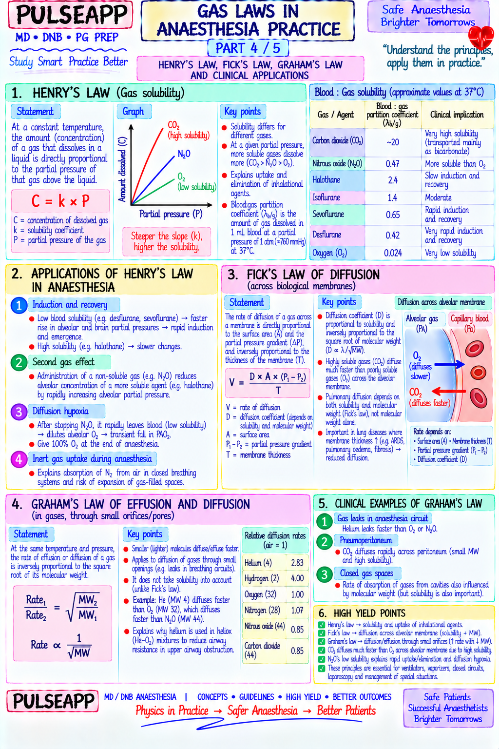
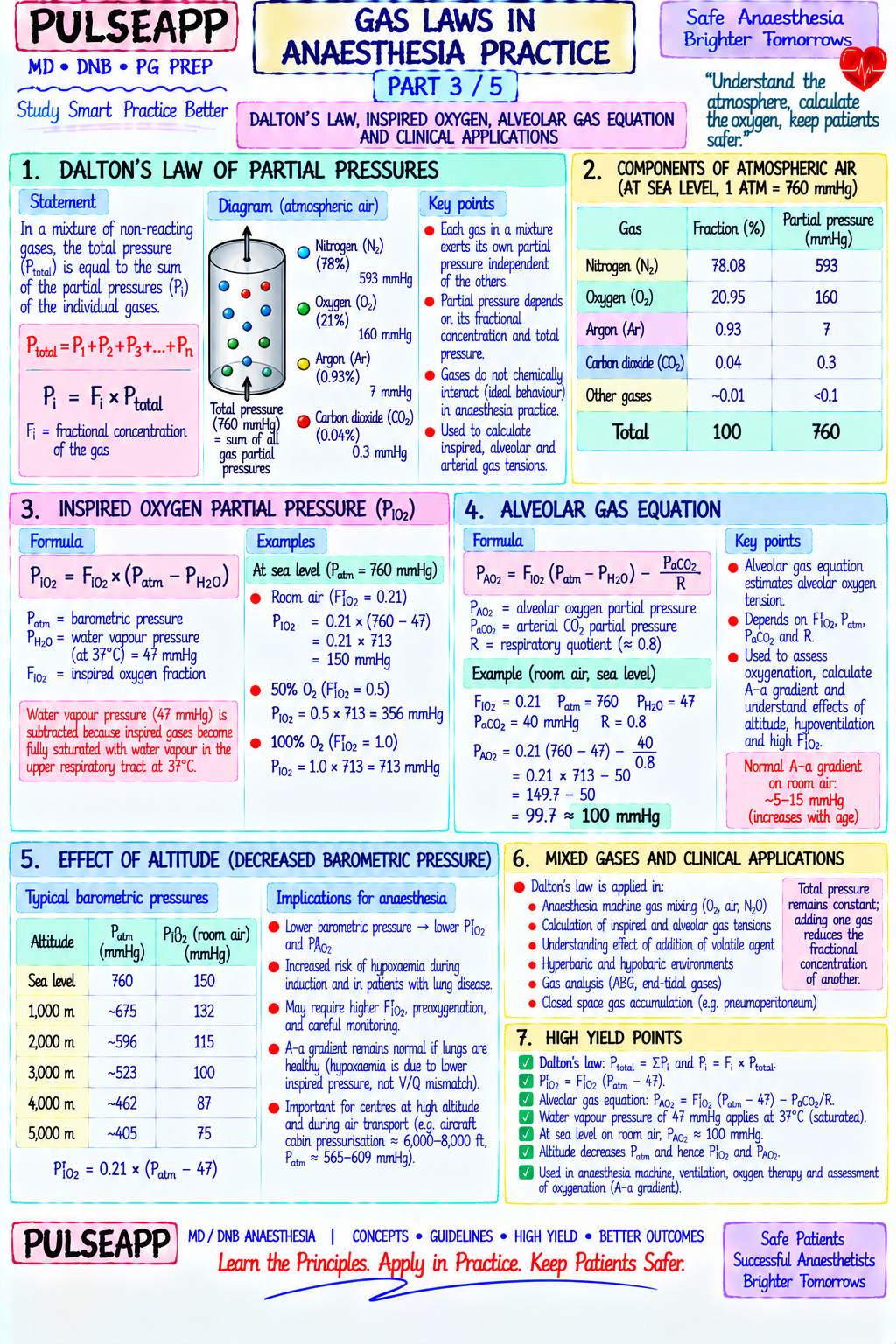
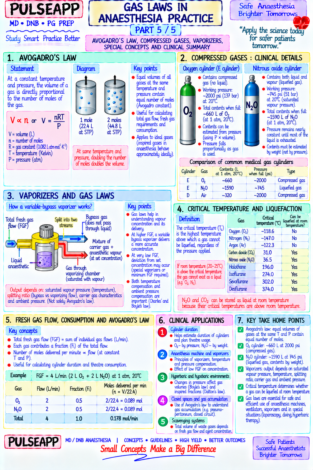
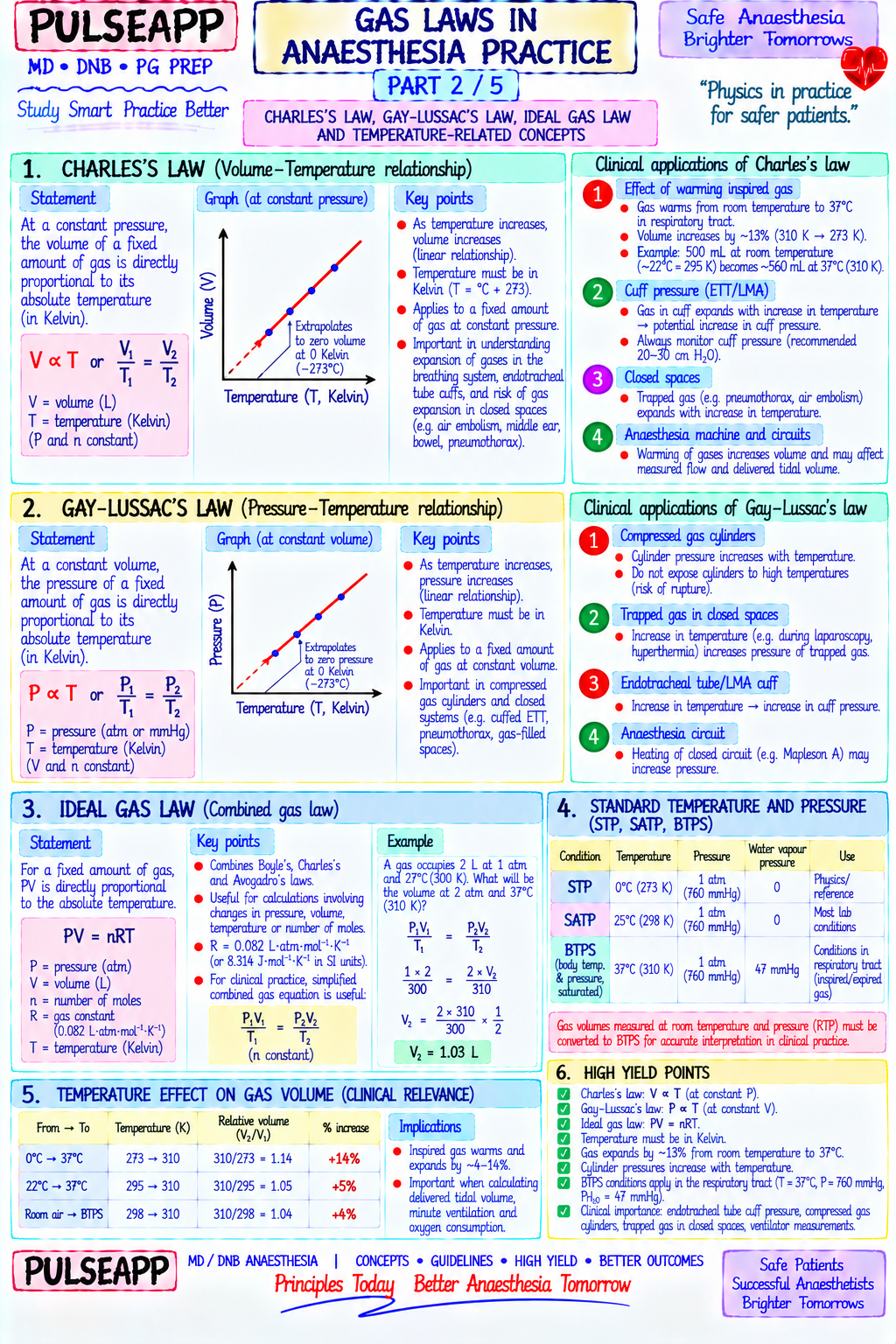
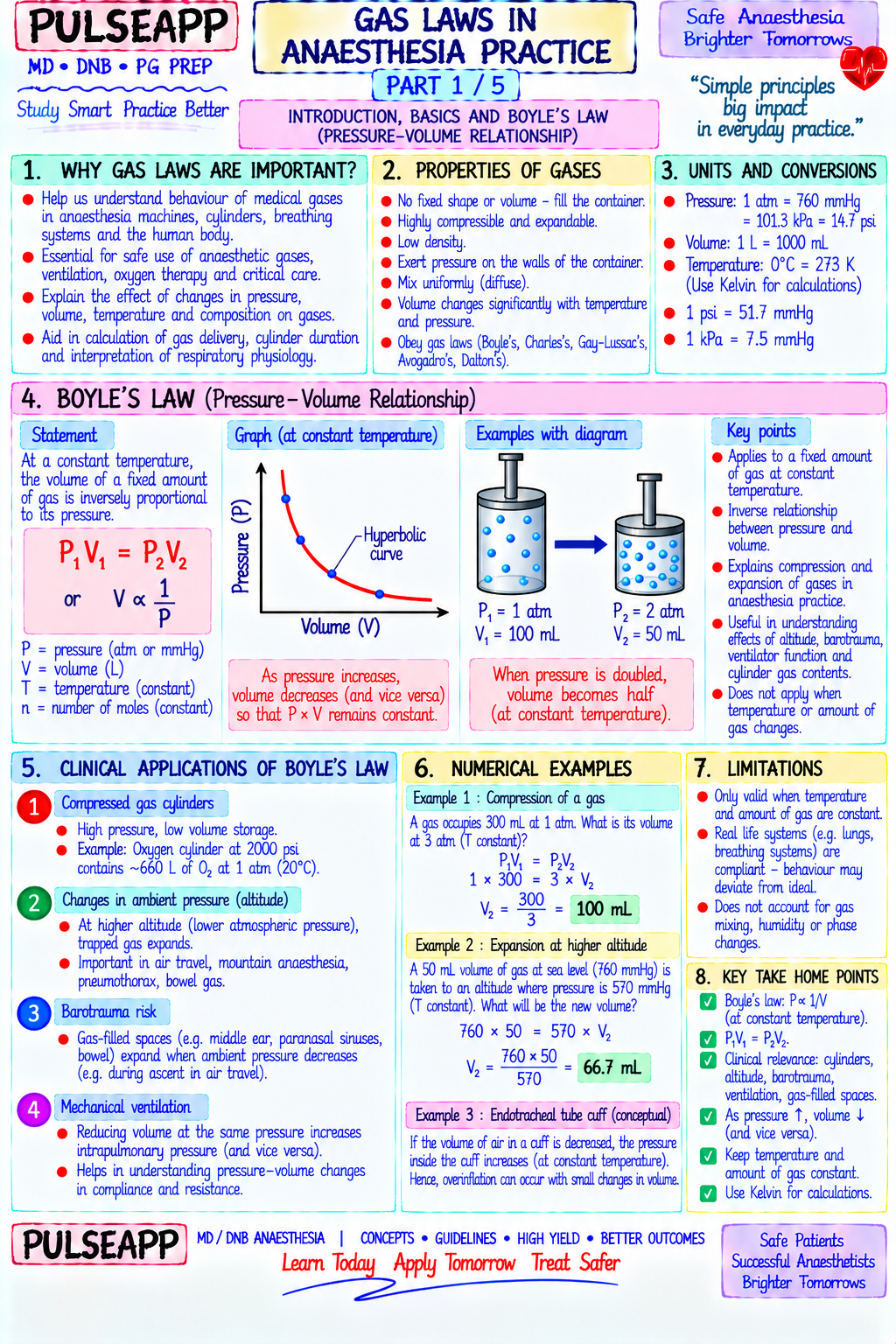
State and explain the gas laws relevant to anaesthesia practice. Apply each law to a specific clinical or equipment situation encountered in the operating theatre.

collections Question Diagrams & Reference Images (1)
Question Reference Diagram
zoom_in View Image
description Clinical Response
⚙ Core Concept
Every piece of equipment in the anaesthetic workstation — from the gas cylinder to the vaporizer to the flowmeter to the breathing circuit — operates according to specific physical gas laws. Every clinical scenario involving a gas-containing body cavity or compressed gas cylinder is governed by these same laws. The anaesthesiologist who understands these laws can predict equipment behaviour, anticipate physiological responses to pressure changes, and explain complications from altitude, diving, or N₂O use in closed spaces — all from a few elegant mathematical relationships. (Miller's Anaesthesia 9th Ed; Dorsch JA — Understanding Anaesthesia Equipment; Nunn's Applied Respiratory Physiology; Al-Shaikh B — Essentials of Anaesthetic Equipment)
A. Boyle's Law — Pressure-Volume Relationship2 marks
✅ Statement: At constant temperature, the volume of a fixed mass of gas is inversely proportional to its pressure — P × V = constant (k)
Anaesthetic Explanation Application Compressed O₂ cylinders at 137 bar contain gas at 137× atmospheric pressure in a fixed cylinder volume; when gas is released into the circuit, pressure falls as gas cylinders volume effectively increases to the room; remaining cylinder content is DIRECTLY proportional to pressure reading (gauge pressure ∝ remaining gas) — a half-full O₂ cylinder reads 68 bar. This linear relationship allows precise content calculation: V_remaining = P_gauge/P_full × V_cylinder N₂O cylinders N₂O is stored as a LIQUID at cylinder temperature; gauge pressure reflects the vapour pressure of liquid N₂O (~52 bar at 15°C) — which remains (DIFFERENT) constant regardless of how much N₂O remains in the cylinder as long as liquid is present; the pressure gauge does NOT fall until ALL liquid N₂O is vaporized and only gaseous N₂O remains; therefore, N₂O cylinder content cannot be determined from pressure alone — must be weighed Pneumothorax During ascent (e.g., aeromedical evacuation), atmospheric pressure falls; by Boyle's law, a fixed mass of gas in a pneumothorax expands as pressure at falls; a 20% pneumothorax at sea level (1 atm) will expand to 40% at cabin altitude equivalent of 0.5 atm; reason to drain pneumothorax before altitude/ascent aeromedical transport Entonox (50% At low temperatures, N₂O liquefies out of the mixture (Poynting effect); the remaining gas becomes O₂-rich at first then hypoxic N₂O-rich; cylinder must O₂/N₂O) be stored above −6°C and inverted/warmed before use
B. Charles's Law — Volume-Temperature Relationship1 mark
✅ Statement: At constant pressure, the volume of a fixed mass of gas is directly proportional to its absolute temperature — V/T = constant
Flowmeters (rotameters): flowmeters are calibrated at room temperature (15–20°C); gas passing through a flowmeter at body temperature (37°C) has a higher absolute temperature → larger volume → flowmeter may slightly underestimate actual gas volume delivered to the patient Spirometers and respirometers: measure expired gas volume at body temperature (37°C, BTPS — Body Temperature, ambient Pressure, Saturated with water vapour); when compared to volumes measured at standard conditions (STPD — Standard Temperature, Pressure, Dry), correction factors must be applied; the Wrights respirometer measures at BTPS; volumes are larger than at STPD by the temperature correction factor
C. Gay-Lussac's Law — Pressure-Temperature Relationship1 mark
✅ Statement: At constant volume, the pressure of a fixed mass of gas is directly proportional to its absolute temperature — P/T = constant
Hot cylinders are dangerous: a compressed gas cylinder (fixed volume) exposed to fire, direct sunlight, or elevated ambient temperature — pressure rises proportionally with absolute temperature; at high enough temperatures, pressure may exceed the cylinder's burst pressure → catastrophic cylinder rupture → cylinder becomes a projectile; hence the warning against heating gas cylinders and the requirement to store them away from heat sources Clinical relevance: explains why fire in an oxygen-enriched environment (as can occur in theatre fires) is so hazardous — the O₂ cylinder itself may explode; also explains why autoclaving cannot be used to sterilise compressed gas cylinders
D. Dalton's Law — Partial Pressures in Gas Mixtures2 marks
✅ Statement: The total pressure of a gas mixture equals the sum of the partial pressures of each component gas — P_total = P₁ + P₂ + P₃ + ...
where P_n = F_n × P_total Application Detail Alveolar PAO₂ = PiO₂ − (PaCO₂/RQ); PiO₂ = FiO₂ × (P_atm − P_H₂O) = FiO₂ × (760 − 47) mmHg; each gas contributes its fractional share of total alveolar pressure; gas water vapour (P = 47 mmHg at 37°C) "dilutes" all other alveolar gases equation O₂/N₂O In a 50% O₂/50% N₂O mixture at atmospheric pressure: P_O₂ = 0.5 × 760 = 380 mmHg; P_N₂O = 380 mmHg; each contributes half the total pressure; the mixtures anaesthetic potency of N₂O depends on its partial pressure (MAC of N₂O = 105% — requiring hyperbaric conditions for complete surgical anaesthesia) Altitude At altitude, P_atm falls; each component gas contributes the same fraction but at a lower absolute partial pressure; at 5000 m, P_atm = 405 mmHg; FiO₂ air effects = 0.21; P_O₂ inspired = 0.21 × (405 − 47) = 75 mmHg — explaining altitude hypoxia even with the same oxygen fraction as sea level Pipeline The oxygen proportioning system (Link-25) uses Dalton's law — it ensures O₂ contributes at least 25% of the total fresh gas flow, regardless of N₂O flow — gas mixing maintaining a minimum P_O₂ in the inspired mixture
E. Henry's Law — Gas Dissolution in Liquids2 marks
✅ Statement: At constant temperature, the amount of gas dissolved in a liquid is directly proportional to the partial pressure of that gas above
the liquid — [gas dissolved] = k_H × P_gas Application Explanation N₂O in closed N₂O has 34× greater blood solubility than N₂; blood in contact with closed gas spaces (pneumothorax, pneumoperitoneum, middle ear, bowel, gas spaces pneumocephalus) delivers N₂O rapidly down its partial pressure gradient into the gas space; N₂O enters faster than N₂ can leave (N₂ is less soluble) → gas space EXPANDS by up to 3× in volume if N₂O is continued; hence N₂O is contraindicated in pneumothorax, bowel obstruction, middle ear surgery, and any closed gas-containing space Blood-gas Describes the solubility of volatile anaesthetic agents in blood — applies Henry's law principle; agents with high blood-gas partition coefficient partition (halothane λ = 2.4) dissolve more in blood per unit partial pressure → blood acts as a "sponge" → slower rise of alveolar concentration → slower coefficient induction; agents with low λ (desflurane 0.42, sevoflurane 0.65) have limited blood dissolution → faster rise of PA → rapid induction; this is WHY λ (Ostwald) determines induction speed Decompression Divers breathe compressed air at depth; by Henry's law, increased pressure dissolves more N₂ into blood and tissues; rapid ascent reduces pressure sickness → N₂ comes out of solution faster than it can be transported to lungs → N₂ bubble formation in tissues → decompression sickness ("the bends") (diving) Oxygen Dissolved O₂ in plasma = 0.003 × PaO₂ mL/dL; at sea level PaO₂ 100 mmHg → 0.3 mL/dL dissolved O₂ (minor); at hyperbaric 3 atm on 100% O₂: therapy PaO₂ 2280 mmHg → 6.8 mL/dL dissolved O₂ — enough to meet basal tissue O₂ demand without haemoglobin → basis of hyperbaric O₂ therapy in severe anaemia
F. Graham's Law — Diffusion Rate1 mark
✅ Statement: The rate of diffusion of a gas is inversely proportional to the square root of its molecular weight — Rate ∝ 1/√MW
O₂ vs CO₂ across the alveolar membrane: CO₂ (MW = 44) vs O₂ (MW = 32); by Graham's law alone, O₂ should diffuse faster (lower MW); however, CO₂ is 20× more soluble in alveolar fluid/plasma than O₂; the net diffusion rate = solubility / √MW; CO₂ net diffusion rate is approximately 20× higher than O₂ — explaining why CO₂ diffuses rapidly across the alveolar membrane even in the presence of mild alveolar disease, while O₂ diffusion may fail (diffusion block presents as hypoxia without hypercapnia) Flowmeters calibration: rotameter flowmeters are calibrated for specific gases (each gas has a different density and viscosity affecting laminar/turbulent flow physics through the flowmeter tube); a flowmeter calibrated for O₂ will give a different true flow rate if used with another gas; rotameters cannot be simply interchanged between gases without recalibration
🎤 Viva Corner
Q. Why does N₂O cause pneumothorax to expand during general anaesthesia, and what is the specific mechanism based on gas laws?
The expansion of a pneumothorax during N₂O anaesthesia is explained by applying Henry's law and the principle of diffusion along partial pressure gradients. The gas within a pneumothorax is essentially room air — approximately 78% N₂, 21% O₂, and trace gases. When N₂O anaesthesia is commenced, N₂O builds up in the alveolar gas and consequently in the arterial blood (where its partial pressure rises progressively as more N₂O is inhaled). Blood now circulates to the tissues adjacent to the pneumothorax with a high partial pressure of N₂O. At the interface between the blood and the pneumothorax gas space: N₂O partial pressure in blood > N₂O partial pressure in the pneumothorax (near zero initially) → N₂O diffuses from blood into the pneumothorax gas space down its partial pressure gradient. The rate of this diffusion is determined by N₂O's solubility in blood — which is approximately 34 times greater than N₂. This means N₂O enters the pneumothorax 34 times faster than N₂ can leave (N₂ leaves the pneumothorax down its own partial pressure gradient, but its low blood solubility means it is carried away from the pneumothorax much more slowly). The net result: gas is being delivered to the pneumothorax (as N₂O) much faster than it can be removed (as N₂), causing progressive expansion. A 20% pneumothorax can double in size within 10–15 minutes of N₂O anaesthesia, potentially converting to a tension pneumothorax. This is why N₂O is absolutely contraindicated in known pneumothorax, as well as in other closed gas-containing spaces such as bowel obstruction, middle ear surgery, pneumocephalus, and certain retinal surgery with intraocular gas.
Q. A patient's O₂ cylinder gauge reads 50 bar (full cylinder = 137 bar, cylinder volume 10 L). How much O₂ remains, and for how long can it sustain a flow of 3 L/min?
By Boyle's law, the volume of gas remaining in the cylinder is proportional to the gauge pressure. Full cylinder: 137 bar × 10 L = 1370 L of gas at atmospheric pressure (this is the total gas content at STP when fully pressurised). At 50 bar: remaining gas = (50/137) × 1370 = 500 L of O₂ at atmospheric pressure. Duration at 3 L/min flow: 500 L ÷ 3 L/min = 167 minutes ≈ 2 hours 47 minutes. This calculation assumes constant flow rate and does not account for any dead volume in the cylinder valve or pressure regulation system. Practically, you should replace or have a backup cylinder ready when the pressure falls to approximately 30–40 bar (220–290 L remaining — approximately 1.5–2 hours at 3 L/min) rather than waiting for complete exhaustion, as pressure regulation becomes less reliable at very low pressures. For N₂O cylinders, this calculation DOES NOT APPLY — N₂O is stored as liquid and the gauge pressure remains constant at ~52 bar until all liquid is consumed; content must be determined by weighing the cylinder (tare weight marked on the cylinder body; full cylinder weight − tare weight = N₂O content in kg; 1 kg N₂O = 500 L at STP).
Q. Using Henry's law and the blood-gas partition coefficient, explain why desflurane produces much faster induction of anaesthesia than halothane. The speed of inhalational anaesthetic induction is determined by how rapidly the alveolar partial pressure of the anaesthetic rises to reach equilibrium with the brain partial pressure — and this rate is critically dependent on how much of the inhaled drug is "captured" by the blood versus remaining in the alveoli. Henry's law tells us that the amount of drug dissolved in blood is directly proportional to the partial pressure of the drug AND the drug's blood-gas partition coefficient (its Henry's constant for blood). Halothane has a blood-gas partition coefficient (λ) of 2.4 — this means for every unit of anaesthetic partial pressure in the alveolus, 2.4 units dissolve in each unit volume of blood passing through the pulmonary capillaries. Blood therefore acts as a very efficient "sponge" for halothane, absorbing large amounts of the drug from the alveolus with each cardiac cycle. This high uptake means the alveolar halothane concentration rises slowly — the alveolus is continuously being depleted by blood absorption faster than the inspired gas can replace it, so achieving an alveolar concentration equal to the inspired concentration (the FA/FI ratio reaching 1.0) takes many minutes. Desflurane, by contrast, has a blood-gas partition coefficient of only 0.42 — blood dissolves approximately 6× less desflurane per unit partial pressure than halothane. Blood is a poor sponge for desflurane; it absorbs very little with each pass through the alveolus; the alveolar concentration rises rapidly toward the inspired concentration because blood uptake barely depletes the alveolar reservoir. Therefore, the FA/FI ratio for desflurane rises to 0.9 within 5–10 minutes, while halothane takes 20–30 minutes — a direct consequence of the six-fold difference in their blood-gas partition coefficients and Henry's law governing gas dissolution.
★ Examiner's Pearl
State each law as a mathematical equation AND immediately link it to a specific clinical/equipment anaesthetic application — examiners reward integration of physics with clinical practice, not abstract physics alone. The N₂O cylinder vs O₂ cylinder difference (liquid storage vs compressed gas — cannot determine N₂O content from pressure, must weigh) is a classic examiner question. The N₂O expansion in closed gas spaces mechanism (Henry's law: N₂O solubility 34× N₂ → enters space faster than N₂ leaves → expansion) must be stated with the specific solubility ratio to score full marks.
Dorsch JA, Dorsch SE. Understanding Anaesthesia Equipment, 5th Ed, Chapters 1–3. Al-Shaikh B, Stacey S. Essentials of Anaesthetic Equipment, 4th Ed. Nunn's Applied Respiratory Physiology, 9th Ed. Miller RD et al. Miller's Anaesthesia, 9th Ed, Chapter 26. Butterworth JF, Mackey DC, Wasnick JD. Morgan & Mikhail's Clinical Anesthesiology, 6th Ed.
lock Protected Clinical Document • Pulse App • Screenshots Strictly Prohibited
QUESTION 28 person Asked by .
bookmark_add

Classify the Bain circuit within the Mapleson breathing system classification. Describe its coaxial design and the functional differences from the standard Mapleson D. State fresh gas flow requirements for spontaneous and controlled ventilation, advantages, disadvantages, and specific safety hazards including inner tube disconnection.

collections Question Diagrams & Reference Images (2)
Question Reference Diagram
zoom_in View Image
Question Reference Diagram
zoom_in View Image
description Clinical Response
⚙ Core Concept
The Bain circuit is the most widely used non-rebreathing circuit in anaesthesia practice — its elegant coaxial design solves the problem of delivering warm, humidified gas to the patient while maintaining the simplicity and portability of a Mapleson D circuit. However, its single most dangerous feature is the invisibility of the inner tube within the outer corrugated tube — an inner tube disconnection at the machine end creates a catastrophic failure mode (effectively converting the circuit to a dead-end bag) that can go undetected for several minutes while the patient rebreathes exhaled gas. Understanding this failure mode and how to detect it before use is a fundamental safety competency for every anaesthesiologist. (Miller's Anaesthesia 9th Ed; Bain JA, Spoerel WE — Anaesthesia 1972; Dorsch JA — Understanding Anaesthesia Equipment; Al-Shaikh B)
A. Classification in the Mapleson System2 marks

The Mapleson classification (1954) categorises non-rebreathing circuits by the relative position of three key components: the fresh gas flow (FGF) inlet, the reservoir bag/APL valve, and the patient connector Bain circuit = Mapleson D (coaxial modification): the FGF enters near the patient end (the machine-end inner tube delivers FGF to the patient connector); the reservoir bag and APL valve are at the MACHINE END — both features defining Mapleson D; the Bain circuit is simply a coaxial version of the Mapleson D, with the inner tube carrying fresh gas and the outer corrugated tube carrying expired gas Mapleson Classification FGF Location Bag/APL Location Rebreathing Efficiency A (Magill's) Near bag (machine end) Near patient (patient end) Most efficient for spontaneous breathing (FGF ≈ MV); least efficient for IPPV B Near patient Near patient Moderate efficiency C (Waters to-and-fro) Near patient Near patient Moderate efficiency D (Bain) Near patient (via inner tube) Machine end Most efficient for IPPV; needs higher FGF for SB than Mapleson A E (Ayre's T-piece) Near patient Open end (no bag) For paediatric use; low resistance F (Jackson-Rees) Near patient Open-ended bag Paediatric standard; allows manual IPPV

B. Structure — The Coaxial Design2 marks

The Bain circuit consists of a narrow inner tube (typically 7 mm diameter) running centrally within a wider corrugated outer tube (22 mm diameter). Fresh gas flows: from the FGF inlet at the MACHINE END → along the inner tube → to the PATIENT END (patient connector) — delivering fresh anaesthetic gas close to the patient's airway. Expired gas flows: from the patient → into the outer corrugated tube (surrounding the inner tube) → back toward the MACHINE END where the reservoir bag and APL valve are located.

Thermal exchange advantage: the warm expired gas flowing backward through the outer tube warms the incoming fresh gas in the central inner tube — a counter-current heat exchange effect that helps warm and humidify the fresh gas before it reaches the patient; reduces respiratory heat and moisture loss compared to standard Mapleson D

Length: standard adult Bain circuit is 1.8 metres (some circuits 1.5 m); this length provides adequate reservoir of expired gas in the outer tube while maintaining low resistance

Introduced by: Bain and Spoerel in 1972 in Anaesthesia — hence the eponym

C. Fresh Gas Flow Requirements3 marks
Spontaneous Breathing

In the Bain/Mapleson D during spontaneous breathing, FGF must be high enough to prevent rebreathing of expired CO₂ from the outer tube

During expiration: expired gas enters the outer tube and fills the corrugated reservoir; during inspiration, if FGF is insufficient, the patient may inhale expired gas from the near end of the outer tube — rebreathing occurs Minimum FGF for spontaneous breathing = 2–3 times the minute volume (MV); for an adult with MV = 5 L/min: FGF = 10–15 L/min; this is higher than Mapleson A (which only needs FGF ≈ MV) — making Bain less efficient than Mapleson A for spontaneous breathing

Controlled Ventilation (IPPV)

Mode Required FGF Rationale Spontaneous 2–3× MV = 10–15 L/min Must flush expired CO₂ from the outer tube reservoir before the next inspiration; high FGF requirement breathing (adult) Controlled 70–100 mL/kg/min — During IPPV, gas is actively delivered by the ventilator/hand ventilation, and the pattern of gas flow is more ventilation (IPPV) approximately 5–7 L/min for predictable; partial rebreathing is acceptable and controlled; this lower FGF makes Bain more economical — normocapnia average adult than for spontaneous breathing Paediatric (per 1000 mL/min for infants + 100 Paediatric circuits use Mapleson E/F (Ayre's T-piece, Jackson-Rees) rather than Bain for most children <25– weight) mL/min/kg (simplified Mapleson 30 kg E/F paediatric rule)

D. Advantages of the Bain Circuit1 mark

Lightweight and portable — no heavy CO₂ absorber or unidirectional valves

Suitable for head and neck surgery — the long circuit keeps the machine away from the surgical field Inspired gas warming by counter-current heat exchange (reduces respiratory heat and moisture loss)

Easy to clean and sterilise — no valves or complex components

Low resistance — suitable for spontaneous breathing (no unidirectional valves to open)

Scavenging easy — single exhaust port at the APL valve

E. Disadvantages & Specific Safety Hazards2 marks
⚠ The Critical Safety Hazard — Inner Tube Disconnection
The inner tube, which carries fresh gas, runs inside the outer corrugated tube and is therefore INVISIBLE throughout most of its length. If the inner tube disconnects at the MACHINE END (where it attaches to the FGF inlet): fresh gas no longer flows toward the patient; instead, the open inner tube at the patient end becomes a dead end into which expired gas is sucked; the circuit now functions as a blind loop — the patient rebreathes 100% expired gas with rising CO₂ and falling O₂; this causes progressive hypercapnia and hypoxia. The outer tube still connects the patient to the bag at the machine end — so manual ventilation APPEARS possible, creating a false sense of security while the patient is being ventilated with 100% CO₂-rich expired gas. Pethick's Test — Detecting Inner Tube Disconnection Before Use Step 1: Connect the Bain circuit to the FGF outlet of the anaesthetic machine; set a moderate FGF (4–6 L/min) Step 2: Occlude the patient end of the circuit completely with your thumb Step 3: Observe the reservoir bag at the machine end — it should INFLATE as the FGF fills the circuit with the patient end blocked; if the bag inflates → the inner tube (FGF pathway) is intact and delivering gas toward the patient end Step 4: Release the thumb occlusion — the bag should deflate (the Venturi effect from FGF flowing through the inner tube at high velocity creates a negative pressure in the outer tube, entraining and deflating the bag) If the bag does NOT inflate when the patient end is occluded: the inner tube is disconnected — fresh gas is not reaching the patient end (it is escaping somewhere proximally); the circuit is unsafe and must NOT be used High FGF requirement: 10–15 L/min for spontaneous breathing → expensive, environmental pollution, rapid depletion of gas supplies No CO₂ absorption: unlike the circle system, the Bain does not recycle expired gas — all gas is wasted; environmentally unfriendly for long cases Risk of barotrauma if APL valve is accidentally closed during controlled ventilation
🎤 Viva Corner
Q. During a Bain circuit pre-use check using Pethick's test, the reservoir bag does NOT inflate when you occlude the patient end with your thumb. What does this indicate, and what do you do?
Failure of the reservoir bag to inflate when the patient end is occluded during Pethick's test indicates that fresh gas is NOT being delivered to the patient end of the circuit — the inner tube has disconnected or is kinked at some point between the FGF inlet and the patient connector. When the patient end is blocked, all the FGF should have nowhere to go except back up the inner tube and into the circuit — filling the outer tube and inflating the reservoir bag. If the bag does not inflate, it means the inner tube pathway is broken; fresh gas is escaping through the disconnection point (usually at the machine-end connection of the inner tube to the FGF inlet) rather than travelling to the patient end. This circuit is UNSAFE and MUST NOT be used. Immediate actions: do not use this circuit on a patient; disconnect it from the anaesthetic machine; replace with a new, checked Bain circuit or switch to an alternative circuit (circle system, Mapleson A); perform the complete pre-use check on the replacement circuit; report the defective circuit for inspection and repair; document the finding in the anaesthetic pre-use checklist. If this is discovered mid-case (the patient has been breathing from a circuit with an inner tube disconnection): switch to manual ventilation with 100% O₂ via the new circuit immediately; assess the patient for CO₂ retention (ETCO₂ will be rising or already elevated), hypoxia, haemodynamic compromise from hypercapnia; increase minute ventilation to wash out the accumulated CO₂; inform the surgical team; consider ABG to assess the degree of CO₂ retention.
Q. Why does the Bain circuit require a higher fresh gas flow for spontaneous breathing than for IPPV, when intuitively IPPV seems to require more gas?
The apparent paradox resolves when we understand the mechanism of CO₂ rebreathing in the Bain circuit and how the pattern of gas flow differs between spontaneous and controlled ventilation. During spontaneous breathing: the pattern of gas flow is determined entirely by the patient's respiratory effort; during expiration, expired gas fills the outer corrugated tube; during the subsequent inspiration, the patient actively draws gas from the proximal (patient-end) portion of the outer tube — if the FGF has not flushed this expired gas away during the expiratory pause, the patient inhales expired CO₂ from the nearest portion of the outer tube; the FGF must be high enough to flush the outer tube during expiration to prevent this rebreathing; because expiratory time is limited and flow patterns are passive, this requires a high FGF (2–3× MV = 10–15 L/min) to reliably prevent CO₂ rebreathing. During IPPV: a mechanical ventilator actively delivers a controlled tidal volume and rate; during the expiratory phase, the FGF has the entire expiratory time to flush expired gas away from the patient end of the outer tube before the next mechanically-delivered breath; the controlled, predictable flow pattern allows the FGF to efficiently flush the circuit; additionally, in IPPV, some degree of controlled rebreathing can be deliberately accepted and managed by adjusting the ventilation rate and volume to maintain normocapnia; this makes the Bain circuit much more FGF-efficient during IPPV, requiring only 70–100 mL/kg/min (approximately 5–7 L/min for an adult) — less than half the flow required for spontaneous breathing.
Q. Why is the Mapleson A (Magill) circuit most efficient for spontaneous breathing but least efficient for IPPV, while the Bain (Mapleson D) is the opposite?
The efficiency of each Mapleson circuit in a particular ventilatory mode is determined by which gases vent through the APL valve at the end of expiration — circuits where predominantly expired CO₂-rich gas vents and fresh gas is preferentially retained are more efficient. In the Mapleson A (Magill) circuit: the FGF inlet is near the reservoir bag (machine end); during spontaneous expiration, expired gas travels from the patient toward the bag, but it first encounters the APL valve (which is at the patient end in Mapleson A); the expired alveolar gas (CO₂-rich) vents preferentially through the APL valve before reaching the bag; fresh gas from the FGF fills the tube nearest the bag for the next inspiration; very little fresh gas needs to be wasted, and FGF ≈ alveolar ventilation (3–5 L/min) is sufficient to prevent rebreathing. During IPPV with Mapleson A: the positive pressure of IPPV drives gas toward the bag first (away from the patient-end APL valve); fresh gas and expired gas mix in the bag; the high-pressure ventilation forces gas out through the APL valve in a mixed composition (not pure CO₂) → more fresh gas is wasted; FGF must be 2–3× MV to maintain normocapnia → very inefficient for IPPV. For Bain (Mapleson D): FGF arrives at the patient end; during expiration, expired gas fills the outer tube moving toward the machine end; fresh gas from the inner tube flushes expired gas toward the APL valve at the machine end during expiration; controlled ventilation produces a predictable, efficient flushing pattern; for spontaneous breathing, the patient may inhale expired gas from the proximal outer tube before FGF can flush it away → requires high FGF (2–3× MV) → inefficient for spontaneous breathing. The two circuits are therefore complementary — Mapleson A is the spontaneous breathing circuit of choice; Bain/D is the IPPV circuit of choice.
★ Examiner's Pearl
State the FGF requirements with specific numbers: spontaneous breathing = 2–3× MV (10–15 L/min adult); IPPV = 70–100 mL/kg/min (5–7 L/min adult) — these specific numbers are tested in DNB written papers. Pethick's test must be described step-by-step (occlude patient end → bag should inflate → release → bag deflates) with the interpretation (no inflation = inner tube disconnected = unsafe). The Mapleson efficiency comparison (A = most efficient for SB; D/Bain = most efficient for IPPV; reverse for each) is a classic examination table that must be reproduced with the mechanistic reasoning, not just memorised facts.
Bain JA, Spoerel WE. A streamlined anaesthetic system (Can Anaesth Soc J 1972;19:426-435). Mapleson WW. The elimination of rebreathing in various semi-closed anaesthetic systems (BJA 1954;26:323-332). Dorsch JA, Dorsch SE. Understanding Anaesthesia Equipment, 5th Ed, Chapter 7. Al-Shaikh B, Stacey S. Essentials of Anaesthetic Equipment, 4th Ed. Miller RD et al. Miller's Anaesthesia, 9th Ed, Chapter 26.
lock Protected Clinical Document • Pulse App • Screenshots Strictly Prohibited
QUESTION 29 person Asked by .
bookmark_add

A 2 kg neonate presents on Day 1 of life with gastroschisis. Describe the distinction from omphalocele, preoperative stabilisation priorities, anaesthetic technique for surgical repair, and postoperative challenges including ventilatory management.

collections Question Diagrams & Reference Images (1)
Question Reference Diagram
zoom_in View Image
description Clinical Response
⚙ Core Concept
Gastroschisis is a neonatal surgical emergency that demands simultaneous management of three catastrophic physiological threats: (1) massive evaporative heat and fluid loss from exposed bowel — the neonate is essentially losing the equivalent of 30–50% body surface area insensible losses; (2) progressive bowel ischaemia from vascular compromise and inflammatory peel; (3) intra-abdominal pressure crisis when the bowel is reduced back into a markedly underdeveloped abdomen. The anaesthesiologist and surgeon must act in parallel — neither waiting for perfect stabilisation before beginning nor rushing to surgery before the immediate life threats are controlled. (Miller's Anaesthesia 9th Ed; Cote CJ — Practice of Anaesthesia for Infants and Children; Adzick NS — Paediatric Surgery; Stringer MD)
A. Gastroschisis vs Omphalocele — Critical Distinction2 marks

Feature Gastroschisis Omphalocele (Exomphalos) Location of Para-umbilical — ALWAYS to the RIGHT of the umbilicus (very rarely THROUGH the umbilicus — the umbilical ring fails to close; umbilical cord defect left); the umbilical cord is intact and normally inserted inserts at the apex of the sac Sac/covering NO SAC — bowel is completely bare, directly exposed to amniotic fluid; SAC PRESENT — peritoneal sac covered by a membrane; sac may rupture bowel appears thickened, matted, and covered with an inflammatory but initially contains bowel and possibly liver fibrinous "peel"

Associated Rare — isolated bowel malrotation; NO chromosomal associations; 50% associated with chromosomal anomalies (Trisomy 18, 13, 21); cardiac anomalies 15% intestinal atresia (from intrauterine vascular accident) defects in 30%; Beckwith-Wiedemann syndrome (organomegaly, hypoglycaemia, macroglossia)

Surgical EMERGENCY — bare bowel exposed → progressive ischaemia, Semi-elective — sac provides protection; allows 24–48 hours for urgency inflammation, bacterial contamination; surgery within hours cardiac/chromosomal evaluation if stable; ruptured sac = emergency

Liver Liver rarely herniates — too large to exit small paraumbilical defect Liver herniates in ~50% of large defects — makes primary closure much more herniation difficult Bowel Foreshortened, inflamed, matted, no normal peristalsis; prolonged ileus If sac intact: bowel usually normal appearance and function; if sac ruptured: condition at expected post-operatively (weeks); TPN required similar to gastroschisis surgery

B. Preoperative Stabilisation — Immediate Priorities3 marks
Bowel Protection (First 5 Minutes)

Wrap eviscerated bowel immediately in warm, saline-moistened gauze swabs; then cover with transparent cling film (occlusive dressing) — reduces evaporative loss of heat and fluid; maintains bowel warmth; allows visual inspection of bowel colour and perfusion without repeated dressing changes

Positioning: right lateral decubitus — the weight of the bowel falls to the right, preventing kinking of the mesenteric vessels at the bowel-abdominal wall junction; reduces vascular compromise of the herniated gut Never squeeze, compress, or attempt to reduce the bowel manually at the bedside — forced reduction without anaesthesia dramatically raises intraabdominal pressure → inferior vena cava compression → cardiac arrest

Fluid Resuscitation — The Most Urgent Physiological Priority

Evaporative losses from the exposed bowel in a 2 kg neonate are 3–5× normal insensible losses; neonates have a very small total blood volume (~80–90 mL/kg = 160–180 mL for this 2 kg baby) and limited cardiovascular reserve

IV access: umbilical venous catheter (UVC) — fastest and most reliable access in the neonate; the umbilical vein is large, catheterisation is quick; position tip at the inferior vena cava/right atrial junction (confirmed by X-ray or ETCO₂ monitoring); peripheral IV as backup

Initial fluid bolus: 10–20 mL/kg of normal saline or Ringer's lactate over 10–15 minutes; reassess perfusion; repeat as needed; target urine output >1 mL/kg/hr, improving capillary refill (<3 seconds), improving HR toward 120–160 bpm, restoring MAP ≥40 mmHg in a neonate

Maintenance: 5–10% dextrose in 0.45% NaCl at 4–6 mL/kg/hr; glucose infusion rate 4–6 mg/kg/min to prevent hypoglycaemia (neonatal glycogen stores limited, especially in a stressed, unwell gastroschisis neonate); monitor glucose 1-hourly

Other Immediate Measures

NGT decompression: decompress the stomach and bowel via NGT on free drainage — reduces bowel distension, decreases the volume that must be reduced at surgery

Thermal management: overhead radiant warmer; warming mattress; all IV fluids warmed; the neonate's large surface area:body mass ratio makes hypothermia extremely rapid without active warming

Antibiotics: broad-spectrum IV antibiotics immediately (ampicillin + gentamicin ± metronidazole) — the exposed bowel is an open portal for bacterial contamination AVOID mask ventilation/CPAP/bag-mask ventilation before intubation — gas insufflation would distend the bowel, making reduction at surgery even more difficult

C. Anaesthetic Management for Surgical Repair3 marks
Induction — RSI in the Neonate

Full stomach/aspiration risk from GI obstruction and NGT (stomach not empty) → modified RSI Atropine 20 mcg/kg IV (minimum 100 mcg) before induction — neonates are highly vagotonic; laryngoscopy without prior atropine can cause severe reflex bradycardia and cardiac arrest

Induction: propofol 2–3 mg/kg IV or thiopentone 4–5 mg/kg IV; ketamine 1–2 mg/kg is an alternative (maintains cardiovascular stability in haemodynamically compromised neonate)

Muscle relaxant: suxamethonium 2 mg/kg IV for RSI (neonates require relatively higher doses than adults for equivalent block); alternatively rocuronium 1.2 mg/kg IV with sugammadex 16 mg/kg available

Intubation: use uncuffed ETT (neonatal airway is funnel-shaped — the narrowest point is the subglottis where the cricoid ring constricts the airway; a correctlysized uncuffed tube provides an airtight fit at this level); size: weight/10 + 3.5 = (2/10) + 3.5 = 3.7 → use 3.5 mm ID ETT; depth: weight + 6 = 8 cm at lip

Maintenance

Sevoflurane or isoflurane in O₂/air (avoid N₂O — distends bowel); TIVA with propofol/remifentanil is alternative

Fluid management: maintenance + replacement of surgical losses; gastroschisis surgery involves significant "third-space" losses into the peritoneum as the bowel is handled; typical additional intraoperative losses 10–20 mL/kg/hour; use crystalloid (Ringer's lactate) and colloid (albumin 4.5% if significant haemodynamic instability) Warm all gases and fluids; maintain temperature ≥36.5°C; operating theatre temperature 28°C (neonate) AVOID 100% O₂ — use minimum FiO₂ to maintain SpO₂ 94–98% in preterm; hyperoxia causes retinopathy of prematurity

The Intra-Abdominal Pressure (IAP) Challenge
⚠ Primary vs Staged Closure — The Critical Surgical-Anaesthetic Decision
The bowel of a gastroschisis neonate has never been in the abdomen — the abdominal cavity is markedly underdeveloped and small. Forcing all the bowel back into this cavity (primary closure) may raise intra-abdominal pressure to dangerous levels: IAP >20–25 mmHg → inferior vena cava compression → ↓ venous return → cardiovascular collapse IAP >25 mmHg → diaphragmatic splinting → respiratory failure; peak airway pressures rise dramatically IAP >30 mmHg → mesenteric ischaemia → bowel infarction Intraoperative monitoring: monitor peak airway pressure; bladder pressure (reflects IAP) via urinary catheter; maintain adequate MAP; if peak pressures rise >25 cmH₂O or MAP becomes unsustainable → surgeon must abandon primary closure → silo placement (bowel contents returned to a spring-loaded silo hung over the abdomen and gradually reduced over days to weeks).
D. Postoperative Management2 marks

Post-operative ventilation: almost all gastroschisis neonates require mechanical ventilation post-operatively; primary closure raises IAP → diaphragmatic splinting → impaired spontaneous ventilation; plan ICU admission and continued ventilation for 24–72 hours minimum

Prolonged ileus: the inflamed, peel-covered bowel of gastroschisis recovers function slowly — bowel sounds and function may not return for 2–6 weeks; parenteral nutrition (TPN) via a central line is essential until bowel function is confirmed (absence of bile in NGT, passage of stool, tolerance of enteral feeds)

Analgesia: morphine infusion (10–20 mcg/kg/hr) — provides analgesia and reduces respiratory effort against the ventilator; regional analgesia (caudal or spinal) is rarely feasible in the immediate post-operative period given the abdominal pathology

NEC surveillance: increased risk of necrotising enterocolitis post-gastroschisis (bowel ischaemia from vascular compromise, bacterial colonisation of inflamed bowel, immature gut immunity); monitor for abdominal distension, bloody stools, rising inflammatory markers

🎤 Viva Corner
Q. During primary closure of a gastroschisis repair, the peak airway pressure suddenly rises from 18 cmH₂O to 35 cmH₂O after the surgeon begins reducing the bowel. What do you do?
A sudden rise in peak airway pressure from 18 to 35 cmH₂O during bowel reduction indicates critical elevation of intra-abdominal pressure — the bowel cannot be safely returned to the underdeveloped abdominal cavity without causing respiratory and cardiovascular compromise. Immediate actions: inform the surgeon immediately — "peak airway pressure is 35 cmH₂O, we cannot safely proceed with primary closure at this pressure." Simultaneously: check the abdomen for bladder pressure (if a urinary catheter is in place, transduce the bladder to measure IAP — correlates reasonably with intra-abdominal pressure); reassess cardiovascular status: HR, MAP, SpO₂, capillary refill; check that the ETT is not kinked and that there is no pneumothorax (bilateral breath sounds, ETCO₂ waveform). If the pressure rise is purely from abdominal compression (not from a mechanical airway problem): the surgeon must STOP attempting primary closure; return the bowel to a position outside the abdomen; place a SILO (a spring-loaded plastic or Dacron bag/silo placed over the abdominal defect, into which the bowel contents are suspended); the silo allows gradual reduction of the bowel contents over 5–10 days as the abdomen stretches; daily reduction sessions in the ICU under sedation/analgesia; formal closure once IAP is acceptable. This staged approach is the preferred strategy when primary closure is not achievable, and has equivalent or better outcomes compared to forced primary closure in most series.
Q. Why is positioning the gastroschisis neonate in the right lateral decubitus position specifically important, and what happens if this is not done?
In gastroschisis, the bowel herniation is through a right paraumbilical defect. The mesentery supplying the herniated bowel must pass through this narrow defect — and the mesenteric vessels (the superior mesenteric artery and vein) travel through this same narrow opening from their origin in the retroperitoneum to supply the eviscerated gut. When the neonate is supine, the weight of the herniated bowel (which may be several times the volume of the abdominal cavity) pulls downward under gravity — this downward traction on the bowel causes the mesenteric vessels to kink at the point where they exit through the narrow fascial defect, creating a functional volvulus or mesenteric vessel occlusion. This produces progressive intestinal ischaemia from vascular compromise — which may cause bowel infarction within hours if not corrected. In the right lateral decubitus position, the weight of the bowel falls toward the right side — in the same direction as the defect — rather than pulling the mesentery at an angle across the defect; this removes the kinking stress on the mesenteric vessels, maintaining blood flow to the herniated gut. Additionally, this position prevents the bowel from falling across the midline and rotating, which would further compromise the mesenteric blood supply. The clinical urgency of correct positioning is real: a neonate left supine with gastroschisis for 30–60 minutes before an IV line is placed and transfer to the operating theatre is arranged may arrive with significantly more ischaemic bowel than if correctly positioned from the moment of diagnosis.
Q. Distinguish the anaesthetic implications of gastroschisis from those of omphalocele — what specific additional steps must you take for omphalocele that may not be needed in gastroschisis?
The fundamental anaesthetic difference stems from the dramatically different associated anomaly profiles of the two conditions. Gastroschisis is almost an isolated bowel defect — the key additional evaluations needed are focused on the bowel (assessing for intestinal atresia, bowel perfusion, length of viable gut) and the physiology of acute fluid and heat loss. Omphalocele, by contrast, carries a 50% risk of chromosomal anomalies (Trisomy 13, 18, 21) and a 30% risk of congenital cardiac defects — making a complete pre-anaesthetic evaluation mandatory before proceeding: echocardiography to define the cardiac anatomy, chromosomal analysis, assessment for Beckwith-Wiedemann syndrome (hypoglycaemia from pancreatic hyperplasia — monitor glucose hourly; macroglossia — airway management considerations; organomegaly), and assessment for other VACTERL-type associations. The airway may be more challenging in omphalocele due to Beckwith-Wiedemann macroglossia. Cardiac defects may require specific modifications to the anaesthetic (avoidance of agents that further reduce cardiac output, potentially maintaining a right-to-left shunt via the ductus arteriosus in duct-dependent circulations). For Trisomy 18 specifically, palliative care discussions should occur with parents before surgical intervention, given the very high early mortality. For gastroschisis: the urgency means there is rarely time for full chromosomal evaluation before surgery — focus on the surgical emergency and the immediate physiological priorities; the bowel pathology itself is more severe (inflamed, peelcovered, possibly atretic) and postoperative TPN/ileus management is more prolonged than omphalocele.
★ Examiner's Pearl
The gastroschisis vs omphalocele distinction table is the most tested single comparison in paediatric surgical anaesthesia — reproduce all five distinguishing features (location, sac, anomalies, urgency, liver herniation). The IAP monitoring strategy during bowel reduction — peak airway pressure as proxy for IAP, threshold of 25–30 cmH₂O for abandoning primary closure and placing a silo — is the specific clinical decision point examiners test. Atropine before induction in neonates (vagotonia → severe reflex bradycardia with laryngoscopy) is a mandatory specific neonatal safety fact.
Cote CJ, Lerman J, Anderson BJ. A Practice of Anaesthesia for Infants and Children, 6th Ed. Adzick NS et al. Correction of gastroschisis in utero — controversies. Miller RD et al. Miller's Anaesthesia, 9th Ed, Chapter 93 (Paediatric Anaesthesia). Stringer MD, Subramaniam R. Pediatric Surgery and Urology: Long-Term Outcomes. Collins S et al. Gastroschisis — anaesthetic considerations (Cont Ed Anaesth Crit Care Pain 2010;10:165-169).
lock Protected Clinical Document • Pulse App • Screenshots Strictly Prohibited
QUESTION 30 person Asked by .
bookmark_add

Describe CSF production, circulation, and absorption. Classify hydrocephalus. Outline the anaesthetic management for ventriculoperitoneal (VP) shunt insertion and endoscopic third ventriculostomy (ETV), emphasizing ICP control and neurophysiological monitoring.

collections Question Diagrams & Reference Images (3)
Question Reference Diagram
zoom_in View Image
Question Reference Diagram
zoom_in View Image
Question Reference Diagram
zoom_in View Image
description Clinical Response
⚙ Core Concept
CSF circulation is a precisely balanced dynamic system — production and absorption must match exactly to maintain normal ICP of 10–15 mmHg. Hydrocephalus represents a failure of this balance, producing progressive ventricular enlargement and raised ICP with all its consequences. The anaesthesiologist managing a patient with raised ICP from hydrocephalus must simultaneously optimise cerebral perfusion pressure (MAP − ICP ≥ 60 mmHg), prevent secondary injury from hypoxia or hypercapnia, and provide conditions for safe neurosurgical intervention. (Miller's Anaesthesia 9th Ed; Cottrell JE — Neuroanesthesia; Bhardwaj A — Handbook of Neuroanesthesia)
A. CSF Production, Circulation & Absorption3 marks

Parameter Value Total CSF volume (adult) ~140–150 mL; neonates: 10–60 mL Production rate 500 mL/day (0.35 mL/min); produced continuously by choroid plexus (70%) + ependymal cells (30%) Normal ICP (lateral recumbent) 10–15 mmHg (7–10 mmHg in neonates with open fontanelle) Lateral ventricles (choroid plexus production) → Foramen of Monro (interventricular foramina) → Third ventricle → Aqueduct of Sylvius (most common site of obstruction → non-communicating hydrocephalus) → Fourth ventricle → Foramina of Magendie (median) + Luschka (lateral ×2) → Subarachnoid space (basal cisterns → cerebral convexities) → Arachnoid granulations (Pacchionian bodies) → Superior sagittal sinus (venous absorption)

Drug effects on CSF production: ↑ production: vasodilators, volatile anaesthetics (halothane most, sevoflurane/desflurane least), N₂O; ↓ production: acetazolamide (carbonic anhydrase inhibitor), furosemide, steroids; ↑ absorption: mannitol (raises osmotic gradient), furosemide

Monroe-Kellie doctrine: total intracranial volume = brain + blood + CSF = constant (skull is a rigid box); ↑ in any one component must be compensated by ↓ in another; compensation buffers ICP initially (CSF displaced to spinal subarachnoid space, venous blood expelled); once compensatory mechanisms exhausted → exponential ICP rise

B. Hydrocephalus Classification2 marks

Type Mechanism CSF Pressure Common Causes Obstructive Block WITHIN the ventricular system — CSF cannot flow from High Aqueduct stenosis (most common), tectal glioma, Chiari (non- ventricles to subarachnoid space; all ventricles proximal to the malformation, colloid cyst of 3rd ventricle, posterior fossa communicating) block are dilated tumours Communicating Block OUTSIDE the ventricular system — CSF flows from High (usually) Post-meningitic fibrosis of arachnoid granulations, postventricles to subarachnoid space but absorption at arachnoid SAH blood products blocking granulations, carcinomatous granulations is impaired; all four ventricles dilated meningitis Normal Communicating hydrocephalus with normal or intermittently normal Normal (10–15 Hakim's triad: Wet (urinary incontinence) + Wacky Pressure ICP; pathogenesis incompletely understood; classic triad: Hakim's mmHg) but (dementia) + Wobbly (gait apraxia/ataxia); idiopathic in Hydrocephalus triad intermittent elderly; responds to VP shunt (NPH) spikes Hydrocephalus Apparent ventricular enlargement from brain tissue loss (atrophy); Normal Alzheimer's disease, chronic ischaemia, post-traumatic; ex vacuo ICP is NORMAL; not true hydrocephalus distinguished from true hydrocephalus by normal ICP and lack of periventricular oedema on MRI

C. Anaesthetic Management — VP Shunt Insertion3 marks

Preoperative Assessment

Clinical ICP assessment: Cushing's triad (hypertension + bradycardia + abnormal respirations) indicates severely elevated ICP with brainstem compression — anaesthetic induction in this state requires extreme care (avoid further ICP elevation from laryngoscopy, coughing, straining) GCS, pupillary reflexes, CT scan (ventricle size, midline shift, evidence of herniation) Full stomach? — urgent VP shunt for acute hydrocephalus → aspiration precautions mandatory

Induction — The Most Critical Phase in Raised ICP

⚠ Avoid ICP Spikes During Induction — Laryngoscopy and Intubation are the Highest-Risk Moments
Preoxygenate adequately; ensure MAP is adequate (CPP = MAP − ICP; if ICP = 30 and MAP = 70 → CPP = 40 mmHg — critically low) Induction: thiopentone (3–5 mg/kg) or propofol (2–3 mg/kg) — both reduce ICP by reducing CMRO₂ and secondary CBF; avoid ketamine (increases CMRO₂, raises ICP) Lignocaine 1.5 mg/kg IV 3–5 minutes before intubation — attenuates the ICP spike from laryngoscopy Rocuronium 0.6–1.2 mg/kg for intubation; suxamethonium (though traditionally avoided due to fasciculation → transient ICP rise — this ICP rise is modest and its use is acceptable for RSI in full-stomach neurosurgery patient) Establish normoventilation immediately after intubation — maintain PaCO₂ 35–40 mmHg; hyperventilation (↓PaCO₂) is used ONLY for acute herniation crisis as a temporary bridge (causes cerebral vasoconstriction → reduces CBV → reduces ICP temporarily) Maintenance TIVA preferred for neurosurgery with raised ICP: propofol + remifentanil; propofol reduces CMRO₂, CBF, and ICP; remifentanil provides excellent analgesia with brief post-infusion effects; no effect on cerebral autoregulation at clinical doses If volatile used: ≤0.5 MAC of sevoflurane or isoflurane (higher doses cause cerebral vasodilation → increase CBV → increase ICP; all volatile agents impair cerebral autoregulation dose-dependently); maintain normocapnia Head position: 15–30° head-up (reduces ICP by improving jugular venous drainage); avoid extreme rotation or neck flexion (obstructs jugular venous drainage → raises ICP) Mannitol 0.5–1 g/kg IV over 15–20 minutes if ICP is acutely elevated intraoperatively (creates osmotic gradient → draws water from brain interstitial fluid → reduces cerebral water content → reduces ICP over 15–30 minutes) ETV (Endoscopic Third Ventriculostomy) — Specific Considerations ETV creates a fenestration in the floor of the third ventricle → allows CSF to bypass the obstructing aqueduct and drain into the basal cisterns; preferred for obstructive hydrocephalus in children >6 months old (lower failure rate than in younger infants) Bradycardia during ETV: manipulation of the floor of the third ventricle is adjacent to the hypothalamus and mammillary bodies — sudden severe bradycardia (even asystole) can occur from distension of the third ventricle with irrigation fluid or from direct hypothalamic stimulation; atropine and ephedrine must be immediately available; if sustained bradycardia occurs, inform the surgeon to cease ventricular irrigation/manipulation immediately Hypothermia from irrigation: large volumes of room-temperature irrigating fluid can cause hypothermia, particularly in children; all irrigation fluid should be warmed to 37°C
D. ICP Control — The Five-Tier Approach2 marks

Tier Intervention Mechanism 1 Head position: 15–30° head-up; neutral neck Improves jugular venous drainage → reduces cerebral venous blood volume → lowers ICP 2 Normocapnia (PaCO₂ 35–40 mmHg); normoxia (SpO₂ >95%); Hypercapnia → cerebral vasodilation → ↑CBV → ↑ICP; hypoxia → cerebral oedema; fever normothermia → ↑CMRO₂ → ↑CBF → ↑ICP 3 Osmotherapy: mannitol 0.5–1 g/kg IV or 3% hypertonic saline Osmotic gradient draws interstitial water out of brain → reduces cerebral volume → 3–5 mL/kg reduces ICP; effect within 15–20 minutes; lasts 3–4 hours 4 Controlled hyperventilation (PaCO₂ 30–35 mmHg) — Hypocapnia → cerebral vasoconstriction → ↓CBV → ↓ICP; effect immediate but tolerance TEMPORARY ONLY develops within 4–6 hours; brain ischaemia risk if prolonged; used ONLY as bridge to definitive treatment 5 Barbiturate coma (thiopentone 3–5 mg/kg boluses, infusion 1– Maximum CMRO₂ reduction (burst-suppression EEG = 50% CMRO₂ reduction); reserves (salvage) 5 mg/kg/hr) targeting burst-suppression EEG; decompressive for refractory ICP not controlled by tiers 1–4 craniectomy

🎤 Viva Corner
Q. During ETV, the surgeon begins irrigation of the third ventricle. The heart rate suddenly drops from 80 to 28 bpm. What is happening, and what do you do?
This is acute bradycardia from hypothalamic/third ventricle stimulation during ETV — a well-recognised and potentially life-threatening complication. The floor of the third ventricle is immediately adjacent to critical hypothalamic structures including the mammillary bodies and the tuber cinereum; distension of the third ventricle from irrigation fluid or direct mechanical stimulation during fenestration can trigger a vasovagal-type reflex causing severe bradycardia (and occasionally asystole). Immediate actions: call the surgeon to stop all irrigation immediately and remove the endoscope if the bradycardia does not resolve within seconds of stopping manipulation — this is the most important single intervention, as the stimulus (mechanical/pressure) must be removed. Give atropine 500 mcg IV if the heart rate is below 40 bpm or the patient is haemodynamically compromised (BP falling); dose may be repeated. Give ephedrine 3–6 mg IV if there is concurrent hypotension from reduced cardiac output. Ensure adequate oxygenation (manual ventilation if needed to maintain SpO₂). Continue ECG monitoring and document the event. If sinus rhythm does not restore within 30 seconds of stopping stimulation and administering atropine, start CPR. Once resolved, discuss with the neurosurgeon about proceeding cautiously versus abandoning and rescheduling. This event should be clearly documented and the patient counselled post-operatively about the intraoperative complication.
Q. Explain the Monroe-Kellie doctrine and why it predicts that ICP rises exponentially rather than linearly once compensatory mechanisms are exhausted. The Monroe-Kellie doctrine states that the total volume within the rigid skull is constant and equals the sum of three compartments: brain tissue (approximately 80% of total volume), blood (approximately 10%), and CSF (approximately 10%). Since the skull cannot expand (after fontanelle closure in infancy), any increase in the volume of one compartment MUST be compensated by a proportional decrease in one or both of the others, otherwise ICP must rise. The compensatory mechanisms are: CSF displacement — CSF can be displaced from the cranial subarachnoid space into the compliant spinal subarachnoid space through the foramen magnum; and venous blood displacement — cerebral venous blood can be squeezed from the highly compliant intracerebral veins and dural sinuses into the systemic venous circulation. These compensatory mechanisms are finite — once all available CSF has been displaced to the spine and all compressible venous blood has been expelled, the intracranial compliance (ΔV/ΔICP — volume that can be absorbed per unit ICP rise) falls to near zero. At this point, even very small additional volume increments (a breath, a cough, a small haematoma) produce enormous ICP spikes — the pressure-volume curve becomes exponential (or rather, the compliance curve becomes a steep exponential fall). This explains why ICP can be maintained near-normal by compensation for relatively large lesions early in their course, then suddenly decompensate catastrophically when the compensatory reserve is exhausted — the "critical pressure" point on the exponential ICP curve. It also explains why even a small increment — 1 mL of additional blood — can raise ICP from 20 to 80 mmHg once compensation is lost. Q. Why is controlled hyperventilation described as "temporary only" for ICP control, and what happens if you continue it beyond 4–6 hours?
Controlled hyperventilation reduces ICP by lowering PaCO₂, which causes cerebral arteriolar vasoconstriction — directly reducing cerebral blood volume (CBV) and therefore ICP. This effect begins within seconds to minutes of reducing PaCO₂. However, the cerebral vasculature adapts to the new PaCO₂ set-point through bicarbonate shifts: over 4–6 hours, the choroid plexus reduces CSF bicarbonate secretion and the blood-brain barrier equilibrates the new pH — the cerebral pH normalises despite the continued low PaCO₂. Once adaptation is complete, the vasoconstriction reverts toward baseline and the ICP reduction effect is largely lost, despite maintaining the same low PaCO₂. This means hyperventilation becomes ineffective at reducing ICP beyond the first 4–6 hours in sustained application. More dangerously: if hyperventilation is then abruptly stopped (returning to normocapnia), the brain cells — adapted to the low PaCO₂ — now see an acute relative hypercarbia, causing rebound cerebral vasodilation and potentially a severe rebound ICP spike that overshoots the pre-hyperventilation level. Additionally, sustained vasoconstriction from prolonged hyperventilation carries a real risk of secondary ischaemia, particularly in areas of the brain with already-compromised perfusion — reducing CBF below the ischaemic threshold. For these reasons, hyperventilation is reserved for the acute management of impending herniation (as a bridge of 30– 60 minutes while definitive treatment is organised) and should not be used as a sustained ICP control strategy; instead, osmotherapy, sedation, and surgical decompression are the appropriate sustained treatments.
★ Examiner's Pearl
The CSF production rate (500 mL/day, 0.35 mL/min) and total volume (140–150 mL) are specific numbers tested in written papers. The flow pathway with the specific site most commonly obstructed (Aqueduct of Sylvius → non-communicating hydrocephalus) must be stated. Hakim's triad (wet, wacky, wobbly = incontinence, dementia, gait apraxia) for NPH is a classic viva question about a specific clinical entity. The five-tier ICP management table must be reproduced in order — examiners award marks for each tier correctly placed and mechanistically explained.
Miller RD et al. Miller's Anaesthesia, 9th Ed, Chapter 70 (Neurosurgical Anaesthesia). Cottrell JE, Young WL. Cottrell and Young's Neuroanesthesia, 5th Ed. Bhardwaj A et al. Handbook of Neurocritical Care. Cote CJ et al. A Practice of Anaesthesia for Infants and Children, 6th Ed. Hakim S, Adams RD. The special clinical problem of symptomatic hydrocephalus with normal cerebrospinal fluid pressure (J Neurol Sci 1965).

Showing 2130 of 337 questions

account_tree

Subcategory Tree

Explore Anesthesia subcategories

folder_special Anesthesia
Main
lock

Category Subscription

Subscribe to Anesthesia to unlock this module and all nested subcategories.

  • check_circle Access Anesthesia & all subcategories
  • check_circle Detailed, Peer-Reviewed Answers
  • check_circle High-yield visual aids & imaging
Get Category Subscription arrow_forward
Secure 256-bit SSL Connection

Anatomical Models

Explore high-fidelity 3D visualizations included in premium modules.

Case Reviews

Real-world clinical scenarios narrated by senior consultants.

Pulse App