Anesthesia | Eklavya Medical
vaccines
medical_services Main Specialty Domain lock Subscription Required

Anesthesia

Clinical anesthesiology, local and general anesthesia delivery systems, monitoring, and perioperative care.

verified Verified Medical Faculty menu_book 337 Q&A Modules
lock Protected Clinical Document • Pulse App • Screenshots Strictly Prohibited
QUESTION 31 person Asked by .
bookmark_add

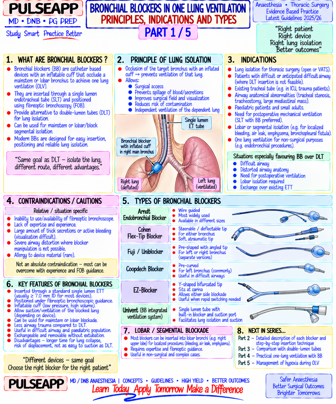
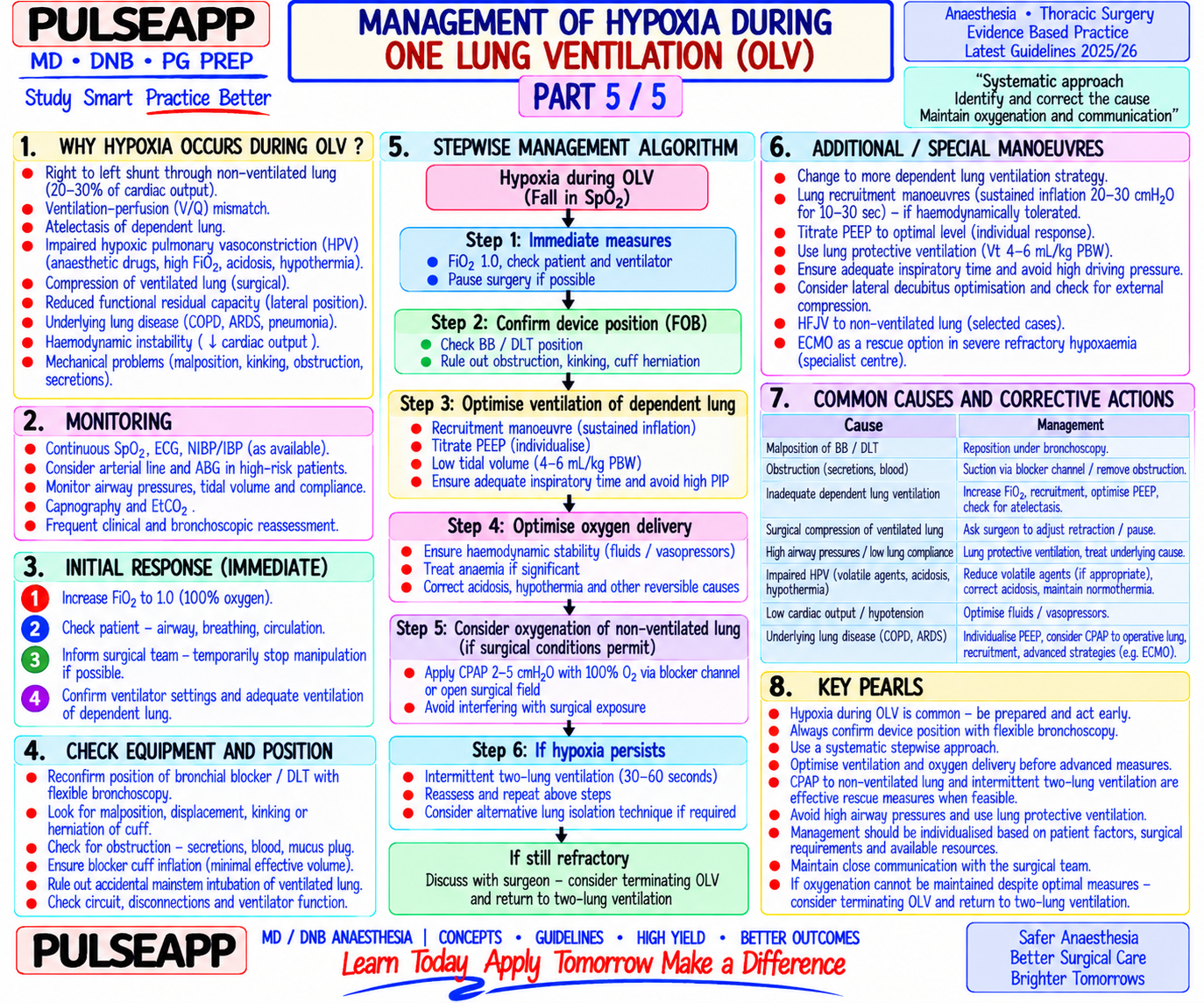
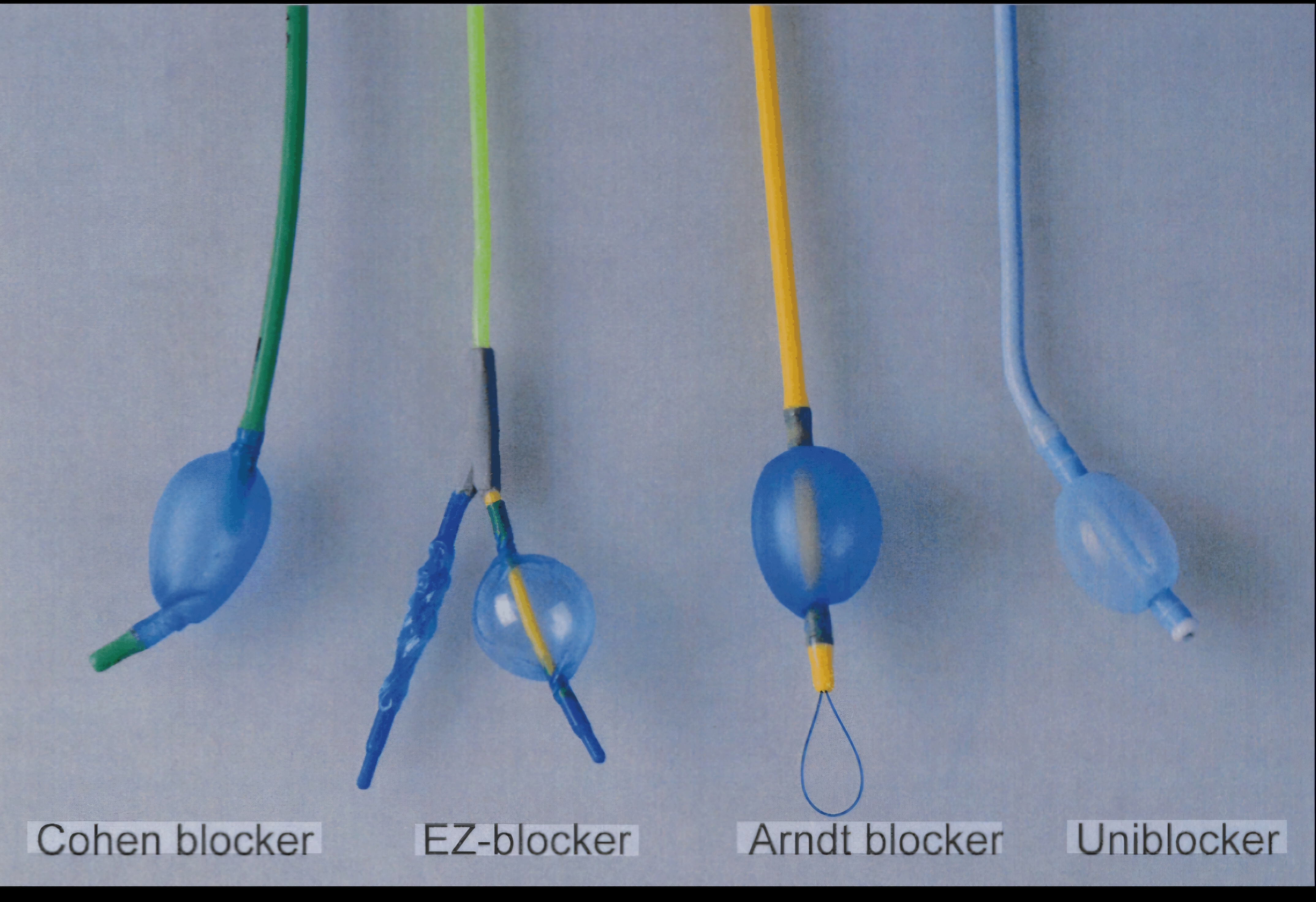
Describe bronchial blockers — types available, insertion technique, advantages and disadvantages compared to double-lumen tubes, and management of hypoxia during one-lung ventilation.

collections Question Diagrams & Reference Images (2)
Question Reference Diagram
zoom_in View Image
Question Reference Diagram
zoom_in View Image
description Clinical Response
⚙ Core Concept
Bronchial blockers provide selective bronchial occlusion and one-lung ventilation (OLV) through a standard single-lumen ETT — avoiding the bulk and rigidity of the double-lumen tube (DLT). They are the device of choice when a DLT is technically difficult or impossible: difficult airway, small children, the patient already intubated with a single-lumen tube, tracheostomy, and upper airway pathology. However, they have specific limitations — slower lung collapse, inability to suction the operative lung, and risk of displacement during surgery that must be understood to use them safely. (Miller's Anaesthesia 9th Ed; Benumof JL — Thoracic Anaesthesia; Campos JH — Bronchial blockers vs DLT)
A. Types of Bronchial Blockers3 marks

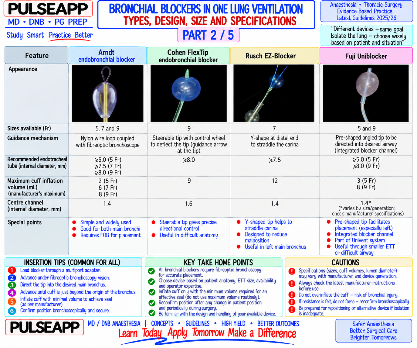
Device Key Feature Sizes Specific Advantage Arndt Wire- Contains a wire loop at the tip that couples to the fiberoptic bronchoscope (FOB) — the FOB 5, 7, 9 Fr Most widely used; the wire-loop coupling Guided is threaded through the loop, then advanced under direct vision into the target bronchus; the makes bronchoscope-guided positioning Blocker blocker follows the bronchoscope and wire; once positioned, the wire is removed leaving the very reliable; 9 Fr has a large central channel lumen allowing CPAP to the operative lung Cohen Tip- A wheel mechanism at the proximal end deflects the blocker tip up to 90° in any direction — 9 Fr More directional control; useful when the Deflecting allows steering into the target bronchus under FOB vision; no need for wire coupling Arndt wire cannot reach the target bronchus; Blocker large central lumen for CPAP Fuji Pre-shaped curved tip; positioned under FOB guidance; no wire; simple design; the curve 5, 9 Fr Simplest design; can be used with any Uniblocker directs the blocker toward the target bronchus when rotated single-lumen ETT ≥7.5 mm ID; lower cost (Coopdech) EZ-Blocker A Y-shaped device with two cuffs — one for each main bronchus; inserted through the ETT Universal Can switch operative side quickly; positioned (Y-shaped) and positioned at the carina; one cuff is inflated to block the operative bronchus; allows at carina (very stable — rarely displaced); switching to block either side without FOB repositioning the Y-shape means it straddles the carina Torque Designed for paediatric use; smaller profile; positioned under FOB guidance Paediatric Provides OLV option in children where DLTs Control sizes are not available (<8 years, <30 kg) Blocker (Vitaid)

B. Insertion Technique2 marks

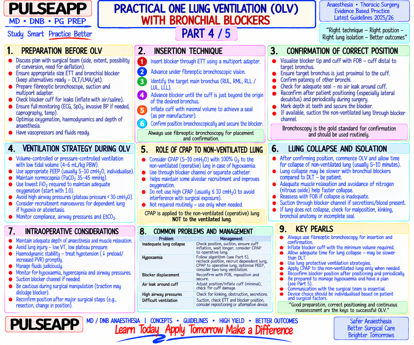
ETT size requirement: a standard single-lumen ETT of at least 7.5–8.0 mm ID is needed to accommodate both the bronchial blocker and the fiberoptic bronchoscope simultaneously (the FOB occupies ~4 mm; the blocker ~5–9 Fr); some centres use a dedicated multiport airway adapter that allows simultaneous passage of the blocker, bronchoscope, and ventilation circuit without circuit disconnection Step-by-step (Arndt wire-guided example): 1. Intubate with a ≥7.5 mm ETT; connect multiport adapter to ETT hub 2. Insert the Arndt blocker through the blocker port of the multiport adapter (blocker wire loop at the tip) 3. Insert the FOB through the bronchoscope port of the adapter; thread the FOB through the wire loop of the Arndt blocker 4. Advance the FOB under direct vision into the TARGET bronchus (left for left-sided surgery; right for right-sided surgery in most cases); once the FOB is in the correct bronchus, the blocker wire loop guides the blocker to follow the FOB into the same bronchus 5. Remove the FOB; inflate the blocker cuff with 5–8 mL air under direct FOB vision (re-insert FOB to confirm blocker position) — cuff inflated in the bronchus creates the seal for OLV 6. Remove the wire; ventilate the contralateral lung through the ETT lumen; the blocker allows the operative lung to collapse through passive resorption of gas through the inflated cuff

C. Bronchial Blockers vs Double-Lumen Tubes — Comparison3 marks

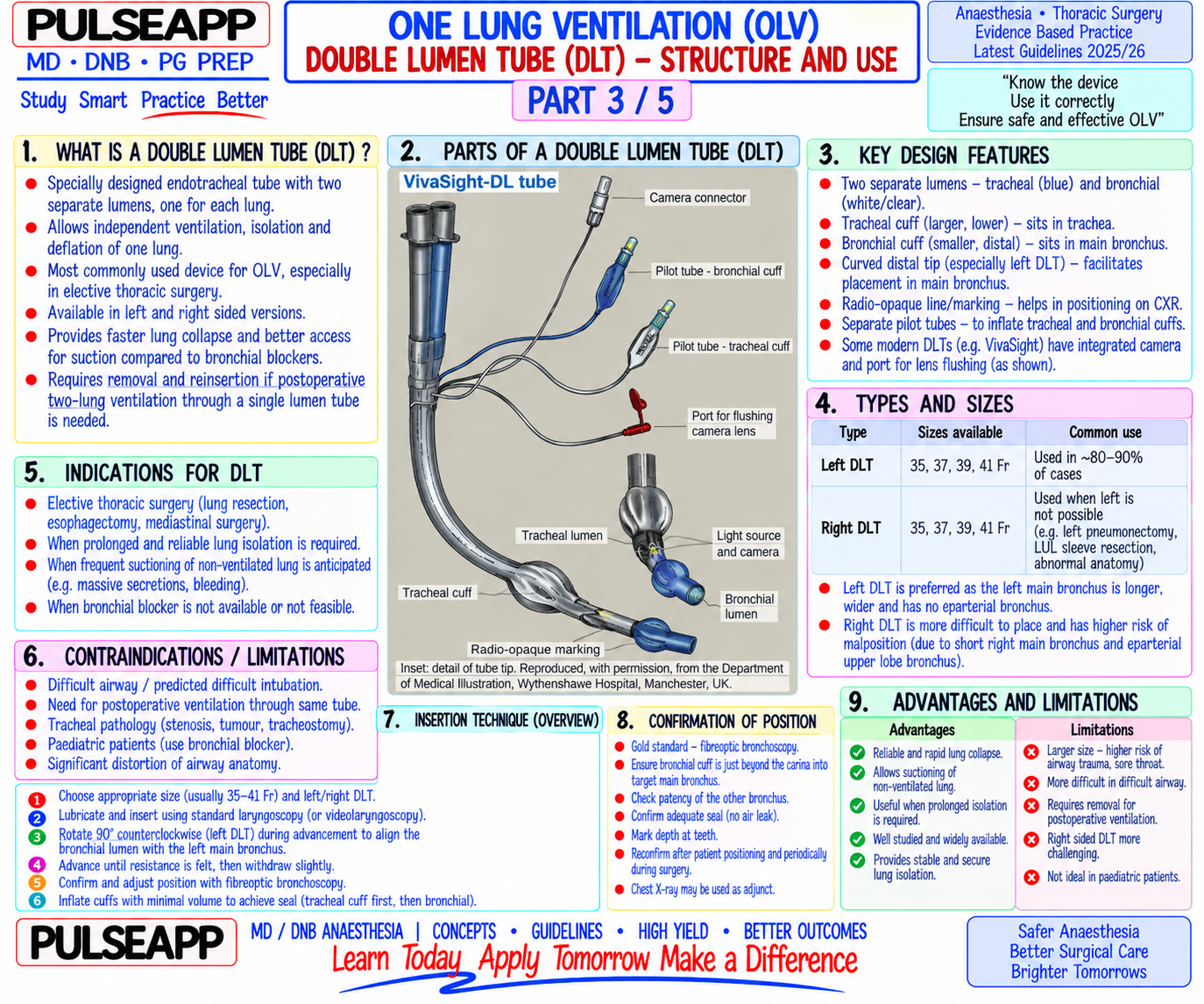
Feature Bronchial Blocker (via SLT) Double-Lumen Tube (DLT)

Ease of Easier overall — DLT requires specific technique; BB allows use of familiar SLT; FOB More complex; requires correctly-sized DLT (left DLT most insertion guidance ensures accuracy common — avoids right upper lobe orifice occlusion); DLT malposition is common Lung Adequate; but lung collapse takes LONGER (10–15 min via passive gas resorption vs 3– Faster lung collapse — the bronchial lumen can be actively isolation 5 min with DLT active venting) suctioned and the lung deflated; better surgical exposure quality more rapidly

Ability to Limited — the central lumen of the blocker is narrow (allows CPAP delivery but not Full suctioning capability through the bronchial lumen; can suction effective suctioning); cannot clear secretions from operative lung clear blood, secretions from operative lung operative lung

CPAP to Possible through the central lumen of larger blockers (9 Fr): apply 5 cmH₂O CPAP to the Possible through the bronchial lumen; same CPAP strategy operative partially collapsed operative lung to improve oxygenation available lung

Risk of HIGHER — the blocker can migrate proximally during surgical manipulation; Lower displacement risk once correctly secured; DLT is displacement displacement mid-surgery restores two-lung ventilation suddenly; displacement distally more rigid and secured at the ETT hub level can cause complete contralateral bronchial occlusion Preferred Difficult airway (use existing SLT); already intubated with SLT; children (no appropriate Elective thoracic surgery; rapid lung collapse needed; need clinical DLT size); tracheostomy; right upper lobe surgery (right-sided DLT risks RUL occlusion) for frequent intraoperative suctioning; most thoracic cases scenarios in adults Post-op Deflate cuff; remove blocker; leave SLT for post-op ventilation; can extubate from the SLT Must exchange DLT for SLT post-operatively if mechanical management directly ventilation needed (DLT is uncomfortable and highresistance for prolonged ventilation)
D. Management of Hypoxia During OLV2 marks
⚠ Stepwise Algorithm for Hypoxia During OLV (SpO₂ <90%)
1. Increase FiO₂ to 1.0 — immediate first step; may resolve mild hypoxia 2. Check blocker/DLT position — FOB to confirm adequate lung isolation and no displacement; re-inflate cuff if needed 3. Apply PEEP 5 cmH₂O to the ventilated (dependent) lung — prevents atelectasis, improves V/Q matching in the ventilated lung 4. Recruitment manoeuvre to the dependent lung — sustained inflation 30 cmH₂O for 30 seconds then resume PEEP 5 cmH₂O 5. Apply CPAP 5 cmH₂O to the operative (non-ventilated) lung — delivers O₂ to the partially collapsed lung without inflating it; most effective rescue manoeuvre short of resuming TLV; use the central lumen of the bronchial blocker for CPAP delivery 6. Switch anaesthesia to TIVA (propofol) — eliminates volatile agent-induced HPV inhibition; may significantly improve SpO₂ 7. Intermittent two-lung ventilation — inform surgeon; periodically re-inflate the operative lung
🎤 Viva Corner
Q. You have inserted an Arndt bronchial blocker for a left VATS lobectomy. The SpO₂ suddenly falls from 97% to 85% 30 minutes into the case. What has happened and how do you manage it?
The sudden SpO₂ fall mid-surgery most likely indicates bronchial blocker displacement — the blocker cuff has migrated proximally out of the left main bronchus back toward the carina. This would simultaneously restore ventilation to the operative (left) lung (blood from the surgical field enters the now-ventilated left lung — aspiration/contamination risk) AND may occlude the trachea or right main bronchus if the cuff migrates to the carina level. Management: immediately increase FiO₂ to 1.0; alert the surgeon to pause surgery; re-insert the FOB through the multiport adapter to visualise the blocker position — if displaced to the carina, the cuff may be partially occluding both bronchi; deflate the cuff immediately; gently advance the blocker back into the left main bronchus under direct FOB vision; re-inflate the cuff under FOB confirmation; confirm left lung re-collapse before resuming surgery. Simultaneously: assess the patient haemodynamically (hypoxia + potential blood aspiration from the operative field); apply SpO₂, ETCO₂ trends to assess degree of shunt. If repositioning is not achievable with the bronchial blocker, convert to a left DLT (exchange the SLT for a left DLT under direct laryngoscopy or video laryngoscopy with a tube exchanger). If DLT exchange is not feasible, place a right DLT and proceed with two-lung ventilation while discussing with the surgeon about alternative surgical access.
Q. Why is a right-sided DLT avoided for most thoracic procedures, and when is it specifically preferred?
A right-sided DLT is avoided for most thoracic procedures because of the anatomical challenge posed by the right upper lobe (RUL) bronchus. The right upper lobe bronchus arises from the right main bronchus only 1.5–2.5 cm below the carina — an extremely short distance. A right-sided DLT, when advanced into the right main bronchus, must have its bronchial cuff positioned to occlude only the right main bronchus while simultaneously allowing the RUL bronchial orifice to remain open for ventilation. Even millimetres of displacement in either direction can either fail to seal the right main bronchus (insufficient seal → inadequate lung isolation) or completely occlude the RUL bronchial orifice (RUL atelectasis → right lung not adequately ventilated). The Murphy eye (a side hole in the bronchial tip of the right DLT) is specifically designed to align with the RUL orifice, but achieving and maintaining this alignment intraoperatively is technically demanding. In contrast, the left main bronchus is approximately 5 cm long before it bifurcates — providing much more margin for error in positioning a left DLT, which is why the left DLT is used for the vast majority of cases regardless of which side is being operated on. When a right-sided DLT is specifically preferred: left pneumonectomy (the left main bronchus will be divided — a left DLT would have its bronchial tip in the surgical field); left sleeve resection; left main bronchial tumour obstructing placement of a left DLT; left thoracic aortic aneurysm repair where the left main bronchus may be compromised. In these situations, the technical challenges of right DLT positioning are accepted because the alternative is worse.
★ Examiner's Pearl
Name at least three specific bronchial blocker types (Arndt, Cohen, EZ-Blocker minimum) with their distinguishing feature — generic answers score less. The DLT vs blocker comparison table is the most frequently tested content in this topic — state the key differences: blocker allows SLT use + easier in difficult airway, but DLT gives faster collapse + better suctioning capability. The right-sided DLT avoidance reason (RUL orifice 1.5–2.5 cm from carina — Murphy eye must align with RUL orifice) is a mechanistic fact specifically tested.
Miller RD et al. Miller's Anaesthesia, 9th Ed, Chapter 68 (Thoracic Anaesthesia). Campos JH. An update on bronchial blockers during lung separation techniques in adults (Anesth Analg 2003;97:1266-1274). Benumof JL. Anesthesia for Thoracic Surgery, 2nd Ed. Lumb AB, Slinger P. HPV physiology and anaesthetic implications (Anesthesiology 2015;122:932). Narayanaswamy M et al. Choosing a lung isolation device (Can J Anaesth 2009;56:867-875).
lock Protected Clinical Document • Pulse App • Screenshots Strictly Prohibited
QUESTION 32 person Asked by .
bookmark_add

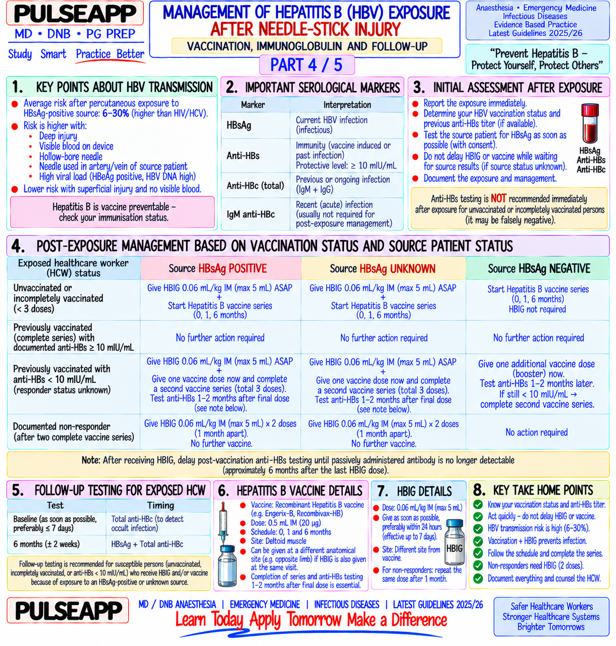
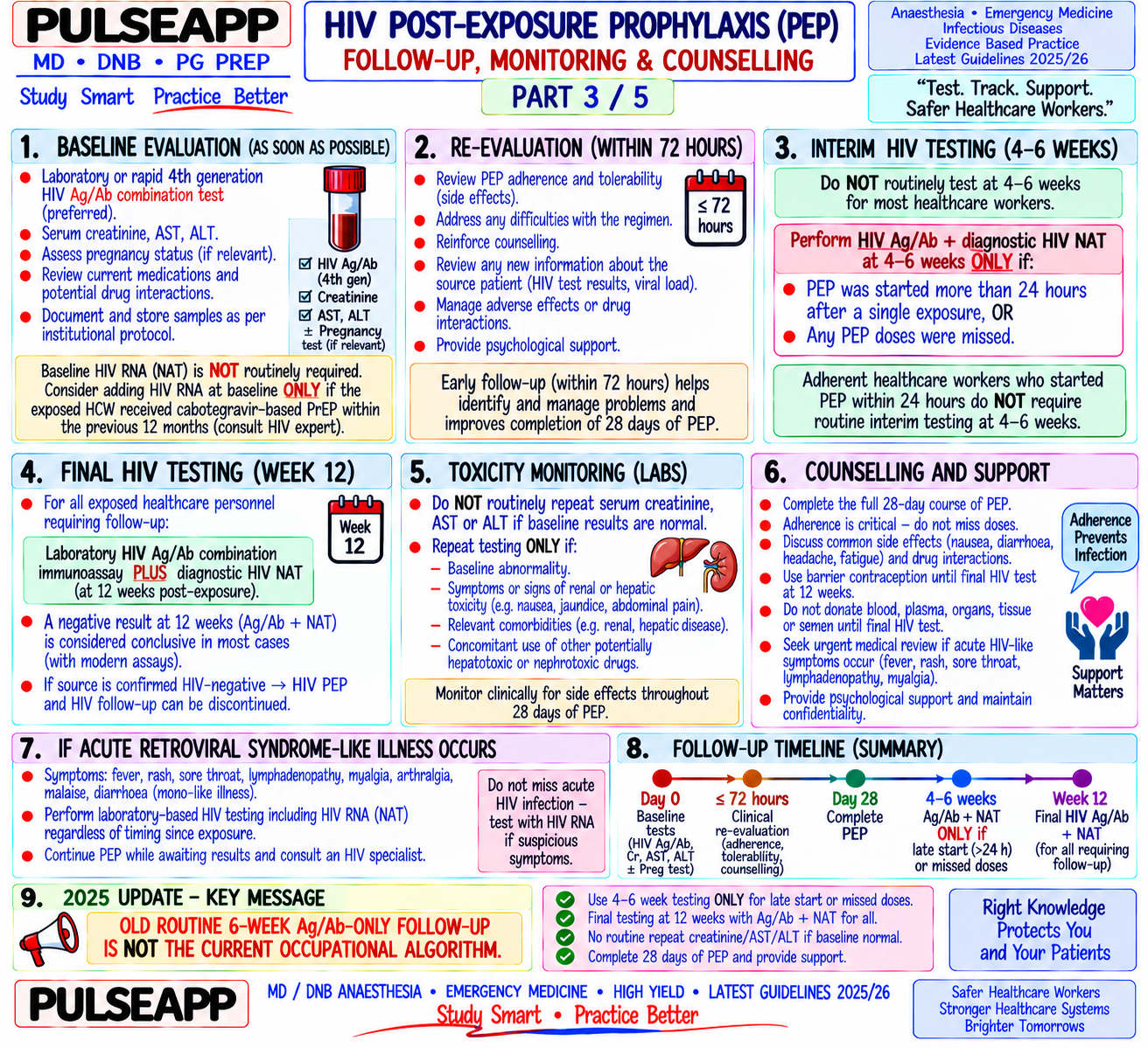
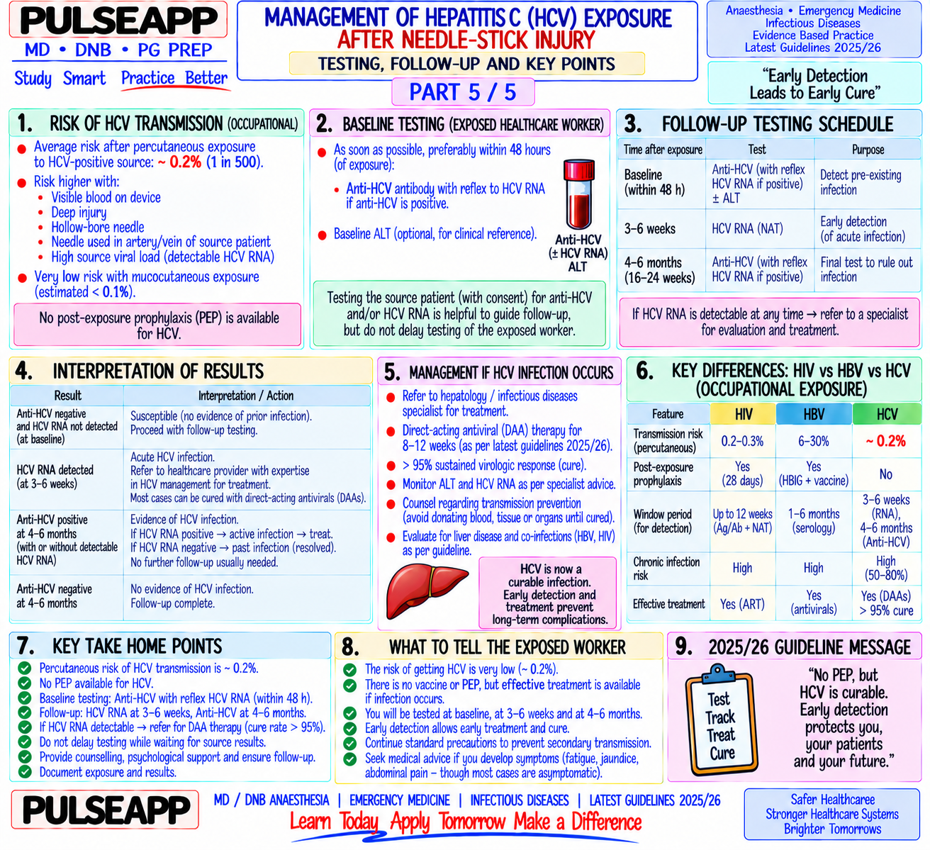
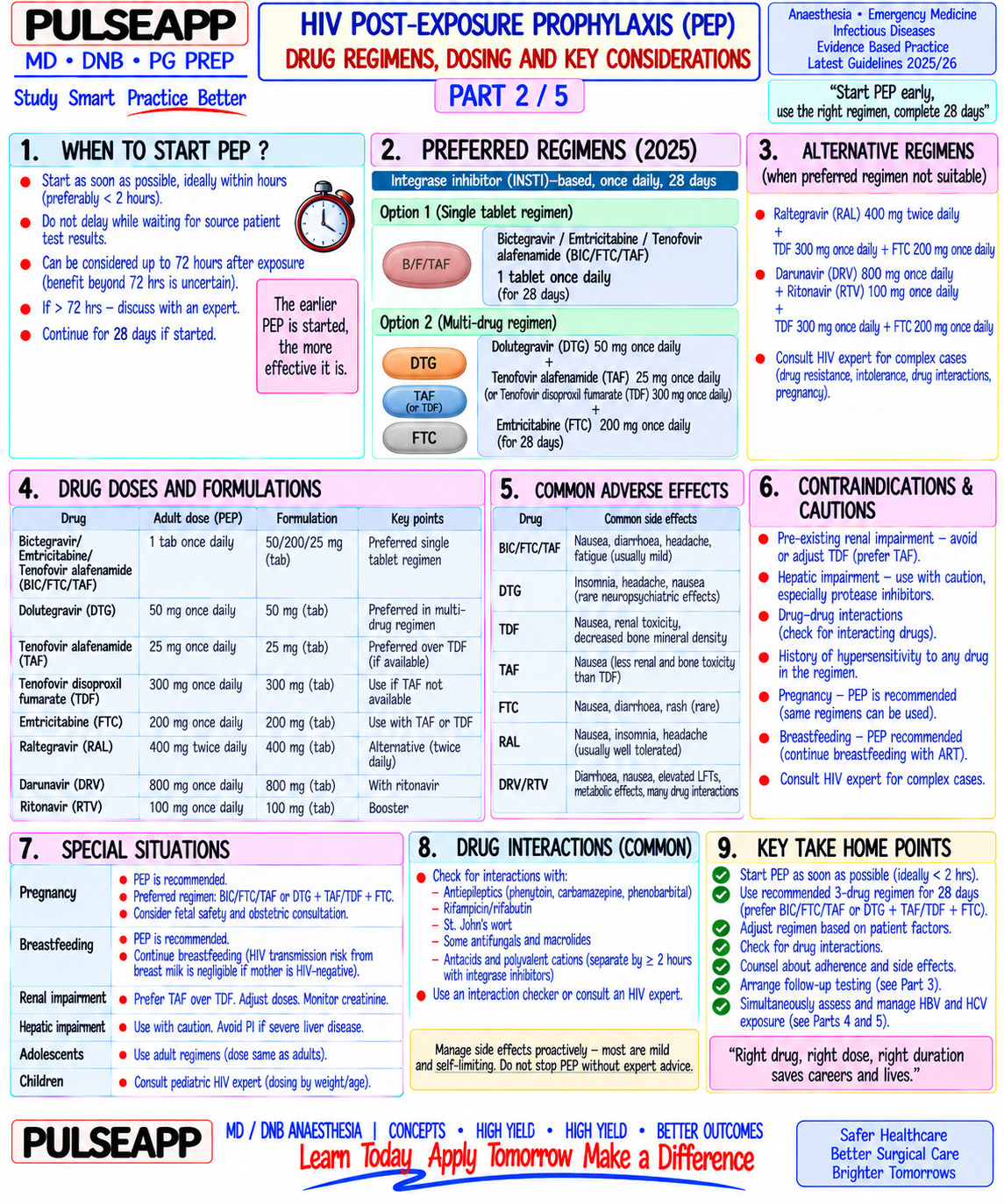
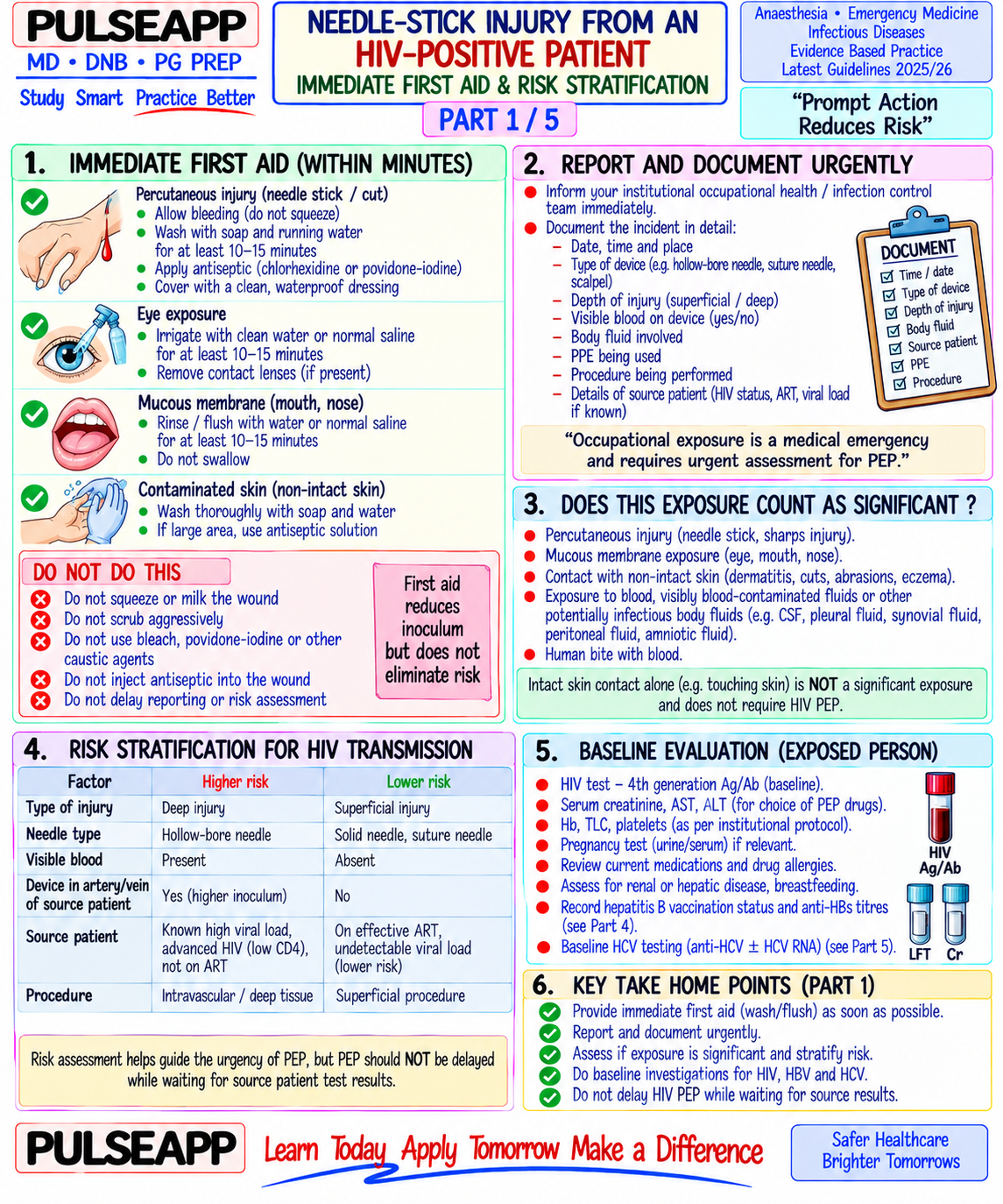
You sustain a needle-stick injury from an HIV-positive patient. Describe the immediate first aid, risk stratification, PEP drug regimen, follow-up protocol, and simultaneous management for HBV and HCV exposure.

description Clinical Response
⚙ Core Concept
Needle-stick injuries (NSI) are a critical occupational hazard in healthcare — the risk of HIV transmission from a hollow-bore needle contaminated with HIVpositive blood is approximately 0.3%; for HBV (unvaccinated, HBeAg-positive source) it approaches 30%; for HCV it is 1.8%. The anaesthesiologist, who regularly handles sharp instruments in close proximity to uncontrolled patient movements, is at particular risk. Time is the most critical variable: PEP must begin within 2 hours for maximum efficacy and is not initiated beyond 72 hours. Understanding the immediate protocol — first aid, risk stratification, drug choice, and follow-up — is a clinical competency and a potential examination question in any anaesthesia or critical care syllabus. (WHO PEP Guidelines 2014; CDC MMWR; NACO India HIV PEP Guidelines; Cardo DM et al. — N Engl J Med 1997)
A. Immediate First Aid — First 5 Minutes2 marks
⚠ TIME IS CRITICAL — PEP must start within 2 hours; maximum 72 hours
Remove gloves immediately; expose the wound Wash the wound with soap and water under running water for 5 minutes — do NOT scrub, squeeze, or suck the wound (increases local trauma and may increase viral inoculation); allow the wound to bleed freely under water initially Apply antiseptic after washing: 70% ethanol, 0.5% chlorhexidine, or 10% povidone-iodine; do NOT apply bleach or caustic agents For mucous membrane/conjunctival exposure: immediately irrigate with large volumes of water or normal saline for 10–15 minutes Do NOT apply tourniquet; do NOT cauterize Report immediately to the occupational health department / designated PEP officer / emergency department — do NOT delay reporting for any reason (embarrassment, busy shift, uncertainty)
B. Risk Stratification2 marks

Risk Factor Higher Risk Lower Risk Device type Hollow-bore needle (higher volume blood inoculation — syringes, IV Solid needle (suture needle, lancet — less blood volume transferred) cannulae, blood gas needles) Depth of injury Deep puncture wound (needle passed completely through tissue) Superficial scratch

Visible blood on YES — visible blood increases viral inoculation volume No visible blood device Source patient High viral load (AIDS, untreated HIV, AIDS-defining illness, CD4 <200) Undetectable viral load on effective ART (extremely low risk — but viral load PEP still recommended) Exposure route Parenteral (needle/sharp); mucous membrane to high-titre blood Intact skin contact (essentially no risk) Bloodborne Transmission Risk (hollow-bore needle) Risk Modifiers Virus HIV ~0.3% (1 in 333) Deep injury + visible blood + high viral load → up to 0.9%; undetectable viral load → much lower but not zero HBV 6–30% depending on HBeAg status (HBeAg positive source = up to Highest risk of all three BBVs; vaccination provides near-complete protection 30%; HBeAg negative = 1–6%) HCV ~1.8% (1 in 55) No effective PEP or vaccine; higher risk if HCV RNA positive source

C. HIV PEP Drug Regimen3 marks
✅ WHO / NACO Recommended PEP Regimen (2014 Guidelines)
Preferred regimen (all resource settings): Tenofovir (TDF) 300 mg + Lamivudine (3TC) 300 mg + Dolutegravir (DTG) 50 mg — once daily for 28 days Alternative (if DTG unavailable): TDF 300 mg + Emtricitabine (FTC) 200 mg + Lopinavir/ritonavir (LPV/r) 400/100 mg BD — 28 days India (NACO preferred 2023): TDF + 3TC + DTG (same as WHO preferred) Drug Class Dose Key Points Tenofovir NRTI (nucleoside reverse 300 mg once daily Renal function monitoring (nephrotoxic — reduce dose if eGFR <50); take with food (TDF) transcriptase inhibitor) Lamivudine NRTI 300 mg once daily Well tolerated; renal dose adjustment if needed; also active against HBV (3TC) (or 150 mg BD) Dolutegravir INSTI (integrase strand 50 mg once daily High barrier to resistance; avoid in first trimester pregnancy (neural tube defect risk historically (DTG) transfer inhibitor) reported, newer data more reassuring); no significant drug interactions in most cases Start PEP as soon as possible — ideally within 2 hours; NEVER beyond 72 hours Duration: 28 days (4 weeks) exactly — shorter courses have higher failure rates; counsel on adherence; common side effects include nausea, fatigue, headache (most resolve in 1–2 weeks) PEP reduces HIV transmission risk by approximately 80% if started within 2 hours and completed for the full 28 days Source patient testing: obtain consent and test the source patient for HIV (rapid test) — if source is HIV-negative (confirmed), PEP can be discontinued; if source refuses testing or is unknown, continue full 28-day course HCW (exposed person) baseline bloods: HIV Ab/Ag, HBsAg, HBsAb, HCV Ab, full blood count, renal and liver function (before starting TDF; repeated at 2 and 4 weeks during PEP)
D. HBV and HCV Post-Exposure Management2 marks

HBV Exposure HCW Vaccination Status Anti-HBs Titre Action Vaccinated and responder Anti-HBs ≥10 No action needed — fully protected; document and reassure mIU/mL Vaccinated but non-responder or <10 mIU/mL or Check anti-HBs titre immediately; if <10: HBIG 0.06 mL/kg IM + HBV booster dose simultaneously (in different unknown titre unknown sites); retest anti-HBs at 1–2 months Unvaccinated N/A HBIG 0.06 mL/kg IM within 24 hours of exposure (most effective within 12 hours) + initiate full HBV vaccination series (0, 1, 6 months) simultaneously HCV Exposure There is NO effective PEP for HCV and NO vaccine available

Management: baseline HCV antibody (anti-HCV) and HCV RNA at time of exposure; repeat HCV RNA at 4–6 weeks (HCV RNA becomes detectable 1–2 weeks after infection — earlier than antibody); repeat anti-HCV and ALT at 3 and 6 months

If HCV infection is confirmed (HCV RNA positive): refer to hepatologist; modern direct-acting antivirals (DAAs — sofosbuvir-based regimens) can achieve >95% SVR (sustained virological response = cure) when treatment is started in the acute phase (within weeks of infection); early treatment during acute HCV is the current management strategy

Follow-up Protocol (All BBVs)

HIV: HIV Ag/Ab (4th generation test) at baseline, 6 weeks, 3 months, 6 months post-exposure; if all negative at 6 months — exposure did not result in HIV infection

HBV: LFTs at baseline and 3 months; if HBV infection occurs — refer to hepatologist

HCV: as above; LFTs at baseline, 6 weeks, 3 months, 6 months

Counsel on risk reduction during the window period: use condoms, avoid blood donation, avoid breastfeeding, avoid sharing needles; advise on the meaning of the window period (infection may be present before tests become positive)

🎤 Viva Corner
Q. The source patient's HIV status is unknown and they refuse testing. The needle-stick was from a hollow-bore needle with visible blood. What do you do?
When the source patient refuses testing and their HIV status is unknown, I treat the exposure as potentially HIV-positive and start PEP without delay — the 2-hour window for maximum PEP efficacy cannot be sacrificed while waiting for consent or alternative information. The risk assessment here supports starting PEP: hollowbore needle with visible blood = higher-risk exposure; unknown source status in a healthcare setting where HIV prevalence may be significant. Immediate action: start TDF 300 mg + 3TC 300 mg + DTG 50 mg immediately — today. Simultaneously: document that the source refused testing; consider the epidemiological context (the underlying risk of HIV in the patient population, any clinical features suggesting HIV/AIDS — oral candidiasis, wasting, known AIDS-defining illness on the medication chart, CD4 count in the notes); if any clinical or contextual evidence suggests a high prior probability of HIV in this patient, continue the full 28-day course. If the source patient later agrees to testing and tests negative (with a 4th generation test that rules out recent infection), PEP can be discontinued. Legal/ethical aspects: in India, under the HIV/AIDS (Prevention and Control) Act 2017, healthcare workers who are exposed occupationally have a right to know the source patient's HIV status; the designated HIV testing centre can advise on the legal framework for disclosure in this context. In the meantime: continue PEP, baseline bloods, and follow-up protocol regardless of the source patient's testing status.
Q. Why is PEP started within 2 hours considered the ideal, and what is the biological basis for the 72-hour absolute cutoff?
The timing of PEP initiation is based on the biology of HIV post-exposure viral replication and the window of opportunity to interrupt establishment of systemic infection. After a needle-stick inoculation: HIV first replicates locally at the site of inoculation within dendritic cells and macrophages in the skin and subcutaneous tissue; this local replication phase lasts approximately 24–48 hours before the virus begins trafficking to regional lymph nodes; systemic dissemination through the lymphatics to the bloodstream begins at approximately 48–72 hours post-exposure. PEP works by introducing antiretroviral drugs (NRTI backbone + integrase inhibitor) that block HIV reverse transcriptase and integrase — key enzymes required for viral replication and integration into host DNA. If PEP is started within 2 hours: the drugs are already at therapeutic plasma concentrations when the virus begins local replication → maximum inhibition of local viral expansion → the very small initial inoculum (0.3% transmission rate reflects that many exposures involve only a few viral particles) may be completely eliminated before systemic seeding; this is when PEP is most effective (~80% risk reduction). If PEP is started between 2 and 72 hours: viral local replication and early lymph node trafficking has already begun; PEP is less effective but still meaningful (perhaps 50–70% risk reduction depending on timing); still strongly recommended. Beyond 72 hours: the virus has almost certainly already established systemic dissemination and begun integrating into CD4 T-cell reservoirs; PEP cannot eradicate established infection and has minimal benefit; the risk of drug toxicity outweighs the minimal possible benefit; PEP is not recommended after 72 hours.
Q. An anaesthesia nurse sustains a needle-stick from a patient known to have chronic HBV (HBeAg positive). The nurse received the full 3-dose HBV vaccine series 5 years ago but has never had their anti-HBs titre checked. What is your immediate management?
This is a high-risk HBV exposure: hollow-bore needle, HBeAg-positive source (transmission risk up to 30%), and unknown vaccination response status. The key uncertainty is whether the vaccine produced protective immunity (anti-HBs ≥10 mIU/mL) — without this information, we must act conservatively. Immediate management: send an urgent anti-HBs quantitative level (can be available within hours at most labs); simultaneously, without waiting for the result — administer HBIG (Hepatitis B Immune Globulin) 0.06 mL/kg IM now. HBIG provides immediate passive immunity by delivering high-titre anti-HBs antibodies; its efficacy is greatest within 12 hours and still meaningful up to 24 hours post-exposure; there is no benefit beyond 7 days. HBIG and vaccine (if needed) are given in different injection sites simultaneously. Once the anti-HBs result returns: if anti-HBs ≥10 mIU/mL → the nurse is fully protected; HBIG already given provides additional protection; no further vaccine needed; document for future reference. If anti-HBs <10 mIU/mL (non-responder or waned immunity) → administer an HBV booster vaccine dose now; recheck anti-HBs at 1–2 months; if still <10 mIU/mL after the booster (true non-responder) → the nurse will need HBIG for any future HBV exposures and cannot rely on the vaccine for protection; consider referring to occupational health for further assessment. Monitor LFTs and HBsAg at 3 and 6 months to detect any breakthrough infection. Document the entire episode with incident reporting as per hospital protocol.
★ Examiner's Pearl
State the HIV transmission risk (0.3% hollow-bore needle) and the PEP drugs (TDF + 3TC + DTG — WHO preferred 2014) with the 28-day duration and the 2-hour/72hour window — all four facts tested in written DNB papers. The HBV vs HCV distinction is critical: HBV has vaccine + HBIG; HCV has NO PEP and NO vaccine (only early DAA treatment after confirmed infection). The HBIG dose (0.06 mL/kg IM) and timing (ideally within 12 hours, up to 24 hours) are specific numbers tested in safety examinations.
WHO. Consolidated Guidelines on the Use of Antiretroviral Drugs 2016 (PEP chapter). CDC. Updated US Public Health Service Guidelines for the Management of Occupational Exposures to HIV (MMWR 2005;54:RR-9). NACO India. National Guidelines for HIV Testing 2023. Cardo DM et al. A case-control study of HIV seroconversion in healthcare workers after percutaneous exposure (N Engl J Med 1997;337:1485-1490). Henderson DK. Management of needlestick injuries (JAMA 2012;307:75-84).
lock Protected Clinical Document • Pulse App • Screenshots Strictly Prohibited
QUESTION 33 person Asked by .
bookmark_add

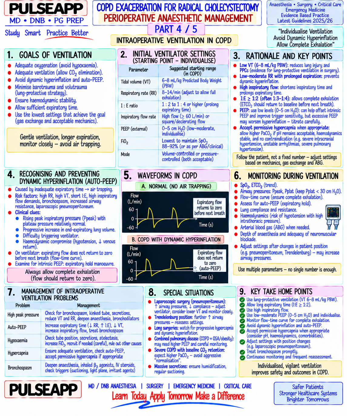
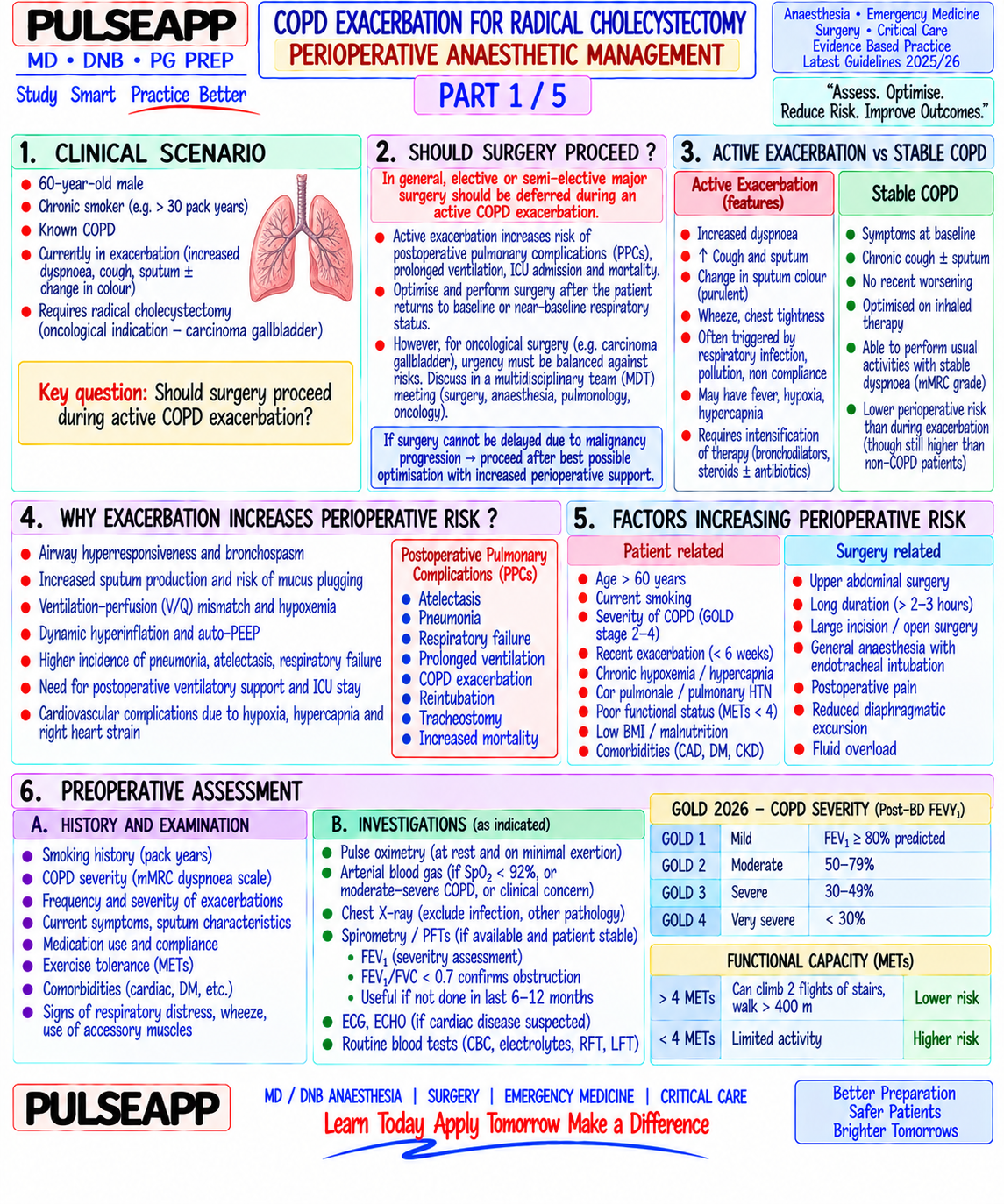
A 60-year-old male smoker with COPD exacerbation requires radical cholecystectomy. Discuss: Should surgery proceed during active exacerbation? Outline the preoperative optimisation, intraoperative anaesthetic strategy, ventilator settings, and postoperative pulmonary complication prevention.

collections Question Diagrams & Reference Images (1)
Question Reference Diagram
zoom_in View Image
description Clinical Response
⚙ Core Concept
COPD represents chronic airflow limitation from a combination of small airways disease and parenchymal emphysema — the anaesthetic consequences of both are profound: hyperinflation, air-trapping, high work of breathing, impaired hypoxic ventilatory drive, pulmonary hypertension, and right ventricular strain. The perioperative pulmonary complication (PPC) rate in moderate-severe COPD undergoing major abdominal surgery can exceed 30–50%. The decision to proceed during active exacerbation, the choice of anaesthetic technique, and the intraoperative ventilator strategy directly determine whether the patient leaves hospital breathing or intubated in the ICU. (Miller's Anaesthesia 9th Ed; GOLD 2023 Guidelines; Qaseem A — Perioperative PPC; Canet J — ARISCAT risk index; Lohser J)
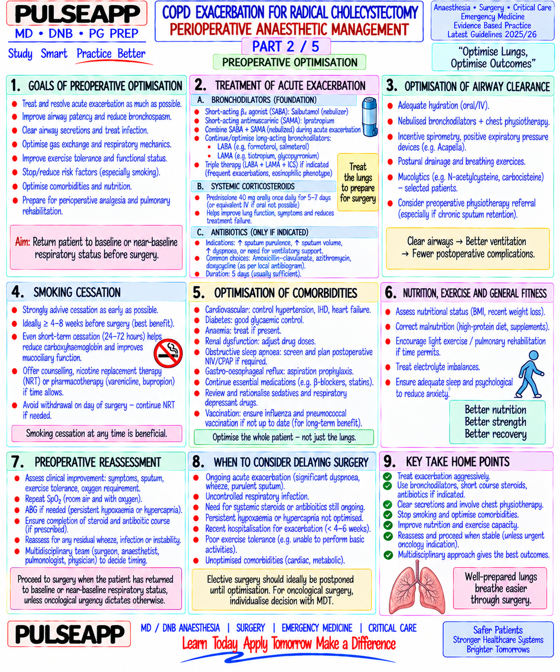
A. First Decision — Should Surgery Proceed During Active Exacerbation?2 marks
⚠ Active COPD Exacerbation + Elective or Semi-Urgent Surgery = DELAY and OPTIMISE
Active COPD exacerbation triples the risk of postoperative pulmonary complications and doubles perioperative mortality. Radical cholecystectomy for cancer = semi-urgent (not immediately life-threatening in most cases). Delay surgery until COPD is maximally optimised: Fever settled and afebrile for ≥48 hours PEF within 80% of patient's personal best (or predicted) Clear/mucoid sputum (not purulent) SpO₂ returning to baseline on room air Minimum optimisation period: 4–8 weeks of treatment before proceeding Exception: if cholecystectomy is being performed for sepsis (gangrenous cholecystitis, perforation) — cannot delay; proceed with aggressive intraoperative management.
B. GOLD Classification of COPD Severity1 mark

GOLD Stage FEV1 (% predicted) Anaesthetic Risk

I — Mild ≥80% Low; managed as routine

II — Moderate 50–79% Moderate; respiratory physiotherapy, bronchodilators, plan for regional if possible

III — Severe 30–49% High; aggressive preoperative optimisation, plan for postoperative HDU/ICU

IV — Very severe <30% Very high; multidisciplinary decision; consider alternatives to general anaesthesia; ICU post-op

C. Preoperative Optimisation3 marks

Intervention Specific Action & Evidence Smoking Immediate cessation reduces carboxyhaemoglobin within 12 hours and improves mucociliary clearance within 2–4 weeks; maximum respiratory cessation benefit at 8 weeks (secretion production normalises); warn against "cutting back" — partial smoking cessation does not reduce PPC risk as much as complete cessation

Bronchodilators Optimise inhaler therapy: SABA (salbutamol MDI 100–200 mcg QID) + LABA (salmeterol 50 mcg BD) + LAMA (tiotropium 18 mcg OD); bronchodilators should be continued until morning of surgery with a sip of water; IV aminophylline if severe bronchoconstriction not responding to inhalers (monitor levels — narrow therapeutic index) Treat infection Course of oral or IV antibiotics if purulent sputum/fever — guided by sputum culture where possible; standard: amoxicillin-clavulanate or cotrimoxazole for community-acquired exacerbation Steroids Short course oral prednisolone 30–40 mg for 5 days for COPD exacerbation (GOLD Guidelines); then continue inhaled corticosteroids (budesonide/formoterol); stress dose steroids perioperatively (hydrocortisone 100 mg IV at induction) for patients on systemic steroids >2 weeks Chest Breathing exercises, incentive spirometry, postural drainage; reduces secretion retention and improves baseline lung function pre-operatively physiotherapy Pulmonary FEV1, FVC, FEV1/FVC ratio; if FEV1 <0.8 L → very high risk of post-op ventilatory failure; 6-minute walk test for functional capacity; ABG for CO₂ function tests retention (PaCO₂ >50 mmHg at rest = severe disease and high risk of post-op hypercapnic respiratory failure) Nutritional COPD + malnutrition = very high PPC risk; optimise nutrition if BMI <18 with supplemental enteral feeding pre-operatively status

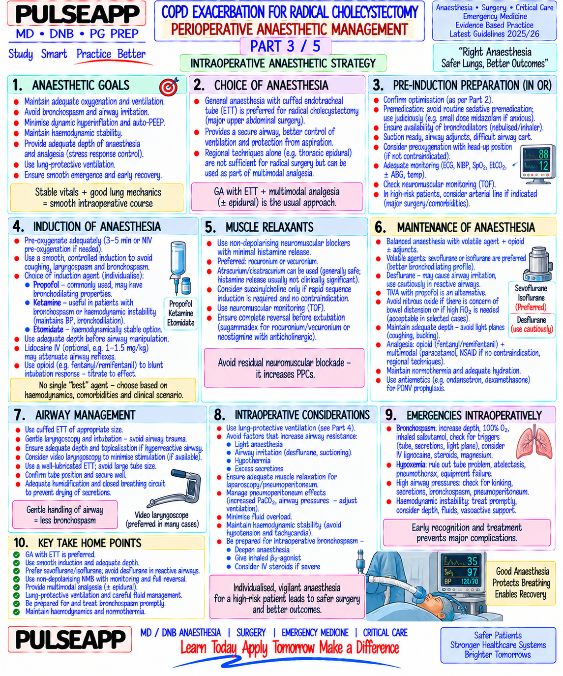
D. Intraoperative Anaesthetic Strategy2 marks

Anaesthetic Technique Choice Regional anaesthesia preferred where feasible — epidural or spinal anaesthesia for abdominal surgery avoids airway manipulation, maintains spontaneous ventilation, provides excellent post-operative analgesia (reducing opioid need and splinting), and is associated with lower PPC rates than general anaesthesia

Laparoscopic radical cholecystectomy: requires GA (pneumoperitoneum + Trendelenburg not compatible with spontaneous ventilation); combine with epidural for analgesia

Open cholecystectomy: thoracic epidural (T6–T8) provides surgical anaesthesia ± sedation as an alternative to GA in selected cases; more commonly combined with GA for better intraoperative control Intraoperative Ventilator Settings for COPD

✅ Key COPD Ventilation Principles — Prevent Air Trapping, Prevent Dynamic Hyperinflation
TV: 6–8 mL/kg IBW (lung-protective); avoid large tidal volumes that further increase air trapping RR: LOW rate — 8–12 breaths/min; a lower respiratory rate lengthens the expiratory time (more time for gas to leave the obstructed airways); prevents gas trapping I:E ratio: 1:3 or even 1:4 (prolonged expiration) — allows complete exhalation before the next breath; standard I:E of 1:2 is inadequate in severe COPD; the characteristic "obstructive pattern" on the flow-volume loop (slow, prolonged expiratory flow) requires extended expiratory time PEEP: controversial in COPD — intrinsic PEEP (auto-PEEP) from air trapping means that extrinsic PEEP may further increase air trapping; generally keep PEEP low (3–5 cmH₂O) or zero if auto-PEEP is present; measure auto-PEEP by inspiratory hold manoeuvre; if auto-PEEP >5 cmH₂O, reduce RR and I:E ratio rather than adding PEEP Permissive hypercapnia: accept PaCO₂ 50–60 mmHg if plateau pressure >30 cmH₂O — forcing normocapnia with high RR in severe COPD causes dynamic hyperinflation and barotrauma; permissive hypercapnia is safer Bronchodilators intraoperatively: inhaled salbutamol via the ETT adapter PRN for bronchospasm; volatile anaesthetics (sevoflurane, isoflurane) provide inherent bronchodilation — preferred over TIVA if bronchospasm is a concern
E. Postoperative Pulmonary Complication Prevention2 marks

Intervention Evidence Epidural / regional analgesia Epidural analgesia reduces splinting → allows deep breathing and coughing → reduces atelectasis and pneumonia; continues for 48– post-operatively 72 hours post-op in major abdominal surgery Early mobilisation Sitting out of bed day 1 post-op → improves FRC and reduces atelectasis; nurse physiotherapist-supervised ambulation Incentive spirometry Pre- and post-operative deep breathing exercises reduce atelectasis rate by 30–50% NIV/CPAP readiness In GOLD III–IV patients: plan for NIV (BiPAP) prophylactically post-extubation; CPAP 5–8 cmH₂O + IPAP 12–16 cmH₂O in pressure support mode; reduces need for reintubation Minimise opioids Opioid-induced respiratory depression + cough suppression directly causes PPC; use multimodal analgesia (paracetamol + NSAIDs + epidural + regional) to minimise systemic opioid requirement ARISCAT risk stratification Use ARISCAT score preoperatively to predict PPC risk: includes age, SpO₂, anaemia, incision site, surgery duration, emergency status, respiratory infection

🎤 Viva Corner
Q. Intraoperatively, the ventilator shows rising peak airway pressures (from 18 to 35 cmH₂O) and the ETCO₂ waveform shows a shark-fin appearance with an upward-sloping plateau. What is happening and how do you adjust the ventilator?
The rising peak airway pressures and characteristic shark-fin capnograph (prolonged upward-sloping alveolar plateau reflecting heterogeneous alveolar emptying from different degrees of airway obstruction — slow alveoli with high CO₂ empty late, continuing to raise the measured ETCO₂ through expiration) confirm severe bronchoconstriction/bronchospasm superimposed on the known COPD. The rising peak pressures may also reflect dynamic hyperinflation — air trapping from inadequate expiratory time. Ventilator adjustments: immediately reduce the respiratory rate to 8 breaths/min (from whatever current rate) — this is the most important single adjustment, as it lengthens expiratory time and allows complete exhalation before the next breath; increase the I:E ratio to 1:3 or 1:4; reduce or eliminate PEEP if auto-PEEP is suspected (perform an expiratory hold to measure auto-PEEP — if >5 cmH₂O, reduce RR further). Check plateau pressure (inspiratory hold for 0.5–1 second) — if plateau is <30 cmH₂O, the high peak pressure is from airway resistance (flow resistance) rather than from loss of compliance; if plateau is also high, there is additional parenchymal stiffness or pneumothorax. Pharmacological treatment: increase volatile agent concentration (sevoflurane 1.5–2 MAC — inherent bronchodilation); administer inhaled salbutamol (5 mg via nebuliser through the inspiratory limb); IV hydrocortisone 200 mg IV; IV magnesium sulphate 2 g IV over 20 minutes (bronchodilator); IV aminophylline 250 mg over 30 minutes if not already on it. If refractory: ketamine 0.5–1 mg/kg IV (bronchodilator via catecholamine release and direct smooth muscle relaxation); ensure adequate depth of anaesthesia (light anaesthesia = bronchospasm from surgical stimulation).
Q. What is "auto-PEEP" (intrinsic PEEP) in COPD, how do you measure it, and why is adding extrinsic PEEP potentially harmful in these patients?
Auto-PEEP (intrinsic PEEP, iPEEP) is the positive end-expiratory alveolar pressure that accumulates within the lung due to incomplete gas exhalation before the next breath begins — the hallmark of dynamic hyperinflation in obstructive lung disease. In COPD, the expiratory flow limitation from airway narrowing means gas cannot exit the alveoli as fast as it enters; if the RR is too high or the I:E ratio is too short for the degree of obstruction, each expiratory phase is insufficient to return the lung to its true resting volume (FRC). Gas accumulates progressively with each breath → lung volume ratchets upward → end-expiratory pressure within the alveoli exceeds the set PEEP on the ventilator (the circuit reads zero PEEP at end-expiration, but the alveoli are at a positive pressure of 5–15 cmH₂O). Measurement: perform an expiratory pause manoeuvre — at end-expiration, momentarily occlude the expiratory valve for 0.5–1 second while keeping the inspiratory valve closed (occlusion of both valves allows the pressure in the alveoli to equilibrate with the ventilator circuit); the resulting plateau pressure on the expiratory side represents the auto-PEEP level; normal = 0–1 cmH₂O; concerning >5 cmH₂O; dangerous >10–15 cmH₂O. Harm from adding extrinsic PEEP in auto-PEEP: if autoPEEP is already 10 cmH₂O and you add 5 cmH₂O of extrinsic PEEP, the total end-expiratory alveolar pressure becomes 15 cmH₂O — further hyperinflating the lung; this compresses intra-pulmonary vessels (increasing PVR and RV afterload), compresses the inferior vena cava (reducing venous return and cardiac output), and risks pneumothorax from overdistension. Extrinsic PEEP is only safe to add up to the level of auto-PEEP (it then stents open the collapsed airways responsible for the flow limitation) — beyond that level it is additive and harmful; the correct treatment for high auto-PEEP is reducing RR and lengthening expiratory time.
★ Examiner's Pearl
State the GOLD classification with FEV1 thresholds (I ≥80%, II 50–79%, III 30–49%, IV <30%) — these are tested numerically. The ventilator settings for COPD must include the specific I:E ratio (1:3 or 1:4), low RR (8–12), and the concept of permissive hypercapnia — these three elements together define the COPD ventilatory strategy and are each separately tested. Auto-PEEP: define it, state how to measure it (expiratory pause), state that adding extrinsic PEEP above the auto-PEEP level is harmful — this is the most commonly tested COPD ventilation advanced concept in DNB/MD examinations.
Miller RD et al. Miller's Anaesthesia, 9th Ed, Chapter 41 (Pulmonary Physiology and Pathophysiology). GOLD. Global Strategy for the Diagnosis, Management and Prevention of COPD 2023 Report. Canet J et al. Prediction of postoperative pulmonary complications in a population-based surgical cohort — ARISCAT study (Anesthesiology 2010;113:1338-1350). Lohser J. Evidence-based management of one-lung ventilation (Anesthesiol Clin 2008;26:241-272). Qaseem A et al. Risk assessment for and strategies to reduce perioperative pulmonary complications (Ann Intern Med 2006;144:575-580).
lock Protected Clinical Document • Pulse App • Screenshots Strictly Prohibited
QUESTION 34 person Asked by .
bookmark_add

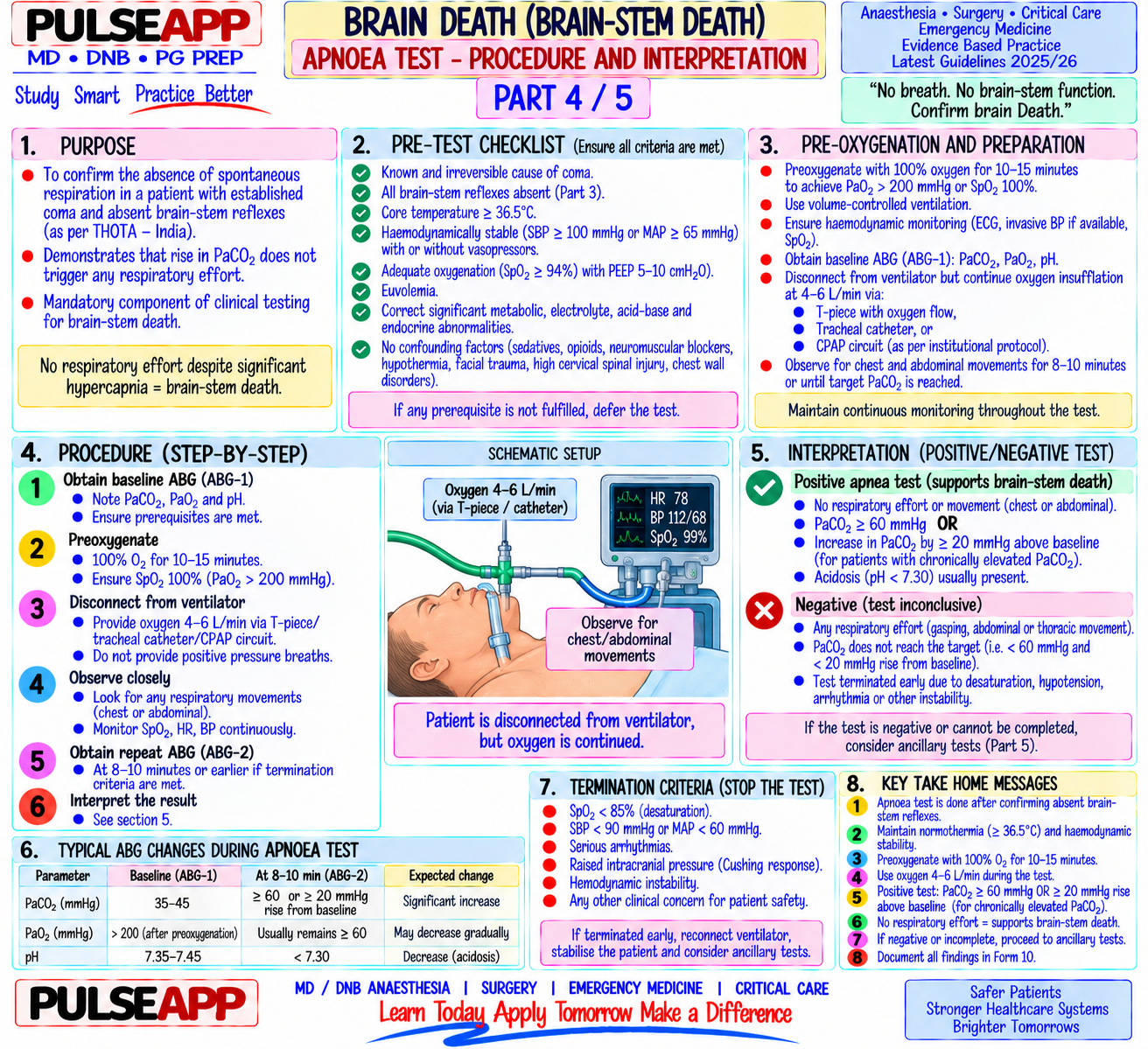
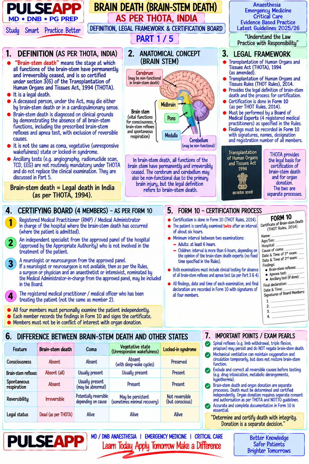
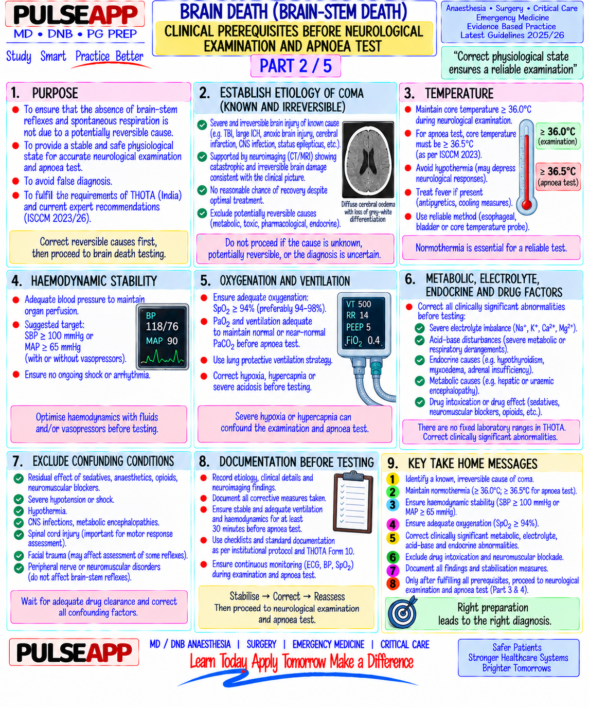
Define brain death as per the Transplantation of Human Organs Act (India). Describe the clinical prerequisites, bedside neurological tests, and confirmatory investigations for certifying brain death.

collections Question Diagrams & Reference Images (1)
Question Reference Diagram
zoom_in View Image
description Clinical Response
⚙ Core Concept
Brain death is the irreversible cessation of all functions of the entire brain including the brainstem. In India, it is legally defined under the Transplantation of Human Organs (THO) Act 1994 (amended 2011) as "brainstem death." Its recognition is not only clinically important — it determines withdrawal of life support and enables organ donation — but is also a medicolegally consequential determination that requires a specific committee, two sets of tests, and strict documentation. (THO Act India 1994/2011; AAN Brain Death Guidelines 2010; Miller's Anaesthesia 9th Ed)
A. Prerequisites Before Testing2 marks

Known cause: irreversible structural brain injury (TBI, massive stroke, anoxic brain injury, hypoxic-ischaemic encephalopathy) — must be established; no unknown or reversible causes

Exclude reversible causes of coma: hypothermia (core temp must be ≥36°C); metabolic derangement (Na, glucose, ammonia within normal limits); drug intoxication (sedatives, neuromuscular blockers, barbiturates, alcohol — must have cleared; if uncertain, drug levels should be measured and/or a minimum of 4–5 half-lives must have elapsed)

Haemodynamic stability: MAP ≥60 mmHg; patient must not be in cardiovascular shock

Duration: in India, observation period of at least 6 hours from the onset of coma before first testing is recommended

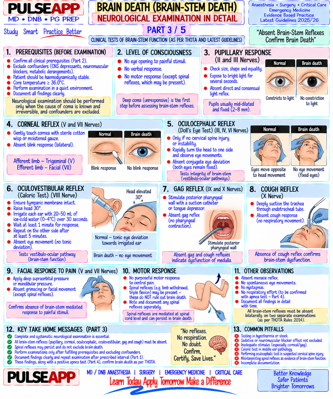
B. Clinical Tests for Brain Death4 marks

Test Method Brain Dead Response GCS Standard assessment GCS = 3 (E1V1M1) — no eye opening, no verbal, no motor response to any stimulus Pupillary light Bright light in each eye Pupils fixed and dilated (4–9 mm); NO constriction to reflex light; tests CN II (afferent) and CN III (efferent — parasympathetic to iris sphincter) Corneal reflex Touch cornea gently with sterile cotton wisp No blink; tests CN V (afferent trigeminal) and CN VII (efferent facial nerve to orbicularis oculi) Oculocephalic Rapidly rotate head side-to-side (ONLY after cervical spine cleared); contraindicated if C- In brain death: eyes remain fixed and do NOT move — reflex (Doll's spine injury they move WITH the head (absent reflex); normally eyes eye) lag behind head movement (brisk reflex = intact brainstem) Oculovestibular Irrigate 50 mL ice cold water into each external auditory meatus (with head 30° elevated); Brain dead: no eye movement; normally produces tonic reflex (Cold observe for 1 minute deviation of eyes toward the irrigated side (brainstem caloric) intact); most sensitive brainstem test Gag reflex Stimulate posterior pharynx with suction catheter No gag; tests CN IX (afferent glossopharyngeal) and CN X (efferent vagus) Cough reflex Pass suction catheter to carina via ETT No cough — complete absence of cough response to tracheal suctioning Pain response Apply painful stimulus (supraorbital pressure, nail bed pressure) No motor response; spinal reflexes (limb withdrawal) may be present — these are spinal cord reflexes, NOT brainmediated, and do NOT exclude brain death Apnoea test Pre-oxygenate with 100% O₂ for 10 minutes; confirm PaCO₂ 35–45 mmHg; disconnect No respiratory effort at PaCO₂ ≥60 mmHg = confirms (most ventilator; deliver passive O₂ via catheter in trachea at 6 L/min; observe for respiratory absent respiratory drive = confirms brainstem death; important movements for 10 minutes; check ABG at end — PCO₂ must rise to ≥60 mmHg (or 20 abort test if SpO₂ <85%, haemodynamic instability, or single test) mmHg above baseline) cardiac arrhythmia

C. Indian Legal Framework — THO Act 19942 marks

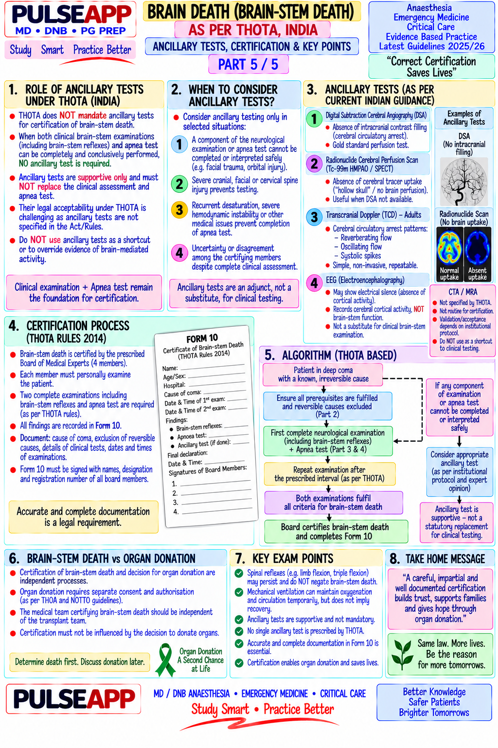
Brain Death Certification Committee (India) Under THO Act, brain death must be certified by a panel of four doctors: 1. Medical Administrator/Registered Medical Practitioner nominated by the hospital 2. Neurologist or Neurosurgeon 3. The treating doctor/intensivist 4. An independent doctor (not from the treating team)

Two sets of tests must be performed — first set, then a second set after an observation interval (minimum 6 hours from first set for adults; 24 hours for neonates/children). Both sets must confirm brain death. The time of death is certified at the time the second set of tests confirms brain death. The death certificate is then issued, and organ donation can proceed with family consent.

D. Confirmatory Investigations (When Clinical Tests are Uncertain)2 marks

Investigation Finding in Brain Death When Used

EEG Electrocerebral silence — isoelectric (flat) EEG at maximum sensitivity When apnoea test cannot be performed (severe pulmonary (Electroencephalogram) for ≥30 minutes disease); drug intoxication suspected; medico-legal requirement CT/MRI Angiography Absence of intracranial blood flow — no filling of cerebral vessels Most specific — absence of cerebral circulation is definitive; above the skull base preferred confirmatory test in many guidelines Transcranial Doppler Reverberating flow or absent flow in the major intracranial arteries Bedside, non-invasive; widely available; reverberating flow = net (TCD) (MCA, ACA, PCA) — systolic spikes only, no forward diastolic flow zero flow = no cerebral circulation Radionuclide brain "Hollow skull sign" — isotope does not cross the blood-brain barrier into Highly specific; demonstrates absent cerebral blood flow and scan (99mTc HMPAO) brain parenchyma; only scalp uptake cellular metabolic activity SSEP (Somatosensory Bilateral absence of N20 cortical response (the cortical component is Useful when EEG unreliable; tests cortical function Evoked Potentials) absent bilaterally)

🎤 Viva Corner
Q. During the apnoea test, the patient's SpO₂ falls to 82% and BP drops to 70/40 after 5 minutes. What do you do?
The apnoea test must be immediately aborted — the haemodynamic and oxygenation criteria for safe test conduct have been violated. Reconnect the ventilator immediately on 100% FiO₂ and full ventilation support; treat the hypotension with IV fluid bolus and vasopressors (phenylephrine or noradrenaline) as required; allow SpO₂ to recover to >95%. The apnoea test is aborted but does NOT by itself exclude brain death — it simply means the test could not be completed safely. After haemodynamic stabilisation, the apnoea test may be re-attempted after thorough pre-oxygenation; alternatively, if the apnoea test consistently cannot be completed safely due to haemodynamic instability or severe lung disease, a confirmatory investigation (radionuclide scan, CT angiography, or TCD) must be used to confirm absence of cerebral blood flow as a substitute for the apnoea test. Document clearly: time of abort, reason, SpO₂ and BP at time of abort, action taken. The fourdoctor committee must be informed, and a decision made about alternative confirmation.
Q. A patient's legs flex and withdraw when you apply painful stimuli during a brain death assessment. Does this exclude brain death?
No — limb withdrawal or other motor responses to painful stimuli (including the "Lazarus sign" — complex spinal motor automatisms that can appear horrifyingly lifelike) do NOT exclude brain death and do NOT represent brain-mediated activity. In brain death, the spinal cord may remain intact and functional — its intrinsic neural circuits can generate reflex motor responses (flexion withdrawal, triple flexion response, even sitting up in the "Lazarus sign") in response to noxious stimulation, completely independently of any input from the brain. The brain death determination assesses the BRAIN (cortex and brainstem) specifically — not the spinal cord. The clinical tests for brain death specifically look for responses mediated by brainstem cranial nerve circuits (pupillary reflex, corneal reflex, vestibulo-ocular reflex, gag, cough, and apnoea test) and cerebral cortical responses. Spinal cord-mediated responses (limb withdrawal, tendon reflexes, abdominal reflexes) are expected to be present in brain death if the spinal cord is intact, and their presence is specifically noted but does not invalidate the diagnosis. This fact must be explained clearly to the family, who may find these movements deeply distressing and interpret them as signs of life.
★ Examiner's Pearl
The four-doctor committee composition under India's THO Act is specifically tested in Indian examinations — state all four categories precisely. The apnoea test specifics (PaCO₂ must reach ≥60 mmHg or rise 20 mmHg above baseline; 100% O₂ pre-oxygenation; passive O₂ delivery during test) are tested with specific numbers. The fact that spinal reflexes DO NOT exclude brain death — and the explanation of why (intact spinal cord, not brain-mediated) — is the most commonly tested "trap question" in brain death examinations.
Transplantation of Human Organs Act, India 1994 (amended 2011). Wijdicks EFM et al. Evidence-based guideline update — determining brain death in adults (Neurology 2010;74:1911-1918). Miller RD et al. Miller's Anaesthesia, 9th Ed. Greer DM et al. Variability of brain death determination guidelines in leading US neurologic institutions (Neurology 2008;70:284-289).
lock Protected Clinical Document • Pulse App • Screenshots Strictly Prohibited
QUESTION 35 person Asked by .
bookmark_add

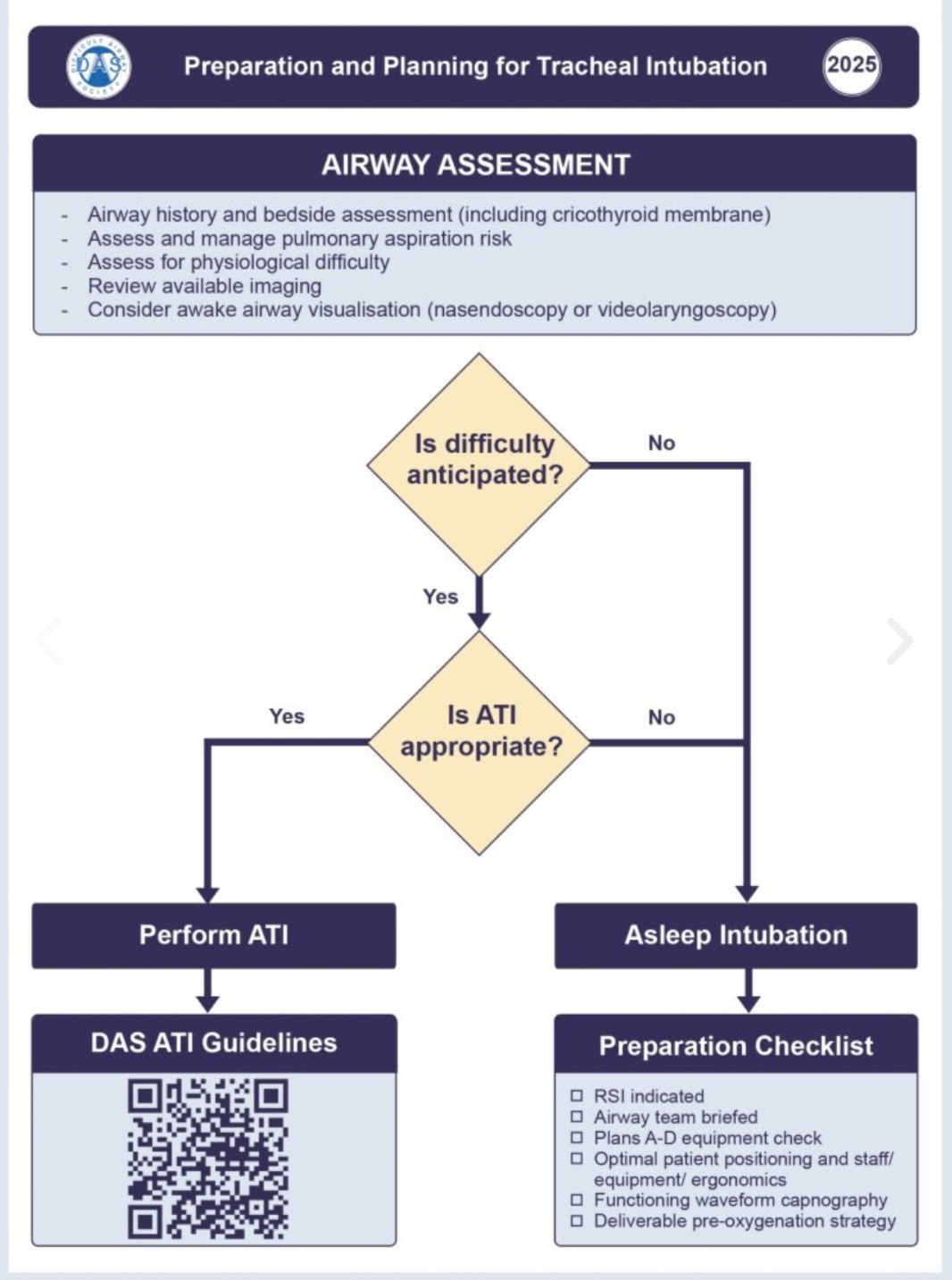
Define difficult airway. Describe the clinical assessment tools for predicting difficult intubation and difficult mask ventilation. Outline the DAS 2015 unanticipated difficult intubation algorithm. Describe the technique and sedation protocol for Awake Flexible Fiberoptic Intubation (AFOI).

collections Question Diagrams & Reference Images (6)
Question Reference Diagram
zoom_in View Image
Question Reference Diagram
zoom_in View Image
Question Reference Diagram
zoom_in View Image
Question Reference Diagram
zoom_in View Image
Question Reference Diagram
zoom_in View Image
Question Reference Diagram
zoom_in View Image
description Clinical Response
⚙ Core Concept
The difficult airway — whether anticipated or unanticipated — remains the leading cause of anaesthesia-related mortality. The ASA Closed Claims analysis consistently identifies airway management failures as the most preventable cause of anaesthetic death. The DAS 2015 guidelines provide a structured four-plan algorithm (Plan A → B → C → D) that reduces cognitive load during a crisis by providing pre-established decision trees. AFOI remains the gold standard for the anticipated difficult airway. (DAS Guidelines 2015; ASA Practice Guidelines for Management of the Difficult Airway 2022; Miller's Anaesthesia 9th Ed)
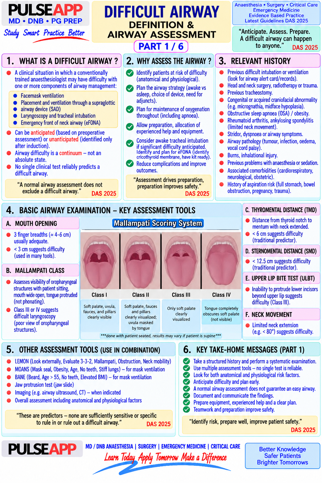
A. Definition — ASA Task Force 20221 mark

Difficult airway: the clinical situation in which a conventionally trained anaesthesiologist experiences difficulty with: (1) face mask ventilation; (2) direct laryngoscopy/intubation; or (3) supraglottic airway placement; or (4) surgical airway (cricothyrotomy)

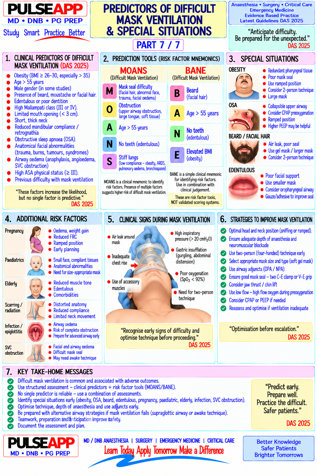
Difficult mask ventilation (DMV): inability to maintain SpO₂ >90% or to prevent/reverse signs of inadequate ventilation during mask ventilation using 100% O₂ and positive pressure

Difficult laryngoscopy: inability to visualise any portion of the vocal cords with conventional laryngoscopy (Cormack-Lehane Grade III–IV)

Failed intubation: proper insertion of the ETT in the trachea cannot be achieved after multiple attempts

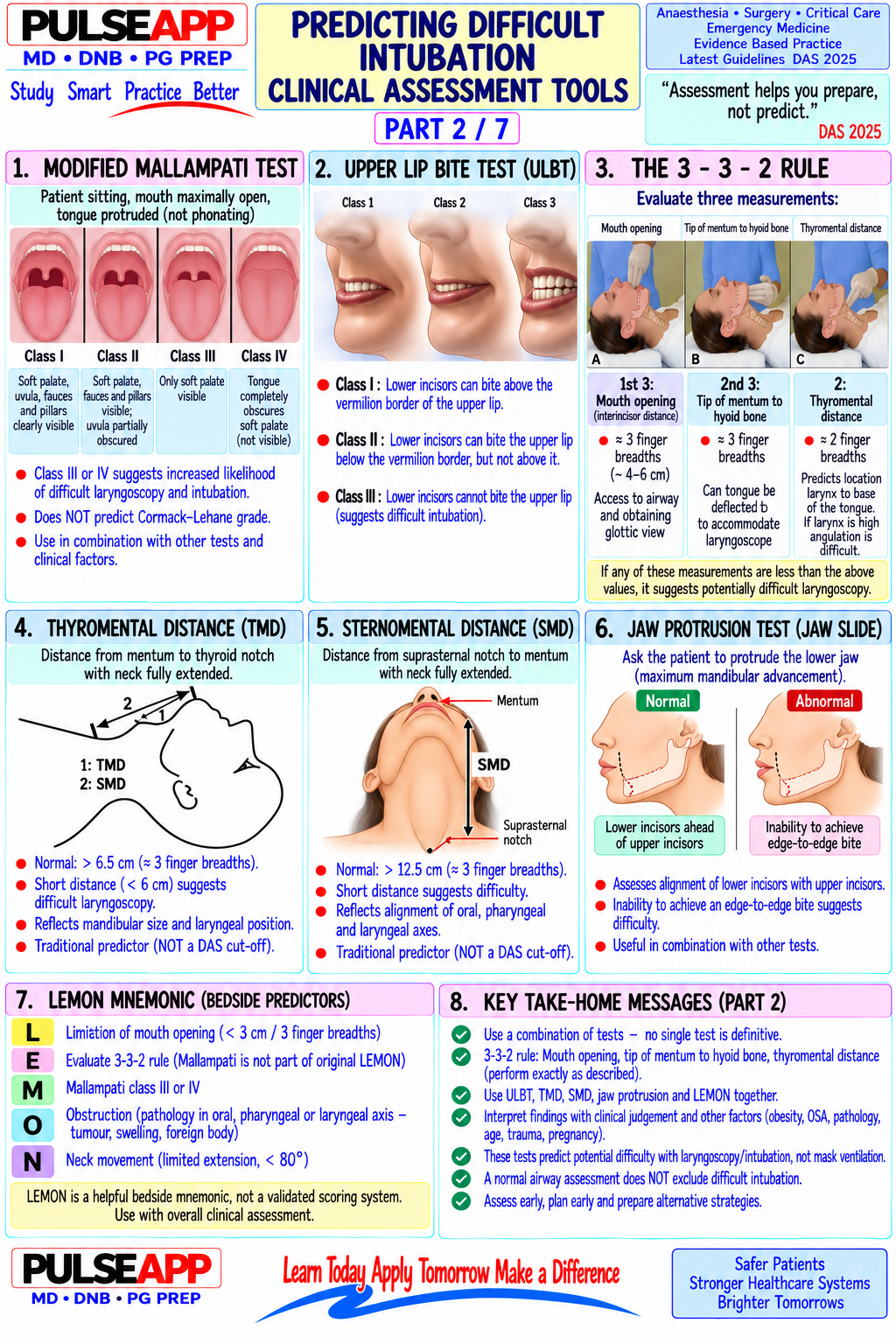
B. Airway Assessment Tools3 marks

Test What It Assesses High-Risk Finding Sensitivity/Specificity Mallampati Pharyngeal space (size of tongue relative to pharyngeal opening); patient Class III (only soft palate visible) or IV Sensitivity ~50%, Specificity score opens mouth and protrudes tongue — no phonation (soft palate not visible): predicts difficult ~85%; poor alone but laryngoscopy valuable in combination Thyromental Distance from thyroid notch to chin in full neck extension; measures space <6.5 cm → difficult laryngoscopy; <6 cm Sensitivity ~50%, Specificity distance available for tongue displacement during laryngoscopy → strongly predictive ~80% (TMD) Sternomental Distance from sternum to chin in full extension; overall neck mobility <12.5 cm → reduced neck extension → Low sensitivity; useful in distance assessment difficult laryngoscopy combination Interincisor Distance between upper and lower incisors in maximum mouth opening <3 cm (two finger breadths) → restricted Specific but insensitive; gap (mouth mouth opening; limits laryngoscope important combined with opening) blade insertion other factors

ULBT (Upper Patient bites their upper lip with lower incisors: Class I = bite above Class III → likely difficult laryngoscopy; Sensitivity ~50%, Specificity Lip Bite Test) vermilion border, Class II = below, Class III = cannot bite at all — assesses better predictor than Mallampati in some ~85% mandibular prognathism and tongue space studies Neck Active flexion/extension range; atlanto-occipital extension; presence of Extension <80° of normal; cervical Important contextual movement collar/fusion/arthritis collar; rheumatoid C-spine instability assessment; cannot be (atlanto-axial subluxation risk) quantified easily LEMON L=Look externally; E=Evaluate 3-3-2 rule; M=Mallampati; O=Obstruction; Any LEMON component positive → Validated in ED setting; easy score N=Neck mobility anticipate difficulty; used in emergency to apply rapidly airway assessment The 3-3-2 Rule (LEMON "E") 3 fingers = interincisor gap (mouth opening); 3 fingers = hyoid-chin distance (mandibular space for tongue); 2 fingers = thyroid-floor of mouth distance (laryngeal height); any measurement less than the finger widths suggests a potentially difficult airway.

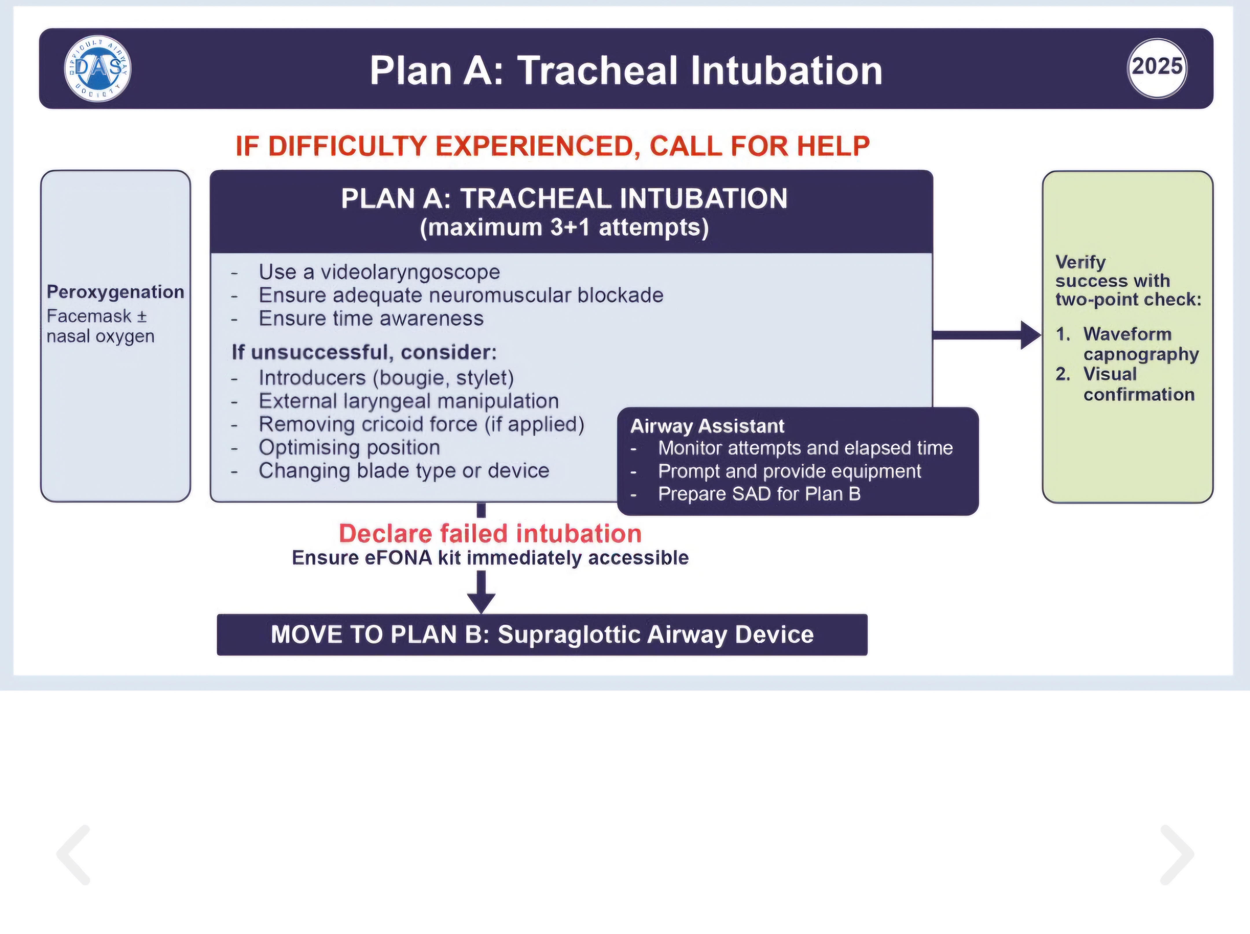
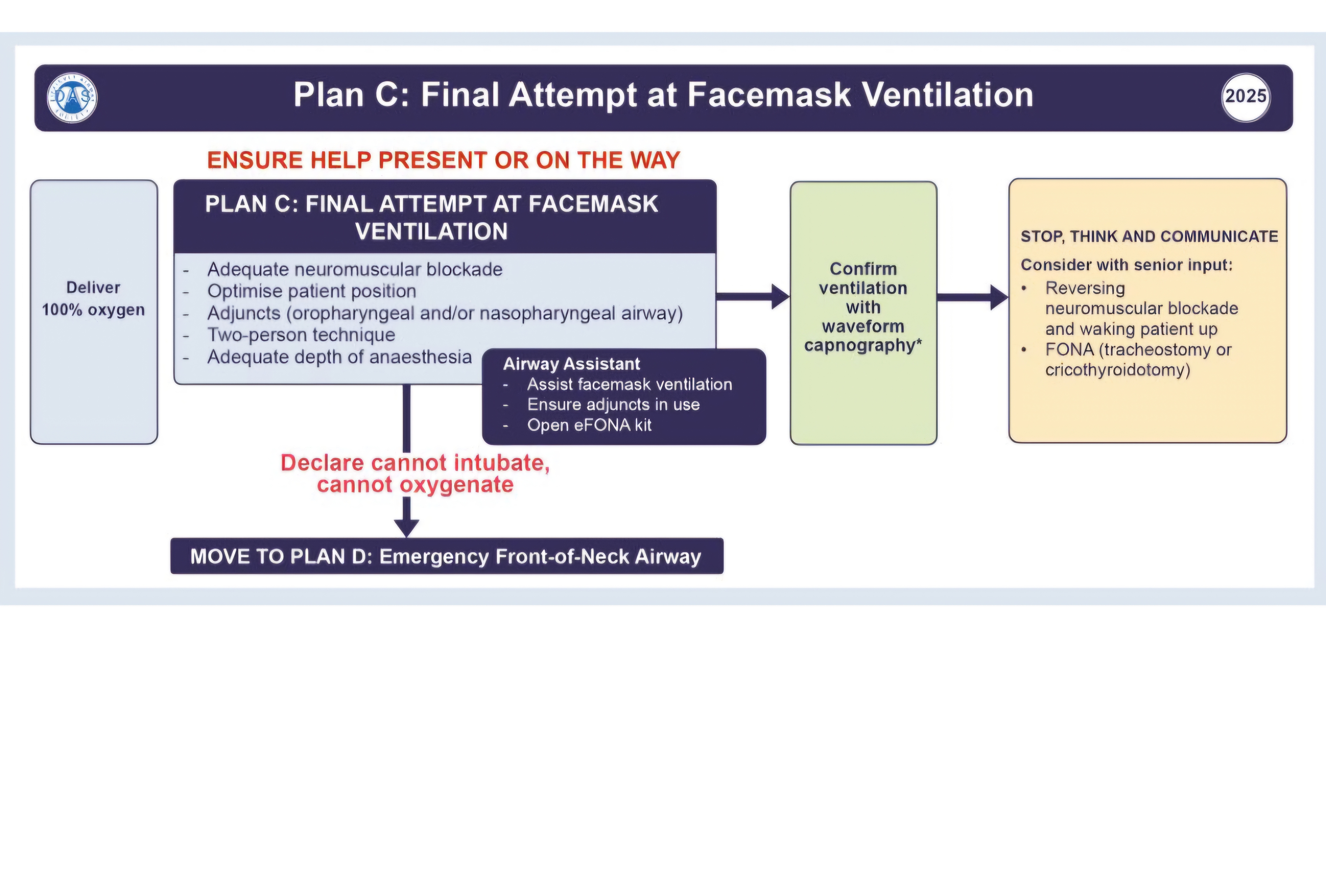
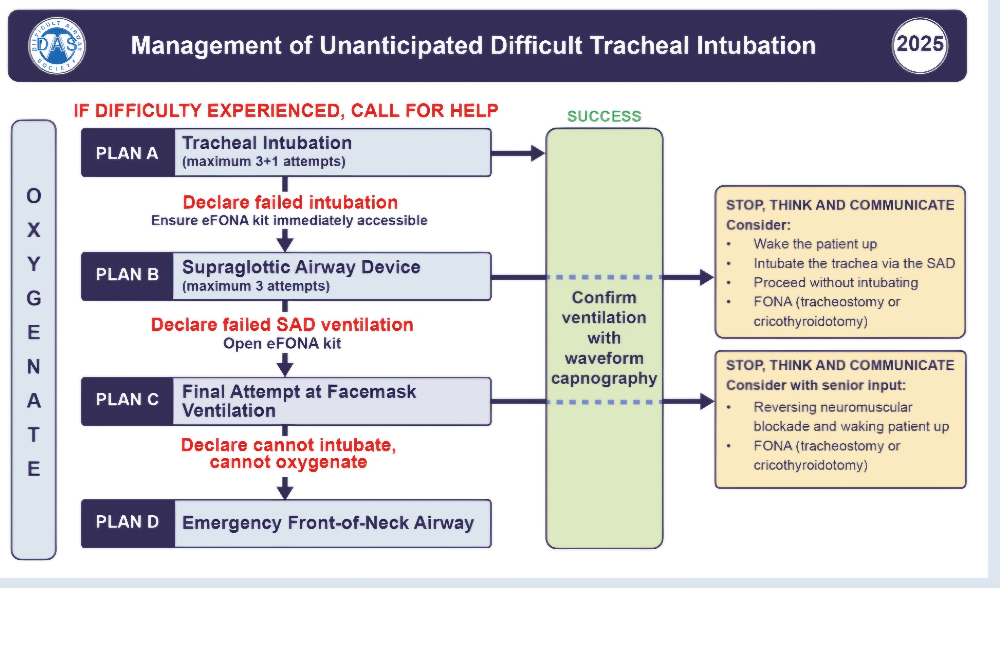
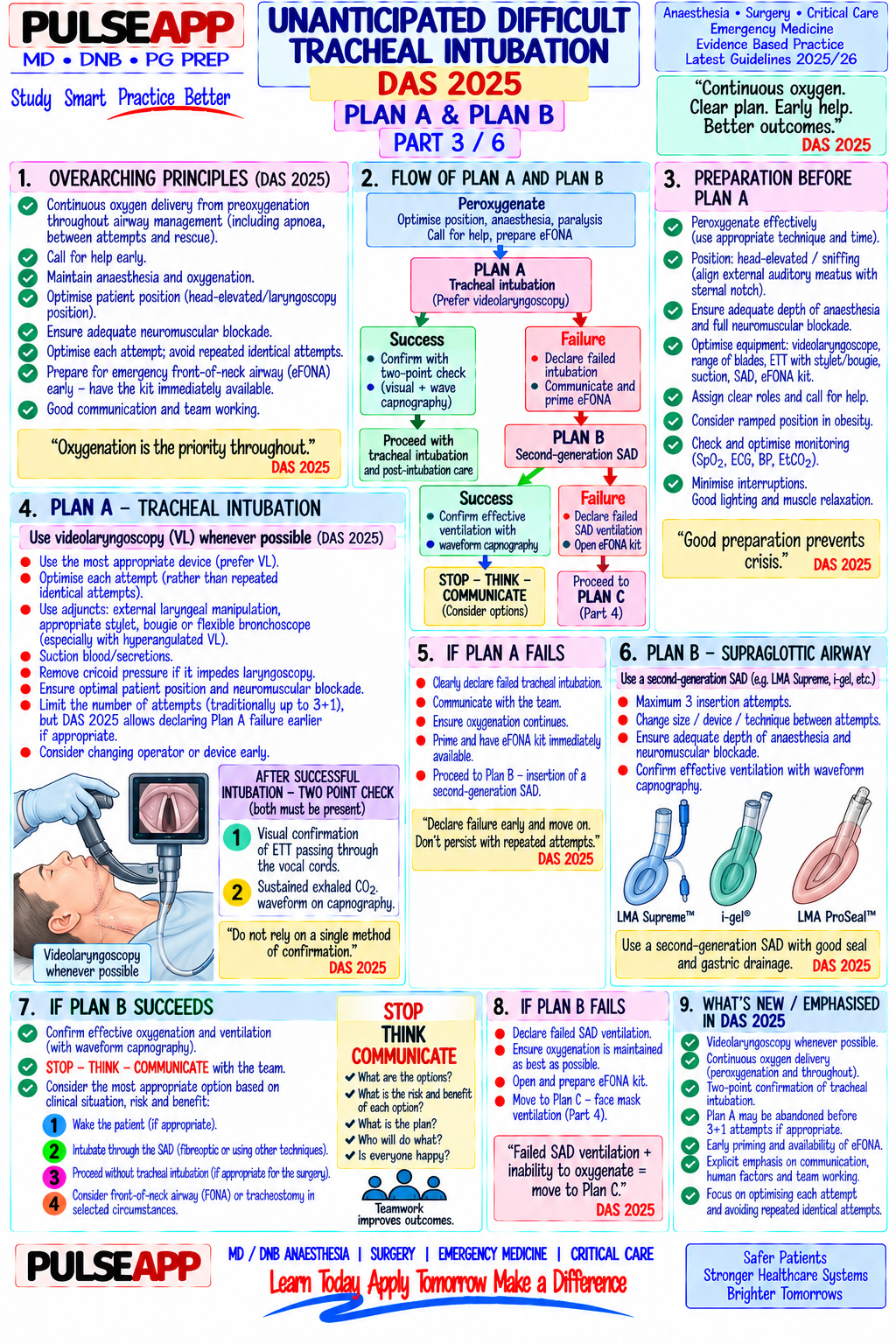
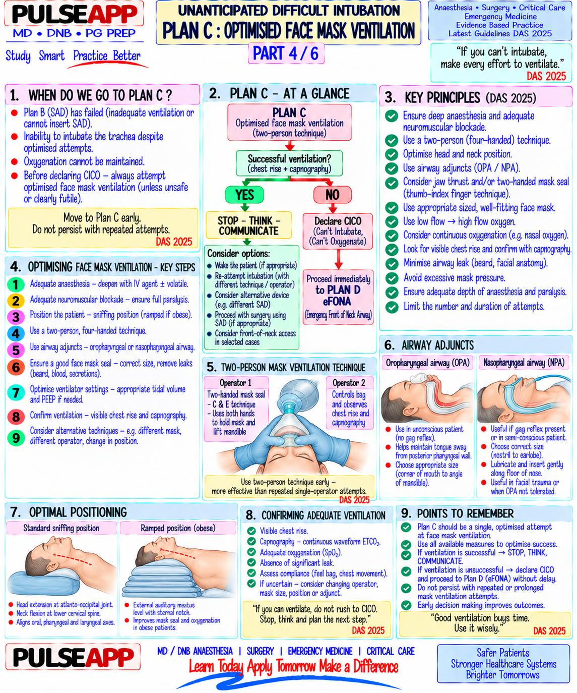
C. DAS 2015 Unanticipated Difficult Intubation Algorithm3 marks

Plan Action Maximum Attempts Proceed If Plan A Direct laryngoscopy (DL) / Video laryngoscopy — optimise: HELP/BURP, 3 attempts maximum (including 1 SpO₂ maintained; can still ventilate bougie, different blade; videolaryngoscope if available; give 100% O₂; with an experienced colleague); after 3 by mask call for help after first failed attempt failed attempts → declare failed intubation → move to Plan B

Plan B Supraglottic airway device (SAD): 2nd generation SAD (i-gel or Proseal 2 SAD insertion attempts; if SAD fails Oxygenation maintained with SAD

LMA — better seal for positive pressure ventilation); use as conduit for → Plan C → use as conduit for FOI; if not → intubation via fibrescope Plan C

Plan C Face mask ventilation — 2-person technique (two-hand grip, jaw thrust, Maximum mask ventilation effort; if can Can maintain SpO₂ ≥90% → wake

Plan C Face mask ventilation — 2-person technique (two-hand grip, jaw thrust, Maximum mask ventilation effort; if can Can maintain SpO₂ ≥90% → wake +oral/nasal airway); attempt to maintain oxygenation while waking the maintain SpO₂ → wake up patient; if up patient; then consider options patient up (if not already paralysed or relaxant has worn off) CANNOT ventilate → CICO → Plan D (awake FOI, tracheostomy, LA technique, abort surgery)

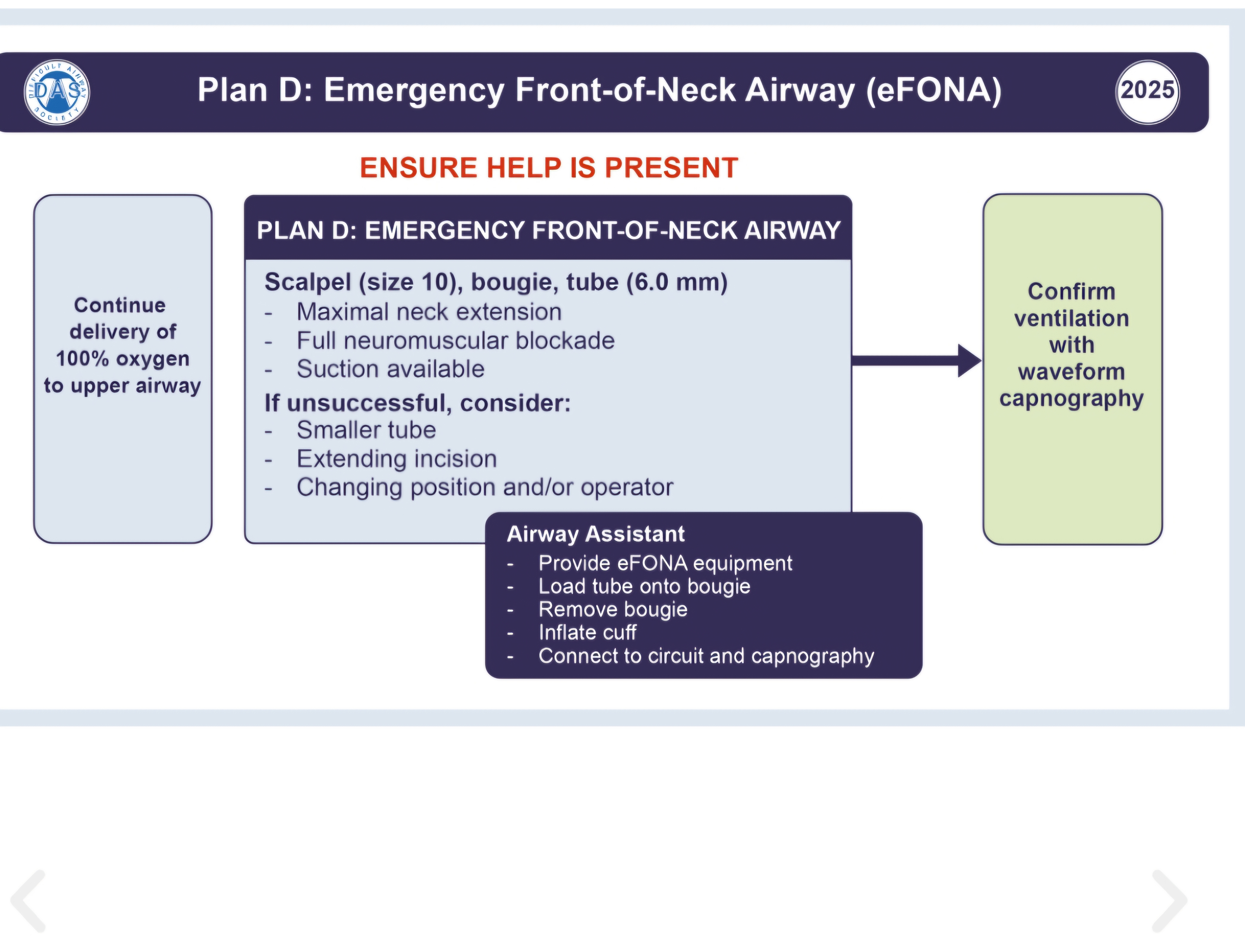
Plan D — Front-of-Neck Access (FONA): scalpel-bougie-tube cricothyrotomy IMMEDIATELY — do NOT delay; this Always — CICO is immediately lifeCICO (DAS recommended technique); simultaneously give sugammadex 16 is a life-or-death emergency threatening; FONA must not be Emergency mg/kg IV if rocuronium was used; 4 mm Melker emergency delayed cricothyrotomy kit (scalpel or needle technique)

⚠ CICO — Cannot Intubate, Cannot Oxygenate — Time to Brain Death = 4–5 Minutes
Declare CICO early; do NOT make repeated futile attempts; FONA simultaneous with sugammadex 16 mg/kg; a scalpel cricothyrotomy (vertical skin incision 4 cm, horizontal membrane incision, bougie, 6.0 ETT) takes <60 seconds in trained hands.
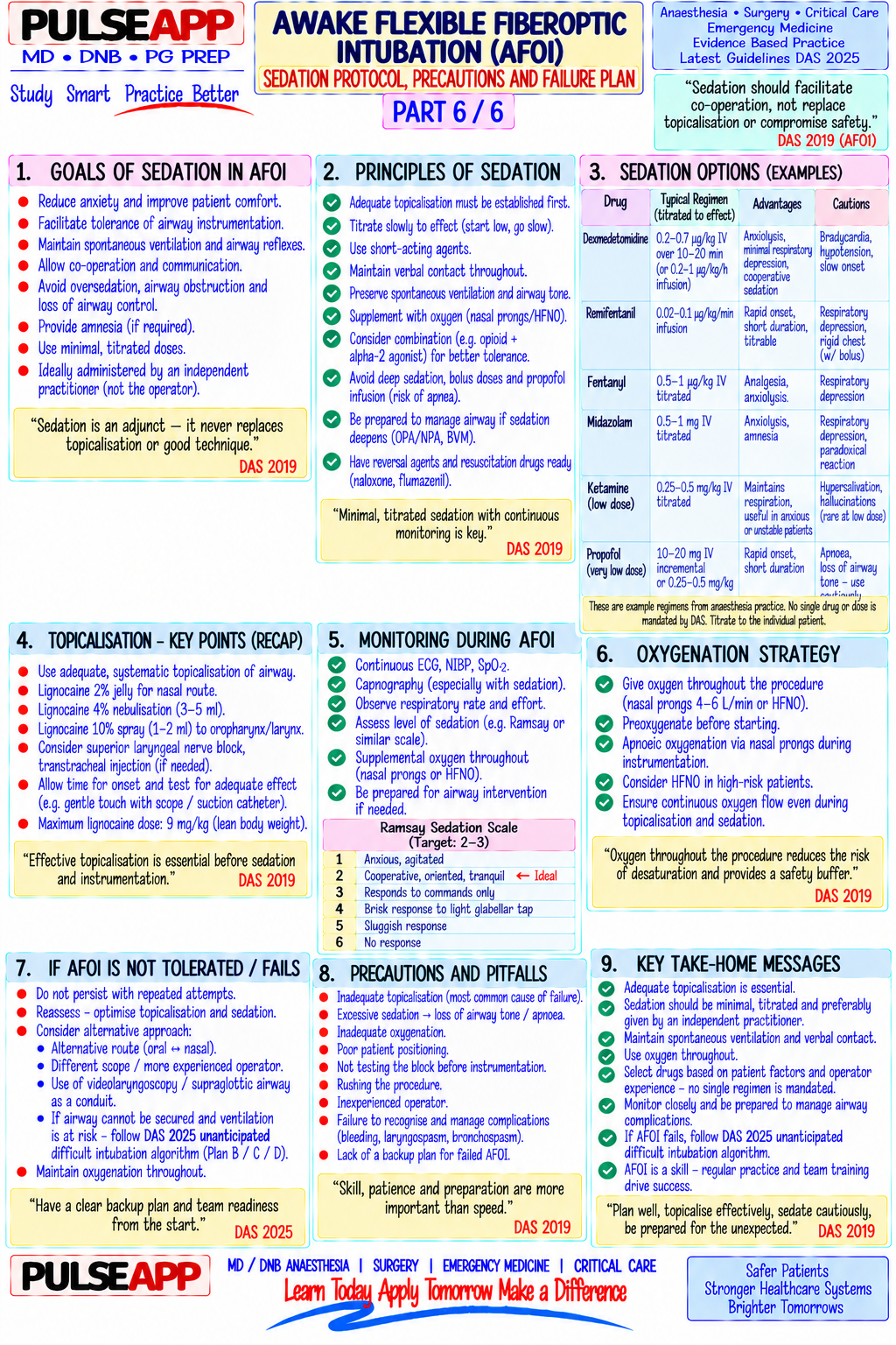
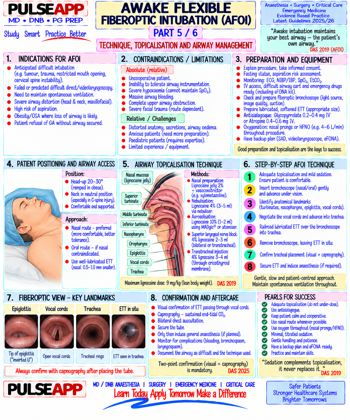
D. Awake Flexible Fiberoptic Intubation (AFOI) — Technique3 marks

Indication: anticipated difficult airway (Mallampati IV, TMD <6 cm, cervical spine instability, obesity with obstructive airway, known previous difficult airway, upper airway mass or tumour); the patient who might lose their airway under GA must be intubated AWAKE Sedation protocol (cooperative sedation, not general anaesthesia): Glycopyrrolate 200 mcg IM 30 minutes before (dries secretions — improves visualisation; secretions coat the lens) Dexmedetomidine 1 mcg/kg over 10 minutes then 0.5–0.7 mcg/kg/hr infusion — produces cooperative sedation without respiratory depression; patient remains rousable and able to follow commands; maintains airway tone and reflexes Midazolam 1–2 mg IV (anxiolysis) ± fentanyl 1 mcg/kg IV (analgesia) — small doses only; avoid over-sedation which abolishes protective reflexes Topical anaesthesia of the airway (most important step):

Oropharynx: lidocaine spray 4% + gargle

Nasopharynx (nasal route): lidocaine gel 2% + vasoconstrictor (xylometazoline) in each nostril; co-phenylcaine spray

Supraglottic: transtracheal injection or spray-as-you-go (SAYGO) — lidocaine 4% 2–3 mL injected through the cricothyroid membrane or through the working channel of the bronchoscope as it advances Total lidocaine dose <9 mg/kg (using 4% — 1 mL = 40 mg; total <400 mg for 70 kg patient)

Technique: pass lubricated FOB through ETT (size 7.0 loaded on the scope); advance the FOB through the nasal or oral route under direct vision; identify the epiglottis, arytenoids, vocal cords; pass the FOB through the cords under direct vision; confirm tracheal rings and carina; railroad the ETT over the FOB into the trachea; remove FOB; confirm placement with ETCO₂ waveform; induce GA (propofol) only after ETT confirmed in trachea

🎤 Viva Corner
Q. After 3 failed laryngoscopy attempts, you insert a 2nd generation SAD (i-gel) and achieve adequate chest rise with SpO₂ recovering to 97%. Your next action?
Excellent — the SAD (i-gel) is ventilating the patient adequately; SpO₂ is recovering. I now have two options: use the SAD as a conduit for fibreoptic-guided intubation through the SAD, or wake the patient up. The priority decision: is this surgery immediately life-saving (cannot be deferred) or elective? If elective/semielective: this is the ideal moment to wake the patient up and plan definitive airway management (awake FOI, tracheostomy under LA, regional anaesthesia technique, or defer surgery). DAS 2015 recommends waking the patient if they can be safely ventilated through the SAD and the surgery is not immediately lifesaving — this is the safest option as the patient wakes with an intact airway. If surgery cannot be deferred (e.g., perforated bowel, obstetric emergency): I will proceed to intubate through the i-gel using a fibrescope — pass a lubricated FOB through the i-gel aperture, visualise the cords, pass the ETT (use a smaller ETT: 6.0 or 6.5 mm to pass through the SAD); alternatively use an Aintree intubating catheter (AIC) as an intermediate step. Once intubated through the SAD, carefully remove the SAD over the ETT while a second person stabilises the ETT at the lips. Throughout all of this: maintain 100% FiO₂; call for senior help; keep the surgical team informed; document every step precisely including times and SpO₂ values.
Q. Name the DAS recommended technique for Front-of-Neck Access and describe it step by step. The DAS 2015 guideline recommends the scalpel-bougie-tube (SBT) technique as the preferred emergency cricothyrotomy method for CICO. Step 1 — Identify the cricothyroid membrane (CTM): the CTM is the soft tissue between the thyroid cartilage (upper) and the cricoid ring (lower); in a thin patient, place the non-dominant thumb on the thyroid notch, index finger on the cricoid ring — the CTM is the recess between them; in an obese or difficult-to-palpate neck, a vertical stab incision through the skin at the estimated CTM location is made first (a scalpel cuts through soft tissue), then the CTM is identified by palpation through the incision. Step 2 — Horizontal incision through the CTM: using a scalpel (size 10 blade), make a horizontal incision through the CTM; hold the trachea/larynx steady with the nondominant hand; the incision should be in the LOWER third of the CTM (to avoid the cricothyroid arteries running along the superior aspect). Step 3 — Dilate and stabilise: insert a tracheal hook (or the handle of the scalpel) through the incision to maintain access and pull the trachea anteriorly; alternatively, insert a tissuespreading clamp or gloved finger. Step 4 — Bougie insertion: pass a gum elastic bougie through the CTM incision into the trachea; confirm tracheal placement (clicks of bougie on tracheal rings; 24–26 cm depth stops at the carina). Step 5 — Railroad ETT: railroad a 6.0 mm cuffed ETT over the bougie into the trachea; inflate the cuff; confirm with ETCO₂ and bilateral breath sounds; secure the ETT. The entire procedure should be completed in <60–90 seconds in a trained operator. Simultaneous with Step 1: if rocuronium was used for induction → give sugammadex 16 mg/kg IV immediately — this may allow the patient to resume spontaneous ventilation within 2–3 minutes, potentially buying time.
★ Examiner's Pearl
The DAS Plan A→B→C→D algorithm with maximum attempt numbers (3 for Plan A) and the specific trigger for moving to CICO must be stated in order. LEMON and ULBT are newer assessment tools that examiners specifically ask about to distinguish candidates who read recent guidelines from those relying on older texts. For AFOI: dexmedetomidine as the preferred sedation agent (cooperative sedation preserving airway reflexes), glycopyrrolate before (dries secretions), and spray-as-yougo lidocaine technique are the three specifically tested points of airway topicalisation.
Frerk C et al. Difficult Airway Society 2015 guidelines for management of unanticipated difficult intubation (BJA 2015;115:827-848). Apfelbaum JL et al. Practice guidelines for management of the difficult airway 2022 (Anesthesiology 2022;136:31-81). El-Orbany M, Woehlck H. Difficult mask ventilation (Anesth Analg 2009;109:1870-1880). Popat M et al. DAS guidelines for AFOI (BJA 2011;107:308-323).
lock Protected Clinical Document • Pulse App • Screenshots Strictly Prohibited
QUESTION 36 person Asked by .
bookmark_add

Describe the classification and colour-coded segregation of biomedical waste as per BMW Management Rules India 2016. Discuss specific waste generated during anaesthesia practice and the additional precautions required for COVID- 19 anaesthesia waste.

collections Question Diagrams & Reference Images (2)
Question Reference Diagram
zoom_in View Image
Question Reference Diagram
zoom_in View Image
description Clinical Response
⚙ Core Concept
The Biomedical Waste Management Rules 2016 (amended 2018) replaced the 1998 rules with a more comprehensive, internationally aligned framework. Anaesthesia generates a disproportionately high volume of biomedical waste per procedure — sharps, IV lines, breathing circuit components, single-use airways, medication packaging, and fluid lines are all regulated waste. COVID-19 added an additional layer of aerosol-generating procedure (AGP) hazard management. (BMW Rules India 2016/2018; MoEF Guidelines; WHO Healthcare Waste Management)
A. BMW Classification & Colour-Coded Segregation (India 2016)4 marks

Colour / Container Waste Category Treatment/Disposal Anaesthesia Examples YELLOW bag Human anatomical waste (tissues, organs); animal Incineration (temperature ≥1200°C) Bloody gauze swabs; blood-contaminated waste; microbiology waste; soiled waste (cotton, at CBWTF (Common Biomedical drapes; expired IV drugs; placenta (obstetric); gauze, bandages, gowns contaminated with Waste Treatment Facility); or deep surgical tissue specimens sent intraoperatively blood/body fluids); expired medicine burial in specific cases

RED bag Non-chlorinated plastic waste: IV tubing, IV bags, Chemical treatment + shredding + IV administration sets; Bain circuit; breathing catheters, single-use gloves (non-contaminated), urine recycling at authorised recycler; circuit components; LMA (single-use); face bags, oxygen masks, nasogastric tubes; any non- autoclave NOT used (risk of dioxin masks; suction catheters; ETT (after use); sharp plastic recyclable from patient care release from chlorinated plastics) syringe bodies (without needles)

WHITE/PUNCTURE- Sharps: needles, syringes with needles, lancets, Autoclave + shredding at CBWTF; or ALL needles (drawing up needles, IV cannula PROOF container blades, broken glass, glass ampoules encapsulation in cement; or needle needles, spinal needles, epidural Tuohy (Sharps bin) destruction in needle cutters needles, nerve block needles); scalpel blades; glass ampoules; broken glass vials BLUE bag/box Glassware (non-sharp), metallic implants Autoclave/microwave then disposal Empty glass drug vials (intact); glass syringes at municipal solid waste site or (if applicable) recycling

BLACK bag General municipal solid waste (non-biomedical): food Municipal solid waste disposal; no Uncontaminated outer drug packaging; waste, packaging materials NOT contaminated with special treatment needed newspaper; administrative paper waste; nonbody fluids, paper, office waste contaminated cardboard drug cartons

KEY RULE — Sharps NEVER in plastic bags; always in puncture-proof container The most common BMW violation in anaesthesia is discarding needles into soft plastic bags or bins — creating a needlestick injury risk for housekeeping staff. All sharps go ONLY into the labelled white puncture-proof sharps container. Fill to 75% capacity only; do not force needles in; never recap with two hands (one-handed scoop technique only).

B. Waste Generated Specifically in Anaesthesia Practice3 marks

Waste Type Container Special Consideration Expired anaesthetic drugs Yellow bag (expired Controlled substances (opioids) require specific documentation and destruction procedures under (propofol, fentanyl, muscle medicine) NDPS Act — witnessed destruction by two staff, signed register; cannot simply discard in yellow bag relaxants) without documentation Residual drug in Yellow bag for liquid, Red for Separate the needle from the syringe body before disposal; needle in white sharps bin; body in red bag syringes/vials plastic syringe body, White for needle

Used breathing circuits, Red bag Single-use circuits: red bag; reusable equipment (laryngoscope blades, handles, fiberscopes) require LMAs, ETTs proper sterilisation before reuse — contaminated items not BMW waste but infection control waste (decontamination) CO₂ absorbent (spent soda Yellow or black bag Soda lime directly in the breathing circuit — contaminated with exhaled patient air and humidity; treat lime) depending on patient contact as patient contact waste; yellow bag preferred level Vaporizer filling devices and Hazardous chemical waste — Spilled or expired volatile agents are hazardous chemical waste (flammable, environmental pollutant); halogenated liquid agent NOT BMW require specific chemical waste handling; NOT to be poured down drain

C. COVID-19 Anaesthesia Waste Management3 marks
⚠ COVID-19 Generates "Category A Infectious Waste" — Highest Risk Category
AGP (Aerosol-Generating Procedures) waste: COVID-19 intubation/extubation generates infectious aerosols; all waste from AGP rooms must be treated as highly infectious; double-bag technique (inner red bag sealed, placed in outer red/yellow bag) before leaving the room PPE disposal: All COVID PPE (N95 masks, gowns, face shields, gloves, shoe covers) after use in COVID anaesthesia go in YELLOW bag (clinical waste); gowns and masks are contaminated with potential infectious aerosol-laden droplets; they CANNOT go in the regular waste stream Breathing circuit: disposable circuits only for confirmed/suspected COVID cases; placed in red bag after use; do NOT reprocess or reuse; HEPA filter at the expiratory limb and between the patient and the breathing circuit (viral filter) Colour marking: all COVID waste bags should be marked "COVID-19" in addition to the standard colour coding to alert CBWTF workers; dedicated collection trolleys and storage area for COVID waste Transport: leak-proof, sealed outer containers for transport; dedicated elevator route where possible; transportation workers in appropriate PPE; no handsorting of COVID waste at any stage
🎤 Viva Corner
Q. Which colour bag does an ETT contaminated with blood go into, and what do you do with the stylet?
A used ETT contaminated with blood: the ETT itself (plastic body) goes into the RED bag (non-chlorinated recyclable plastic waste contaminated with body fluids). If the ETT came with a metallic stylet: the stylet is a sharp metal object and goes into the WHITE puncture-proof sharps container. This illustrates the principle of segregation at the point of generation — a single item (ETT with stylet) may need to be separated into two different waste streams. The practical step: remove the stylet from the ETT before discarding; ETT tube in red bag; stylet bent or cut if possible to prevent reuse (injury prevention) then into the white sharps container. For single-use equipment: never reprocess or reuse any single-use anaesthesia equipment (ETT, LMA, breathing circuit components) — both for infection control reasons and legal compliance; reuse of single-use items in India is a violation of BMW Rules 2016 and can result in prosecution of the hospital.
Q. A nurse finds an uncapped needle on the anaesthetic trolley after a case. What is the immediate action, and what incident reporting is required?
Immediate actions: the nurse should NOT attempt to recap the needle or pick it up by hand; use forceps or a needle-destroyer device to safely transfer the uncapped needle directly into the white puncture-proof sharps container without hand contact; if there has been any possible contact with the needle tip (even if no obvious injury), treat as a potential needle-stick injury immediately — wash thoroughly with soap and water, report to occupational health, begin the PEP assessment process. Incident reporting: an uncapped needle left on the trolley is a serious safety violation — a near-miss (if no injury occurred) or a needlestick incident (if contact occurred). File a near-miss or adverse event report through the hospital's incident reporting system immediately; include: time, location, who found it, which procedure it relates to (if identifiable), and the action taken. The source patient's notes should be identified if possible to document their infection status for PEP decision-making. The anaesthesia team member responsible for the trolley cleanup should review their practice — BMW Rules 2016 require all healthcare workers who generate waste to be responsible for its proper disposal; leaving an uncapped needle is a personal responsibility failure as well as a systems failure. Preventive measures: single-handed needle-recapping technique only; needle-destroyers available at every anaesthetic trolley; training reinforcement for all anaesthetic staff.
★ Examiner's Pearl
The five-colour system (Yellow/Red/White/Blue/Black) with specific waste types for each is the most directly tested BMW content — reproduce the full table including the specific treatment method for each colour. The "sharps NEVER in soft bags" rule is a mandatory safety point. COVID-19 additions (double-bagging, yellow for all PPE, HEPA filters for breathing circuits, COVID-19 label on bags) are a post-2020 addition to the syllabus that examiners specifically include. State that controlled substances (opioids) require NDPS Act documentation for destruction — this is the most commonly missed specific point in anaesthesia BMW answers.
Biomedical Waste Management Rules 2016 and Amendment 2018 (MoEF, Government of India). WHO. Safe management of wastes from healthcare activities, 2nd Ed, 2014. CPCB. BMW Guidelines 2019. MoHFW India. Guidelines for COVID-19 BMWaste Management 2020.
lock Protected Clinical Document • Pulse App • Screenshots Strictly Prohibited
QUESTION 37 person Asked by .
bookmark_add

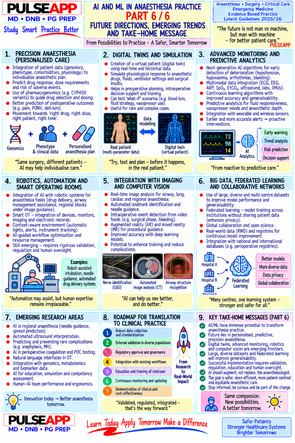
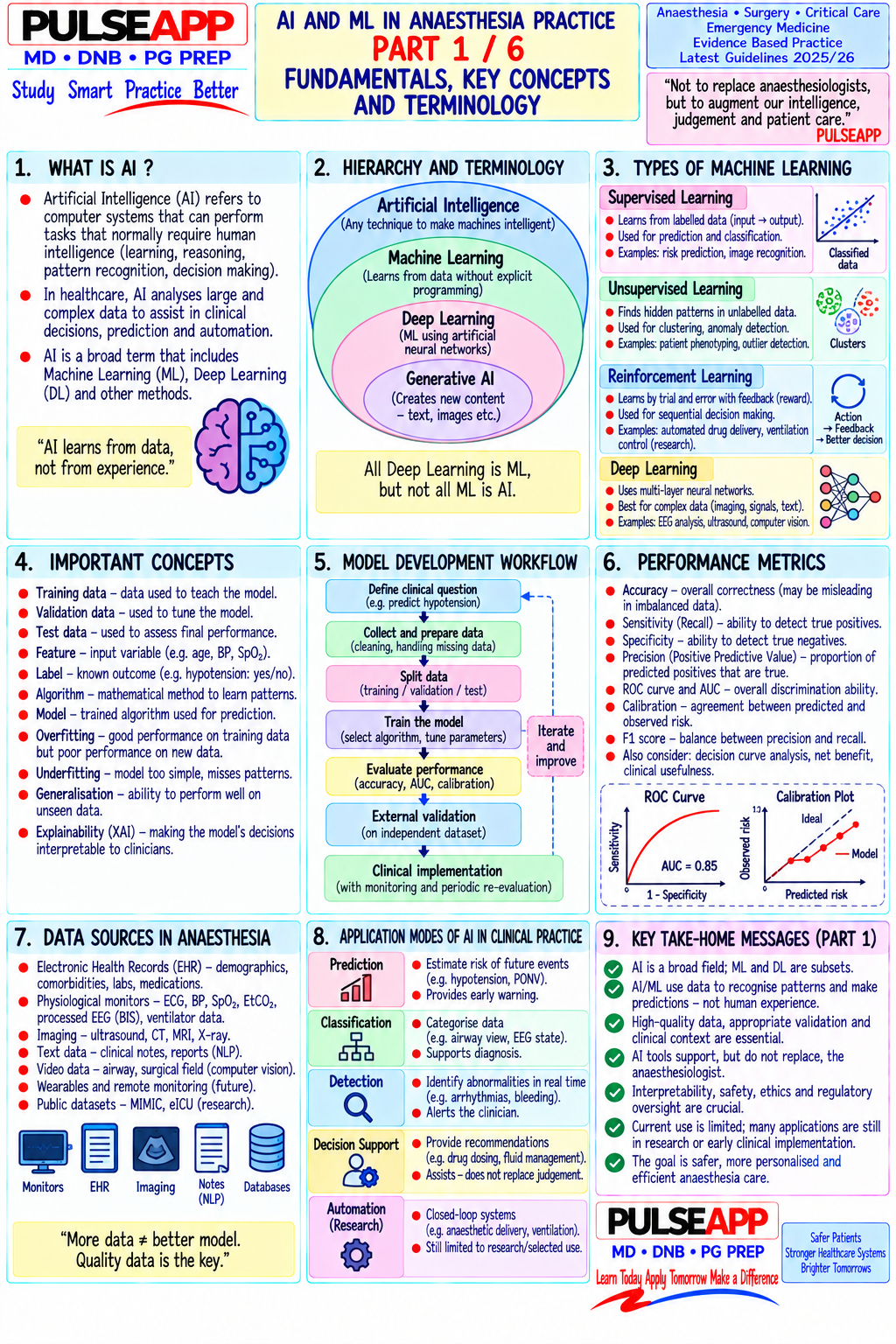
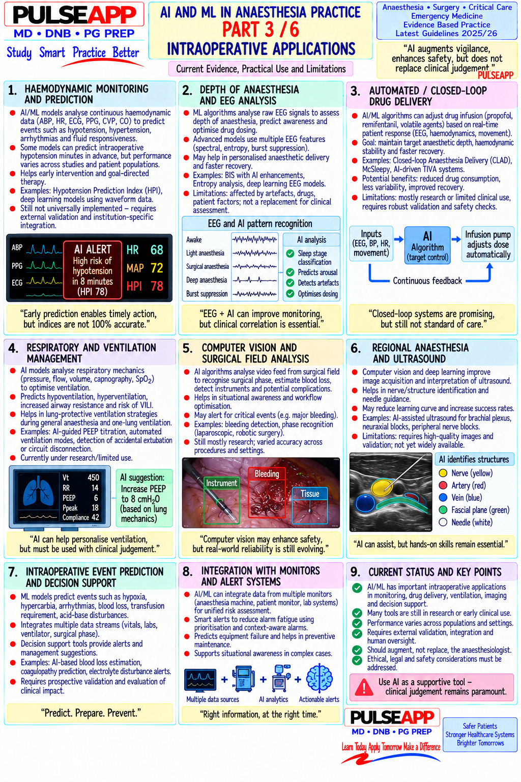
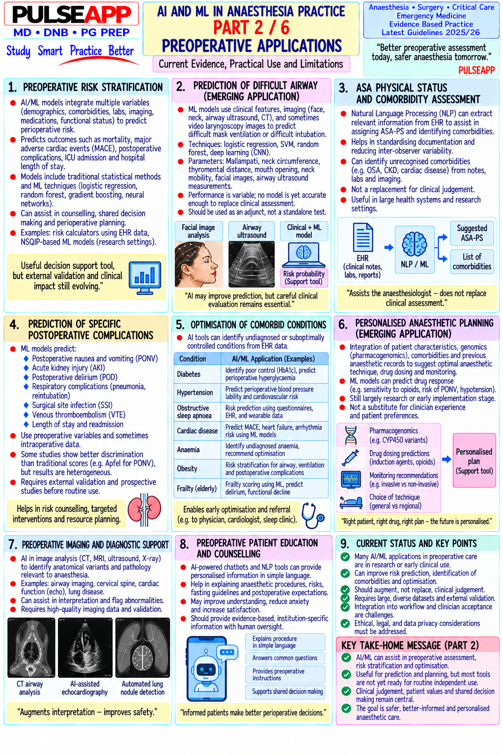
Discuss the definitions, current applications, limitations, and future implications of Artificial Intelligence (AI) and Machine Learning (ML) in anaesthesia practice.

collections Question Diagrams & Reference Images (3)
Question Reference Diagram
zoom_in View Image
Question Reference Diagram
zoom_in View Image
Question Reference Diagram
zoom_in View Image
description Clinical Response
⚙ Core Concept
Artificial Intelligence in medicine — and specifically in anaesthesia — has transitioned from science fiction to regulated clinical practice within a decade. ML algorithms now assist in predicting hypotensive episodes 15 minutes before they occur, in recognising airway images, in automating TIVA drug delivery, and in predicting postoperative outcomes. Understanding the basic principles, validated applications, and current limitations of AI is now a required component of the modern anaesthesiologist's knowledge. (Miller's Anaesthesia 9th Ed; Maheshwari K et al. — HPI; Babar N et al.; Stonemetz J; Hashimoto DA)
A. Definitions2 marks

Term Definition Example in Anaesthesia Artificial The broad field of computer science dedicated to creating systems that perform Computer programs that interpret ECG rhythms, diagnose airway Intelligence tasks that would normally require human intelligence — reasoning, problem- images, or generate anaesthetic plans (AI) solving, learning, perception Machine A subset of AI where algorithms learn patterns from data without being explicitly An algorithm trained on thousands of hypotensive episodes learns Learning (ML) programmed with rules; the algorithm improves its performance with more data to predict the hemodynamic pattern before the event occurs and experience Deep A subset of ML using artificial neural networks with many layers (deep networks) Image recognition of airway anatomy on video laryngoscopy; Learning (DL) — most powerful for image recognition, speech, and complex pattern recognition recognition of EEG burst suppression patterns for depth of tasks anaesthesia monitoring Natural AI subset enabling machines to understand and process human language (text or Automated extraction of preoperative comorbidities from Language speech) unstructured clinical notes to populate risk scores; voice-activated Processing anaesthetic documentation (NLP)

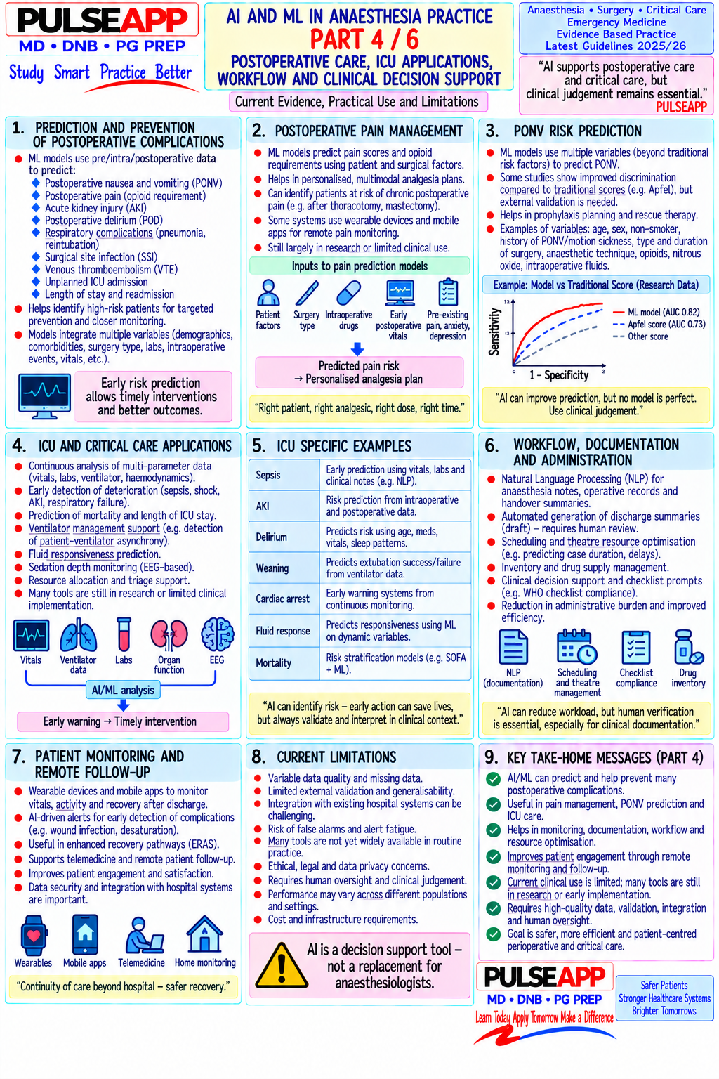
B. Current Clinical Applications in Anaesthesia4 marks

Application System/Tool Evidence/Validation Hypotension Hypotension Prediction Index (HPI — Edwards Lifesciences): an ML algorithm that Multiple RCTs (HYPE trial) show HPI-guided Prediction analyses the arterial waveform in real-time and outputs a probability (0–100) of vasopressor use reduces duration and severity of intraoperative hypotension in the next 15 minutes; uses machine learning on >20 arterial intraoperative hypotension by 50–70%; now cleared by waveform features FDA and CE mark; available in Acumen IQ sensor Closed-loop SEDASYS (now withdrawn from market but concept continues); closed-loop propofol Multiple RCTs show closed-loop TIVA achieves better anaesthesia drug TIVA using BIS feedback — the algorithm automatically adjusts propofol infusion rate to BIS time-in-target than manual titration; closed-loop delivery maintain target BIS 40–60 without manual titration; closed-loop vasopressor systems vasopressor reduces hypotension time in trials (CLMA — phenylephrine titrated by ML algorithm to MAP target) Difficult airway ML algorithms trained on preoperative airway assessment data (Mallampati, TMD, BMI, Proof-of-concept; not yet clinical standard; accuracy prediction etc.) to predict difficult intubation probability; some systems use facial image analysis via superior to single clinical variables but not yet smartphone cameras validated at population level Preoperative risk ML-enhanced versions of ASA, ACS-NSQIP, and APACHE scoring — algorithms trained Multiple validation studies show ML risk scores scoring on large perioperative databases predict individualised mortality, ICU admission, and outperform traditional scoring on discrimination metrics complication probability more accurately than point-score calculators (AUC 0.85–0.92 vs 0.75–0.80 for conventional scores) Depth of Machine learning-based EEG analysis for depth of anaesthesia (Sedline, Masimo); AI AI-enhanced EEG depth monitoring commercially anaesthesia/brain interpretation of processed EEG spectrograms; burst-suppression detection algorithms; available; research phase for real-time awareness monitoring automated alerting for accidental awareness risk detection Postoperative ML algorithms predict PONV, delirium, readmission, and unplanned ICU admission in Several hospital-deployed systems; validation ongoing; outcome real-time during the intraoperative period using intraoperative vital sign patterns; NLP not yet standard practice prediction algorithms extract risk factors from preoperative notes

C. Limitations, Risks & Ethical Concerns2 marks

Black box problem: deep learning algorithms are not interpretable — they produce outputs without explaining their reasoning; clinicians cannot understand why the algorithm recommended a specific action; this reduces trust and makes error analysis difficult

Training data bias: algorithms trained on data from specific populations (predominantly Western, specific hospital systems) may perform poorly in different patient populations (Indian patients, paediatric, rare diseases); biased training data → biased (and potentially harmful) outputs

Regulatory approval: most AI/ML devices in anaesthesia are approved only as decision-support tools, not autonomous treatment systems; the clinician retains legal responsibility for all patient care decisions regardless of AI recommendation

Data privacy: AI systems require large datasets of patient information for training; ethical data governance, consent, and privacy regulations (DPDP Act India 2023; GDPR in Europe) must be complied with

Alert fatigue: AI systems that generate too many false-positive alerts (like all monitoring systems) can lead to alarm fatigue — the very real danger that clinicians learn to ignore AI warnings, including true positives

D. Future Directions2 marks

Fully autonomous closed-loop anaesthesia machines (induction + maintenance + emergence, all algorithm-controlled); currently regulatory frameworks prohibit fully autonomous administration without human oversight in most jurisdictions Preoperative AI-generated anaesthetic plans tailored to individual patient genomics, comorbidities, and predicted drug metabolism Real-time voice-activated anaesthetic documentation — NLP extracts and records events from the spoken intraoperative narrative without manual charting AI-assisted surgical team communication and cognitive load management — real-time situational awareness tools that alert the team to developing physiological trends

🎤 Viva Corner
Q. What is the Hypotension Prediction Index (HPI) and how does it work mechanistically?
The Hypotension Prediction Index (HPI) is a machine learning algorithm developed and validated by Edwards Lifesciences that analyses the continuous arterial pressure waveform in real-time to output a probability score (0–100) indicating the likelihood that the patient will develop a mean arterial pressure below 65 mmHg within the next 15 minutes. The algorithm was trained on a large dataset of arterial waveform recordings from thousands of surgical patients, using supervised machine learning — it learned to identify subtle pre-hypotension patterns in the waveform (changes in pulse pressure variation, arterial waveform morphology features, heart rate trends, and other extracted parameters) that precede a hypotensive episode by 15 minutes. The specific input features include over 20 derived parameters from the arterial waveform, including measures of cardiac preload responsiveness, vasomotor tone estimates, and waveform shape features. An HPI score ≥85 indicates high probability of imminent hypotension — in clinical use, this triggers the anaesthesiologist to pre-emptively administer a vasopressor (phenylephrine or noradrenaline) or fluid before the MAP actually falls. Clinical evidence: the HYPE trial (Wijnberge et al., JAMA 2020, n=68 patients) demonstrated that HPI-guided care significantly reduced the time spent in intraoperative hypotension (MAP <65 mmHg) by approximately 50% compared to standard care — the algorithm predicted hypotension a median of 15 minutes before it occurred, allowing preventive treatment.
Q. What is the "black box problem" in AI, and why does it specifically matter for patient safety in anaesthesia?
The black box problem refers to the inability of deep learning algorithms to explain their reasoning — they produce outputs (predictions, recommendations) based on learned patterns in training data, but the internal mathematical transformations that convert inputs to outputs are not human-interpretable. A clinician cannot ask "why did the algorithm predict hypotension?" and receive a meaningful physiological explanation; the algorithm may respond with "because these 20 waveform parameters showed these numerical values" — which is computationally accurate but clinically uninterpretable. In anaesthesia, this matters specifically for patient safety in three ways: first, when the algorithm is wrong (false positive or false negative), the clinician cannot identify the error in reasoning to correct or override it intelligently — they can only reject the output based on clinical gestalt; second, when an adverse outcome occurs from following an AI recommendation, the medicolegal attribution of responsibility is unclear — the clinician cannot demonstrate that their decision was reasonably based on interpretable information; third, if the algorithm was trained on biased data (predominantly a specific patient population), it may perform poorly on underrepresented groups (elderly, paediatric, pregnant, specific ethnic groups) without any visible warning that it is operating outside its validated domain — a dangerous failure mode that a human clinician would at least recognise as unfamiliar territory.
★ Examiner's Pearl
The four AI/ML definitions (AI, ML, Deep Learning, NLP) with one anaesthesia example for each demonstrate the conceptual framework the examiner tests. The HPI (Hypotension Prediction Index) is the single most examined AI application in anaesthesia — state it by name, state the 15-minute prediction window, and cite the HYPE trial (JAMA 2020). The three limitations (black box, training data bias, regulatory approval as decision-support not autonomous) are the most commonly tested limitation points.
Maheshwari K et al. Hypotension Prediction Index — the HYPE trial (JAMA 2020;323:1052-1060). Hashimoto DA et al. Artificial intelligence in anaesthesiology (Anesthesiology 2020;132:379-394). Stonemetz J et al. Clinical decision support systems in anesthesia (J Clin Anesth 2011;23:658). Miller RD et al. Miller's Anaesthesia, 9th Ed.
lock Protected Clinical Document • Pulse App • Screenshots Strictly Prohibited
QUESTION 38 person Asked by .
bookmark_add

Discuss the carbon footprint of anaesthesia including the global warming potential of volatile agents and N₂O. Outline evidence-based strategies to reduce the environmental impact of anaesthesia practice.

collections Question Diagrams & Reference Images (2)
Question Reference Diagram
zoom_in View Image
Question Reference Diagram
zoom_in View Image
description Clinical Response
⚙ Core Concept
Healthcare contributes 4–5% of global greenhouse gas emissions, and anaesthesia — through volatile anaesthetic agents, nitrous oxide, and single-use equipment — is one of medicine's most carbon-intensive activities. One hour of desflurane at 1 MAC and 2 L/min fresh gas flow is equivalent to driving 235 km in a car. Crucially, the anaesthesiologist can reduce this footprint by over 90% through individual daily practice choices — making this one of the most actionable areas for environmental stewardship in medicine. (Ryan SM et al. — BJA 2010; McGain F — Anaesthesia 2014; NHS Net Zero Report 2020; Evans et al. — SSC 2021; Özelsel TJ)
A. Scale of the Problem — Global Warming Potentials3 marks

GWP (100Agent Atmospheric Lifetime Clinical Equivalent year, CO₂ = 1) Nitrous 265 114 years Also depletes stratospheric ozone; longest-persisting anaesthetic greenhouse gas — 1 L of N₂O delivered = Oxide (N₂O) 265 L CO₂ equivalent; used in huge volumes globally Isoflurane 510 3.2 years Medium impact; 1 L liquid isoflurane at 1 L/min FGF releases ~510 kg CO₂ eq over its lifetime Sevoflurane 130 1.1 years Lowest GWP of halogenated agents; preferred environmentally; short atmospheric lifetime Desflurane 2540 14 years Most potent greenhouse anaesthetic gas; 1 hour at 1 MAC, 2 L/min = driving 235 km; UK NHS banned desflurane from formulary in 2021; Australia, Canada, and several EU nations phasing out Propofol ~3.5 Metabolised — not The environmentally superior anaesthetic choice per MAC-hour; 97% less carbon footprint than desflurane (TIVA) (manufacturing exhaled into for equivalent anaesthesia time + disposal) atmosphere

Other contributors: single-use equipment manufacturing and disposal (anaesthesia circuits, LMAs, ETTs, gloves, syringes — lifecycle analysis shows a reusable LMA has 1/10th the carbon footprint of single-use equivalents over 40 uses); operating theatre energy consumption (3–6× normal hospital areas per m²); patient transport and building heating

N₂O infrastructure leakage: pipeline N₂O systems routinely leak 10–30% of delivered N₂O into the building atmosphere; reducing N₂O infrastructure use reduces both intentional and leak emissions

B. Evidence-Based Strategies for Sustainable Anaesthesia5 marks

Strategy Impact & Evidence 1. Eliminate Single highest-impact action: switching from desflurane to sevoflurane for 1 hour at 2 L/min reduces CO₂ equivalent emissions by ~95%; UK NHS desflurane removed desflurane from national formulary 2021 citing lack of meaningful clinical advantage over sevoflurane combined with catastrophic environmental cost; at the individual clinical level — desflurane provides only marginally faster early emergence (5–10 min) vs sevoflurane, with no difference in actual PACU discharge time or patient outcomes 2. Low-flow / Fresh gas flow (FGF) directly determines volatile agent consumption and atmospheric release; reducing FGF from 4–6 L/min (high-flow) to 0.5–1 L/min minimal-flow (low-flow) in the circle system reduces volatile agent consumption by 70–80%; at metabolic flow (0.35 mL/min), only the patient's metabolic uptake is anaesthesia replaced; requires circle system with CO₂ absorber (not Bain circuit); simultaneously saves cost and reduces operating room pollution 3. TIVA (Total Propofol-based TIVA produces zero volatile greenhouse gas emissions; propofol is metabolised and excreted — not exhaled into atmosphere; the IV environmental cost is propofol manufacturing and packaging, substantially less per MAC-hour than volatile agents; TIVA is the "greenest" general Anaesthesia) anaesthetic option; growing advocacy for TIVA adoption on environmental grounds in addition to its established clinical benefits (reduced PONV, no MH triggering, no HPV inhibition) 4. Eliminate N₂O (GWP 265, atmospheric lifetime 114 years) also depletes stratospheric ozone; clinical alternatives exist for every indication (opioids provide or minimise analgesia; volatile agents provide hypnosis; ketamine provides dissociative anaesthesia); N₂O elimination from routine anaesthesia removes the longestN₂O lasting anaesthetic greenhouse gas; multiple centres have eliminated N₂O from their formulary without impact on patient outcomes 5. Regional Every regional technique (peripheral nerve block, neuraxial) that successfully avoids GA eliminates 100% of volatile agent emissions for that case; the anaesthesia "Regional First" philosophy aligns environmental sustainability with established clinical benefits (reduced opioid consumption, faster PACU discharge, preference lower PONV, superior analgesia); expanding regional anaesthesia is one of the most effective department-level environmental strategies ("Regional First") 6. Volatile Systems (SageTech MARU — Medical Agent Recovery Unit) that capture exhaled volatile agent from scavenging systems and reconstitute it into liquid agent capture form for reuse; pilot programmes in UK and Scandinavia demonstrating both environmental and economic benefit; requires regulatory framework for and recycling recycled agent use (quality assurance); prevents atmospheric release from scavenging exhaust 7. Reduce Where infection risk allows: use reusable LMAs (sterilised between cases — lifecycle analysis shows 90% reduction in carbon footprint vs single-use); single-use metal laryngoscope blades (sterilised) vs single-use plastic; reusable temperature probes; audit single-use vs reusable decisions against infection risk equipment evidence rather than defaulting to single-use for convenience

C. Institutional Framework & Individual Responsibility2 marks

NHS Net Zero commitment (UK 2020): first national health system to commit to net-zero carbon by 2040; explicit desflurane ban (2021); anaesthesia sustainability targets in NHS contracts; green theatre champions programme

ESA Sustainability Task Force: green anaesthesia guidelines (2021); mandatory sustainability education in anaesthesia training programmes

India: desflurane is already rare in most Indian centres (cost-driven); sevoflurane predominates; formalising low-flow protocols and eliminating routine N₂O would meaningfully reduce Indian anaesthesia's carbon footprint without additional cost; TIVA availability expanding with propofol generic manufacturing

Individual anaesthesiologist: each anaesthesiologist makes hundreds of agent selection, FGF, and equipment choices annually — collectively determining a department's carbon footprint; professional responsibility now explicitly includes environmental stewardship in modern anaesthesia training frameworks

🎤 Viva Corner
Q. Your department is reviewing its anaesthetic formulary. Compare the carbon footprints of desflurane, sevoflurane, N₂O, and propofol TIVA per MAC-hour at 2 L/min fresh gas flow. The comparison is striking. At 2 L/min FGF and 1 MAC: Desflurane requires approximately 18% inspired concentration = 360 mL/min of agent vapour; multiplied by its GWP of 2540 and delivered over 1 hour, this produces approximately 60 kg CO₂ equivalent per hour — the same as driving 235 km. Sevoflurane at 1 MAC requires approximately 2.0% inspired = 40 mL/min vapour; multiplied by GWP 130 — approximately 1 kg CO₂ equivalent per hour — 60 times less than desflurane. N₂O at 50% FiO₂ and 2 L/min means 1 L/min of N₂O; N₂O has a GWP of 265 and molecular weight of 44 g/mol — approximately 1.7 g/L at standard conditions; 60 minutes × 60 L = 3,600 L N₂O at standard concentration = substantial CO₂ equivalent contribution. Propofol TIVA: propofol is metabolised in the body and excreted in urine as glucuronide conjugates — it does not reach the atmosphere; the carbon footprint is solely from manufacturing, packaging, and transport of the drug; lifecycle analysis estimates approximately 0.1 kg CO₂ equivalent per MAC-hour of propofol TIVA — approximately 600 times less than desflurane per equivalent anaesthesia time. The formulary recommendation is clear: desflurane should be removed or reserved for extremely rare clinical scenarios where it provides unique benefit; sevoflurane with low-flow technique and TIVA should be the standard options; N₂O should be eliminated from routine use and retained only where specific clinical benefit (relative contraindication to volatile agents, as an analgesic adjunct for specific procedures) justifies the environmental cost. Q. What is the environmental impact of using a Bain circuit vs a circle system for a 4-hour case, and what does this imply for formulary decisions?
The circuit choice directly determines how much volatile agent reaches the patient's lungs vs how much is wasted to atmosphere, because the two circuits operate on fundamentally different gas economy principles. A Bain circuit (Mapleson D) requires a fresh gas flow of at least 5–7 L/min for IPPV to prevent CO₂ rebreathing — because expired gas is vented directly through the APL valve without recirculation or CO₂ absorption; virtually ALL of the volatile agent in that fresh gas flow is used only once (exhaled into the scavenging system and ultimately the atmosphere after one passage through the patient circuit). In contrast, a circle system with low-flow anaesthesia (0.5 L/min FGF) recirculates approximately 85–90% of each breath through the CO₂ absorber — only 0.5 L/min of new gas (with volatile agent) is added per minute, and the remaining ventilation uses re-circulated and re-enriched circuit gas; volatile agent consumption falls by 80–90% compared to a high-flow Bain. Over 4 hours: Bain at 6 L/min = 1440 L total gas flow requiring full vaporizer output; circle at 0.5 L/min = 120 L fresh gas. For sevoflurane specifically: the circle system at low flow saves approximately 30–40 mL of liquid sevoflurane per hour vs high-flow technique — a direct cost saving as well as environmental benefit. The implication: wherever safe to do so (patient appropriate for low-flow, CO₂ absorber fresh), the circle system with low-flow or minimal-flow anaesthesia should be the standard; the Bain circuit should be reserved for cases where a circle system is not appropriate (remote locations, short paediatric cases using Mapleson E/F).
★ Examiner's Pearl
State the four GWP values (N₂O 265, isoflurane 510, sevoflurane 130, desflurane 2540) — these specific numbers are tested. The "1 hour desflurane = 235 km driving" analogy makes the scale vivid and is the most memorable fact examiners use in this topic. The UK NHS desflurane ban (2021) is a specific policy landmark. The hierarchy of environmental preference (propofol TIVA > sevoflurane low-flow > isoflurane > N₂O > desflurane) is the core clinical recommendation.
Ryan SM, Nielsen CJ. Global warming potential of inhaled anaesthetics (BJA 2010;105:760-768). McGain F, Naylor C. Environmental sustainability in anaesthesia (Anaesthesia 2014;69:789-799). NHS. Delivering a Net Zero National Health Service 2020. Xu J et al. Environmental impact of desflurane vs sevoflurane (Anesthesiology 2021;135:853-862). ESA Sustainability Task Force Position Statement 2021. Özelsel TJ et al. The future is now! (Anaesthesia 2019;74:274-278).
lock Protected Clinical Document • Pulse App • Screenshots Strictly Prohibited
QUESTION 39 person Asked by .
bookmark_add

Define sepsis and septic shock per Sepsis- (latest guidelines). Describe the pathophysiology of organ dysfunction in sepsis. Outline the Hour-1 bundle and key recommendations of the Surviving Sepsis Campaign latest guidelines

collections Question Diagrams & Reference Images (2)
Question Reference Diagram
zoom_in View Image
Question Reference Diagram
zoom_in View Image
description Clinical Response
⚙ Core Concept
Sepsis-3 (2016) redefined sepsis away from the SIRS-based model toward a mechanistically accurate definition: life-threatening organ dysfunction caused by a dysregulated host response to infection. The abandonment of SIRS criteria (which lacked specificity) and adoption of the SOFA score (which quantifies organ dysfunction) represents the most significant conceptual shift in sepsis management in three decades. The Surviving Sepsis Campaign 2021 bundled key interventions into an Hour-1 bundle that must begin before the patient leaves the ED. (Singer M et al. — Sepsis-3, JAMA 2016; Evans L et al. — SSC 2021; Hotchkiss RS — Pathophysiology of sepsis; Vincent JL)
A. Sepsis-3 Definitions (Singer et al., JAMA 2016)3 marks

Infection: pathological process caused by microbial invasion of normally sterile tissue

Sepsis: life-threatening organ dysfunction caused by a dysregulated host response to infection; defined clinically as: suspected or confirmed infection + acute increase in SOFA score ≥2 points (indicating acute organ dysfunction); a SOFA score ≥2 is associated with >10% in-hospital mortality in the general ICU population

Septic shock: a subset of sepsis with circulatory, cellular, and metabolic dysfunction that is profound enough to substantially increase mortality; defined as: sepsis PLUS vasopressor requirement to maintain MAP ≥65 mmHg AND serum lactate >2 mmol/L despite adequate fluid resuscitation; hospital mortality for septic shock exceeds 40%

Quick SOFA (qSOFA): a bedside screening tool (not a diagnostic criterion) to identify patients outside the ICU at risk of sepsis: ≥2 of the following: altered mentation (GCS <15), RR ≥22/min, systolic BP ≤100 mmHg; qSOFA ≥2 → high risk of poor outcome → initiate full SOFA assessment and sepsis workup SOFA Parameter Score 0→4 Component Respiratory PaO₂/FiO₂ ratio ≥400 → 0; 300–399 → 1; 200–299 → 2; 100–199 + MV → 3; <100 + MV → 4 Coagulation Platelet count (×10³/ ≥150 → 0; 100–149 → 1; 50–99 → 2; 20–49 → 3; <20 → 4 μL) Hepatic Bilirubin (mg/dL) <1.2 → 0; 1.2–1.9 → 1; 2.0–5.9 → 2; 6.0–11.9 → 3; >12 → 4 Cardiovascular MAP/vasopressors MAP ≥70 → 0; MAP <70 → 1; Dopamine ≤5 or dobutamine → 2; Dopamine 5–15 or NE ≤0.1 → 3; Dopamine >15 or NE >0.1 → 4 CNS GCS 15 → 0; 13–14 → 1; 10–12 → 2; 6–9 → 3; <6 → 4 Renal Creatinine (mg/dL) / <1.2 → 0; 1.2–1.9 → 1; 2.0–3.4 → 2; 3.5–4.9 or UO<500 → 3; >5 or UO<200 → 4 UO

B. Pathophysiology of Organ Dysfunction in Sepsis3 marks

Pathogen (bacteria/virus/fungi) + host susceptibility → Pattern Recognition Receptors (PRRs: TLR-4 for LPS; NOD-like receptors) on innate immune cells → activation of NF-κB transcription factor → massive cytokine release (TNF-α, IL-1β, IL-6, IL-8 — the "cytokine storm") → systemic endothelial activation and injury → four simultaneous cascades producing multi-organ dysfunction: Cascade Mechanism Clinical Consequence Vascular NO overproduction (iNOS) → pathological vasodilation + increased vascular permeability; loss of Distributive shock: low MAP, high CO, low SVR; dysfunction vascular autoregulation; catecholamine resistance from downregulated adrenergic receptors warm extremities; relative hypotension despite high output Coagulation Endothelial injury + TF expression → thrombin generation → microthrombi in capillary beds → Microvascular occlusion → end-organ activation simultaneous consumption of clotting factors → fibrinolysis activation → DIC ischaemia despite normal/high CO; bleeding from factor depletion Mitochondrial Cytokines (TNF-α) + NO → inhibition of mitochondrial electron transport chain (Complex I and Rising lactate in the presence of normal or high dysfunction Complex IV) → cells cannot use oxygen even when delivered → aerobic glycolysis → lactate cardiac output; explains why improving cardiac (cytopathic production → Type B lactic acidosis in well-perfused tissue output does not always resolve lactate in hypoxia) advanced sepsis Immunoparalysis Initial hyperinflammation (cytokine storm) is followed by profound immunosuppression: Patients die of secondary nosocomial infections (late sepsis) lymphocyte apoptosis (programmed cell death), monocyte deactivation, T-cell exhaustion, HLA- (Candida, Aspergillus, reactivated CMV, MDR DR downregulation on monocytes → impaired pathogen clearance → secondary infections bacteria) rather than the original pathogen dominate late clinical course

C. Surviving Sepsis Campaign 2021 — Hour-1 Bundle4 marks
✅ The Hour-1 Bundle — All Five Actions Within the First Hour of Recognition
1. Measure lactate — if initial lactate >2 mmol/L, repeat in 2 hours to assess clearance; if >4 mmol/L → high risk → aggressive resuscitation 2. Obtain blood cultures before antibiotics — at least two sets (aerobic + anaerobic); peripheral + central venous site; do NOT delay antibiotics more than 45 minutes waiting for cultures in critically ill patients 3. Administer broad-spectrum antibiotics — within 1 hour of sepsis/septic shock recognition; empirical antibiotic choice based on likely source and local antimicrobial resistance patterns; anti-MRSA coverage (vancomycin) if risk factors; anti-pseudomonal coverage if immunocompromised or healthcareassociated 4. Administer 30 mL/kg crystalloid IV for hypotension or lactate ≥4 mmol/L — use balanced crystalloids (Ringer's lactate, Plasmalyte) preferentially over normal saline (SSC 2021 strong recommendation based on SMART trial); complete within 3 hours; reassess after each 500 mL for fluid responsiveness (pulse pressure variation, stroke volume variation, passive leg raise response) 5. Vasopressors if MAP <65 mmHg during or after fluid resuscitation — noradrenaline (norepinephrine) is the first-line vasopressor (strong recommendation); vasopressin added as second agent to reduce noradrenaline dose (spares catecholamine effects); adrenaline as third-line or if noradrenaline + vasopressin insufficient; dopamine NOT recommended as first-line (arrhythmia risk) SSC 2021 Key Recommendations Beyond Hour-1 Recommendation Level Use balanced crystalloids over 0.9% NaCl (SMART trial evidence) Weak recommendation Albumin if already received large volumes of crystalloid and remain haemodynamically Weak recommendation unstable Intravenous hydrocortisone 200 mg/day (continuous infusion) if haemodynamically unstable Weak recommendation (ADRENAL trial — did not improve 90-day despite adequate fluids + vasopressors mortality but reduced vasopressor duration) Target glucose 6–10 mmol/L (110–180 mg/dL) using insulin infusion; avoid tight glycaemic Strong recommendation (NICE-SUGAR trial basis) control (hypoglycaemia hazard) Lung-protective ventilation: TV 6 mL/kg IBW; plateau pressure ≤30 cmH₂O; PEEP per ARDSNet Strong recommendation table for ARDS; prone positioning for moderate-severe ARDS CRRT (continuous renal replacement therapy) for AKI in haemodynamically unstable patients Weak recommendation — preferred over intermittent HD DVT prophylaxis; stress ulcer prophylaxis; oral care for VAP prevention; early enteral nutrition Standard ICU care bundle
🎤 Viva Corner
Q. How does Sepsis-3 differ from the previous SIRS-based definition, and why was the change made?
The SIRS-based definition of sepsis (Sepsis-1, 1991; Sepsis-2, 2001) defined sepsis as SIRS (≥2 of: temp >38°C or <36°C, HR >90, RR >20 or PaCO₂ <32, WBC >12,000 or <4,000 or >10% bands) in response to infection. The change to Sepsis-3 was motivated by two critical failures of the SIRS definition: first, SIRS criteria are non-specific — they are present in many non-infectious conditions (post-surgery, burns, pancreatitis, trauma) and are triggered in over 90% of ICU admissions regardless of infection; a definition that applies to 90% of critically ill patients provides no clinically useful differentiation. Second, SIRS criteria do not capture the organ dysfunction that is the actual biological harm of sepsis — a patient with fever and tachycardia from pneumonia has very different prognosis and need for intervention than one with the same vital signs plus renal failure, coagulopathy, and encephalopathy. Sepsis-3 replaced SIRS with the SOFA score — which specifically quantifies the degree of organ dysfunction across six organ systems; a SOFA increase of ≥2 in the context of infection indicates that infection has caused measurable multi-organ harm, which is precisely the dangerous, potentially life-threatening condition that deserves the label "sepsis." This makes the Sepsis-3 definition more specific (fewer false positives) and more clinically meaningful (it identifies the patients who are at genuine risk of death from their infection response). The Sepsis-3 criteria are also prognostically validated — a SOFA score increase of ≥2 predicts >10% in-hospital mortality in large datasets.
Q. Why is noradrenaline preferred over dopamine as the first-line vasopressor in septic shock?
Noradrenaline is the SSC-recommended first-line vasopressor in septic shock on both efficacy and safety grounds. From an efficacy standpoint: noradrenaline's primary action is alpha-1 adrenergic arteriolar vasoconstriction, raising SVR and MAP directly — the specific haemodynamic defect in septic shock (pathologically low SVR from NO-mediated vasodilation) is directly corrected. From a safety standpoint: the pivotal De Backer trial (NEJM 2010, n=1679 patients) compared dopamine vs noradrenaline as first-line vasopressor in shock. The trial showed no difference in 28-day mortality overall, but in the subgroup analysis, dopamine was associated with significantly more arrhythmia events (24% vs 12%) — including atrial fibrillation — and in cardiogenic shock patients, dopamine was associated with higher mortality. Dopamine at the vasopressor dose range (10–20 mcg/kg/min — the α₁-dominant dose) also has beta₁ stimulation causing tachycardia, which increases myocardial oxygen consumption — undesirable in already-stressed hearts. Additionally, dopamine's pharmacology is less predictable than noradrenaline (the dose-dependent receptor selectivity changes at different dose ranges), making it harder to titrate. The SSC 2021 guideline therefore recommends noradrenaline as first-line, dopamine only as an alternative if noradrenaline is unavailable, and vasopressin as the preferred second-line agent to reduce noradrenaline requirements (additive effect via V1 receptor vasoconstriction without catecholamine effects).
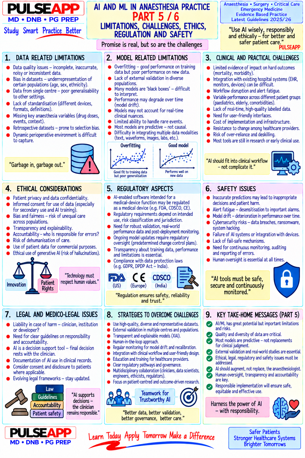
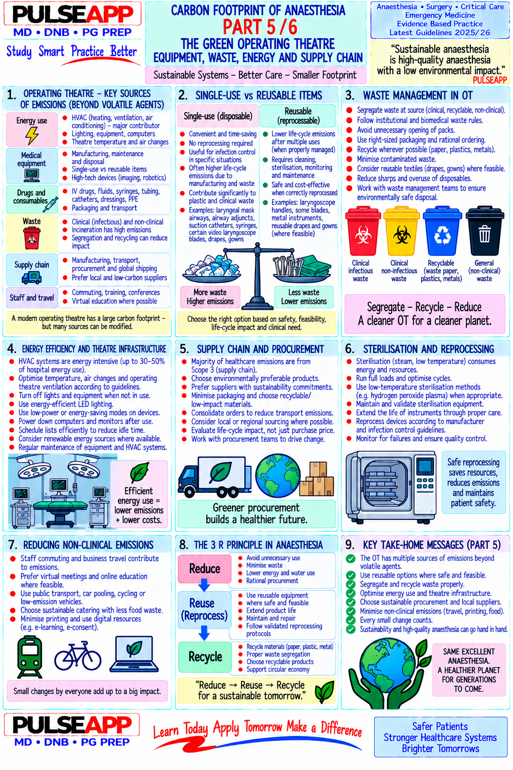
★ Examiner's Pearl
Sepsis-3 definition components must be stated precisely: sepsis = suspected infection + SOFA ≥2; septic shock = sepsis + vasopressor for MAP ≥65 + lactate >2 mmol/L — state all three components of septic shock with their specific numbers. The Hour-1 bundle all five actions (lactate, cultures, antibiotics, 30 mL/kg crystalloid, vasopressor if MAP <65) are separately marked in examinations. First-line vasopressor = noradrenaline with the De Backer trial citation; first-line fluid = balanced crystalloids (SMART trial) — cite both trials.
Singer M et al. Sepsis-3 — the third international consensus definitions for sepsis and septic shock (JAMA 2016;315:801-810). Evans L et al. Surviving Sepsis Campaign 2021 guidelines (Intensive Care Med 2021;47:1181-1247). De Backer D et al. Dopamine versus norepinephrine in shock (NEJM 2010;362:779-789). Semler MW et al. SMART trial — balanced crystalloids vs saline (NEJM 2018;378:829-839).
lock Protected Clinical Document • Pulse App • Screenshots Strictly Prohibited
QUESTION 40 person Asked by .
bookmark_add

Describe the anatomy of the brachial plexus at the costoclavicular space. Detail the ultrasound-guided costoclavicular approach technique, sonoanatomy, advantages over the conventional infraclavicular approach, and complications.

collections Question Diagrams & Reference Images (5)
Question Reference Diagram
zoom_in View Image
Question Reference Diagram
zoom_in View Image
Question Reference Diagram
zoom_in View Image
Question Reference Diagram
zoom_in View Image
Question Reference Diagram
zoom_in View Image

Showing 3140 of 337 questions

account_tree

Subcategory Tree

Explore Anesthesia subcategories

folder_special Anesthesia
Main
lock

Category Subscription

Subscribe to Anesthesia to unlock this module and all nested subcategories.

  • check_circle Access Anesthesia & all subcategories
  • check_circle Detailed, Peer-Reviewed Answers
  • check_circle High-yield visual aids & imaging
Get Category Subscription arrow_forward
Secure 256-bit SSL Connection

Anatomical Models

Explore high-fidelity 3D visualizations included in premium modules.

Case Reviews

Real-world clinical scenarios narrated by senior consultants.

Pulse App