Anesthesia | Eklavya Medical
vaccines
medical_services Main Specialty Domain lock Subscription Required

Anesthesia

Clinical anesthesiology, local and general anesthesia delivery systems, monitoring, and perioperative care.

verified Verified Medical Faculty menu_book 337 Q&A Modules
lock Protected Clinical Document • Pulse App • Screenshots Strictly Prohibited
QUESTION 261 person Asked by .
bookmark_add

Discuss in detail the anatomy of the brachial plexus. Enumerate and describe various blocks used to block the brachial plexus at various levels.

collections Question Diagrams & Reference Images (2)
Question Reference Diagram
zoom_in View Image
Question Reference Diagram
zoom_in View Image
description Clinical Response
"
⚙ CORE CONCEPT

The brachial plexus is the neural network supplying the entire upper limb — formed by the ventral rami of C5–T1 with variable contributions from C4 and T2. Understanding its anatomy — from the interscalene groove to the axilla — is the prerequisite for selecting the correct block for each surgical site, and for avoiding the complications (pneumothorax, intravascular injection, phrenic nerve palsy, Horner syndrome) that differ between approaches. Ultrasound guidance has transformed brachial plexus block from a landmark technique into a direct-visualisation procedure.

(Gray's Anatomy; Neal JM — brachial plexus blocks; Chin KJ — ultrasound-guided regional anaesthesia; Urmey WF — interscalene block; Borene SC — axillary block; Raj P — practical management of pain)
A. Anatomy of the Brachial Plexus
△ BRACHIAL PLEXUS — COMPLETE ANATOMICAL DIAGRAM
NERVE ROOTS (C5-T1) -> TRUNKS -> DIVISIONS (ant/post) -> CORDS -> TERMINAL BRANCHES

C4 (variable) -+
C5 ------------+
               +-- UPPER TRUNK --+-- Anterior ------------------------>
C6 ------------+                 +-- Posterior ----------+
C7 ------------  MIDDLE TRUNK --+-- Anterior ------+     |  POSTERIOR CORD
                                 +-- Posterior -----+     +-- Axillary N. (C5,C6)
C8 ------------+                                     |    +-- Radial N. (C5-T1)
               +-- LOWER TRUNK --+-- Anterior -------+    +-- Thoracodorsal N.
T1 ------------+                 +-- Posterior ------+
T2 (variable) -+                                  MEDIAL CORD
                                                   +-- Ulnar N. (C8,T1)
- Upper trunk (C5,C6): Suprascapular N.           +-- Medial cutaneous N. arm
  + Nerve to subclavius                           +-- Medial cutaneous N. forearm
- All three trunks: anterior divisions -> LATERAL
  and MEDIAL cords; posterior divisions -> POSTERIOR CORD   LATERAL CORD
                                                   +-- Musculocutaneous N. (C5,6,7)
LATERAL CORD (C5,6,7) + MEDIAL CORD (C8,T1) -> MEDIAN NERVE  +-- Lateral root of median N.

LOCATION SEQUENCE (IMPORTANT FOR BLOCKS):
Interscalene groove -> Between anterior and middle scalene muscles
Supraclavicular -> Over 1st rib, behind clavicle, ""corner pocket""
Infraclavicular -> Below clavicle, lateral to coracoid process
Axillary -> Axilla, around axillary artery

ANATOMICAL RELATIONSHIPS AT EACH LEVEL:
Interscalene: TRUNKS; C6 level; surrounded by interscalene fascia
Supraclavicular: DIVISIONS/TRUNKS; superficial, compact, ""corner pocket""
Infraclavicular: CORDS; around axillary artery (posterior, lateral, medial)
Axillary: TERMINAL BRANCHES; within axillary sheath
LevelStructureNerve RootsKey BranchesClinical Supply
ROOTS (C5–T1)Ventral rami exit intervertebral foramina; groove between anterior and posterior scalene tubercles; PREVERTEBRAL FASCIA covers themC5, C6, C7, C8, T1; C4 and T2 variable contributionsDorsal scapular N. (C5); Long thoracic N. (C5,6,7); phrenic N. arises from C4 with contributions C3,C5At root level — deepest and most proximal approach; PARAVERTEBRAL approach targets roots
TRUNKS (3)Upper (C5+C6), Middle (C7), Lower (C8+T1); posterior triangle of neck between anterior and middle scalene muscles; INTERSCALENE GROOVEUpper = C5,C6; Middle = C7; Lower = C8,T1Upper trunk: suprascapular nerve; nerve to subclavius; Upper trunk = ERB'S POINT at C6 levelINTERSCALENE BLOCK targets trunks; excellent for shoulder surgery
DIVISIONS (6)Each trunk divides into anterior (flexor) and posterior (extensor) division; pass BEHIND clavicle to enter axilla; SUPRACLAVICULAR level — compact, superficial, on 1st ribAnterior divisions → lateral + medial cords; Posterior divisions → posterior cordNo named branches at division levelSUPRACLAVICULAR BLOCK targets divisions — most complete upper limb block
CORDS (3)Posterior cord (all posterior divisions); Lateral cord (anterior div of upper+middle trunk); Medial cord (anterior div of lower trunk); named by relationship to AXILLARY ARTERY; INFRACLAVICULARPosterior = C5–T1; Lateral = C5,6,7; Medial = C8,T1Posterior cord: axillary N. + radial N. + thoracodorsal N.; Lateral cord: musculocutaneous N. + lateral root of median; Medial cord: ulnar N. + medial roots of median + medial cutaneous nervesINFRACLAVICULAR BLOCK targets cords — excellent for elbow, forearm, hand; avoids phrenic nerve palsy
TERMINAL BRANCHES (5)Musculocutaneous N., Median N., Ulnar N., Radial N., Axillary N.As abovePlus: medial cutaneous N. arm+forearm from medial cordAXILLARY BLOCK targets terminal branches — safe (no pneumothorax); musculocutaneous often leaves the sheath early → must be blocked separately
B. Brachial Plexus Blocks — Approach-by-Approach
BlockLevelTechnique (US-guided)LA DoseIndicationsComplications
INTERSCALENE BLOCK (ISB)TRUNKS at C5–C6 level; between anterior and middle scalenePatient supine, head rotated away; identify SCM, then scalene muscles; trunks visible as 3 hypoechoic nodules ("traffic light") between scalenes; in-plane needle from posterior20 mL 0.5% ropivacaine or 0.5% bupivacaineSHOULDER SURGERY (best block); clavicle surgery; proximal humerus; NOT suitable for hand surgery alonePHRENIC NERVE PALSY (100% incidence); Horner syndrome; intravascular injection into vertebral artery; pneumothorax (rare); recurrent laryngeal nerve palsy
SUPRACLAVICULAR BLOCK (SCB)TRUNKS/DIVISIONS at supraclavicular fossa ("corner pocket of the body")Identify subclavian artery above 1st rib; plexus = hypoechoic nodules lateral/superficial to artery ("honeycomb"); 25–30 mL deposited in "corner pocket"25–30 mL 0.5% ropivacaine or 0.375% bupivacaineENTIRE UPPER LIMB distal to shoulder — most complete single-injection blockPNEUMOTHORAX (0.5–6% landmark; <0.5% US); phrenic nerve palsy (60–80%); Horner syndrome; intravascular injection
INFRACLAVICULAR BLOCK (ICB)CORDS; below clavicle at coracoid; cords around axillary arteryIdentify axillary artery/vein below clavicle; cords as hyperechoic nodules around artery; single injection behind artery30 mL 0.5% ropivacaineENTIRE UPPER LIMB incl. hand; forearm and hand surgery; avoids phrenic nerve palsy; catheter placement easyPneumothorax (rare); intravascular injection; injury to axillary vessels; failure to block musculocutaneous nerve
AXILLARY BLOCK (AB)TERMINAL BRANCHES in axillary sheath; medial upper armArm abducted 90°, externally rotated; identify axillary artery; nerves around artery (median superficial/lateral, ulnar medial, radial posterior, musculocutaneous separate)5–10 mL per nerve (total 30–40 mL); 0.375–0.5% ropivacaineFOREARM and HAND surgery; SAFEST approach; suitable in anticoagulated patientsMUSCULOCUTANEOUS NERVE MISS (most common failure); intravascular injection; haematoma (compressible)
CERVICAL PARAVERTEBRAL BLOCKNERVE ROOTS at cervical foraminaUS-guided at C5–C7 transverse process level; deepest approach3–5 mL per levelShoulder; neck surgery; rarely usedHIGHEST RISK of serious complications: epidural/intrathecal injection, intravascular injection, phrenic nerve palsy; NOT recommended for routine use
SUPRASCAPULAR NERVE BLOCKBranch of upper trunk; suprascapular notch (posterior shoulder)Identify scapular spine, suprascapular notch, artery; 5–10 mL LA at notch5–10 mL 0.5% ropivacaineSHOULDER PAIN and ROTATOR CUFF SURGERY — covers 70% of shoulder joint innervation; useful adjunct to ISB, avoids phrenic palsyPneumothorax (rare); intravascular; nerve injury (rare)
C. Cutaneous Innervation Summary — Selecting the Right Block
Surgery SiteBest BlockAlternative
Shoulder (glenohumeral joint, rotator cuff)INTERSCALENE BLOCKSuprascapular + axillary nerve block (avoids phrenic palsy)
Clavicle fractureInterscalene (+ superficial cervical plexus block for medial clavicle)Supraclavicular
Entire arm including handSUPRACLAVICULAR BLOCK (most complete single injection)Infraclavicular
Elbow and forearmSupraclavicular or InfraclavicularAxillary (high volume)
Hand and wristAXILLARY BLOCK (safest) or InfraclavicularWrist block for minor procedures
Patient with severe respiratory disease (avoid phrenic palsy)INFRACLAVICULAR or AXILLARY blockSuprascapular + axillary nerve for shoulder
★ EXAMINER'S PEARL

Brachial plexus: ROOTS (C5-T1) → 3 TRUNKS (upper C5+6, middle C7, lower C8+T1) → 6 DIVISIONS (ant+post each) → 3 CORDS (named by axillary artery relationship: lateral, medial, posterior) → 5 TERMINAL BRANCHES (musculocutaneous, median, ulnar, radial, axillary). Mnemonic: "Robert Taylor Drinks Cold Beer." Blocks: INTERSCALENE (trunks, shoulder surgery — 100% phrenic palsy → CONTRAINDICATED in contralateral phrenic palsy); SUPRACLAVICULAR (divisions, "corner pocket," most complete for whole arm, pneumothorax risk); INFRACLAVICULAR (cords around axillary artery, no phrenic palsy risk, good for forearm/hand, catheter placement easy); AXILLARY (terminal branches, SAFEST, no pneumothorax/phrenic palsy, musculocutaneous MUST be blocked separately in coracobrachialis). US guidance = standard of care. Intercostobrachial nerve (T2) = tourniquet pain → always block subcutaneously for arm tourniquet procedures.
References: Neal JM et al. ASRA Practice Advisory on Neurologic Complications in Regional Anesthesia. Reg Anesth Pain Med 2015. Chin KJ, Perlas A. Ultrasonography of the brachial plexus. Anesthesiology 2011. Gray's Anatomy, 41st Ed.
"
lock Protected Clinical Document • Pulse App • Screenshots Strictly Prohibited
QUESTION 262 person Asked by .
bookmark_add

Define ICP and CPP. Discuss CSF circulation in detail. Discuss the various methods used in OT and ICU to manage raised ICP. What are the features of raised ICP?

collections Question Diagrams & Reference Images (1)
Question Reference Diagram
zoom_in View Image
description Clinical Response
"
⚙ CORE CONCEPT

The intracranial compartment is a fixed, rigid vault — the MONRO-KELLIE DOCTRINE states that the total intracranial volume (brain + CSF + blood) is constant; any increase in one component must be compensated by a decrease in another, or intracranial pressure (ICP) will rise. When compensatory mechanisms are exhausted, even small volume additions cause dramatic ICP rises — the "exponential compliance curve." Management of raised ICP is one of the most common and critical challenges in both neurological ICU and neuroanaesthesia.

(Miller RD — Miller's Anaesthesia; Czosnyka M — ICP monitoring review; BTIF TBI guidelines 2023; Rasulo FA — ICP management; Rangel-Castilla L — ICP management)
1. Definitions
TermDefinitionNormal ValuesSignificance
ICP (Intracranial Pressure)The pressure exerted by the contents of the cranial vault (brain parenchyma 80% + CSF 10% + blood 10%) within the rigid skull; measured in mmHg; represents pressure gradient between intracranial contents and atmospheric pressure (transducer zeroed at foramen of Monro)NORMAL: 5–15 mmHg (adults supine); 0–7 mmHg (seated); 1.5–6 mmHg (children); <2 mmHg (neonates); RAISED: >20 mmHg sustained; SEVERE: >40 mmHg critical; >60 mmHg near-fatalSustained ICP >20 mmHg = worse neurological outcomes; treatment threshold: ICP >20–22 mmHg (BTF 2016); ICP >40 mmHg = cerebral perfusion critically compromised
CPP (Cerebral Perfusion Pressure)The pressure gradient driving cerebral blood flow; CPP = MAP − ICP (or MAP − CVP if CVP > ICP); AUTOREGULATION maintains CBF constant over CPP range 50–150 mmHg; PRESSURE-PASSIVE when autoregulation failsNORMAL: 60–80 mmHg; MINIMUM: ≥60 mmHg (adult TBI — BTF); >70 mmHg preferred; PAEDIATRIC: ≥40–50 mmHg; CPP <50 mmHg = cerebral ischaemia likely; CPP <30 mmHg = irreversible injury imminentCPP is the THERAPEUTIC TARGET in neuroanaesthesia — not just ICP alone; CPP 60–70 mmHg target in TBI (BTF 2016); CPP-guided therapy has replaced purely ICP-guided therapy
2. Monro-Kellie Doctrine and Compliance
△ INTRACRANIAL COMPLIANCE CURVE
ICP ^      ,---- DECOMPENSATED PHASE
(mmHg)|    /  (steep: small vol increase = large ICP increase)
 40 --|   /  <- Treatment threshold 20-22 mmHg
      |  /
 20 --|-------------,---'  <- Inflection point (compensation exhausted)
      |      ,------'   COMPENSATED PHASE (flat: CSF + venous displaced)
  5 --|------'  Normal ICP range
      +--------------------------------------------> INTRACRANIAL VOLUME UP

COMPENSATORY MECHANISMS (operate on the flat part of curve):
1. CSF DISPLACEMENT: CSF pushed from cranial to spinal compartment (up to 50 mL)
2. VENOUS BLOOD DISPLACEMENT: cerebral venous blood squeezed into dural sinuses
   and jugular veins (up to 50-75 mL)
3. Once these are exhausted -> EXPONENTIAL ICP RISE (vertical part of curve)
   -> HERNIATION syndromes begin
3. CSF Circulation — Complete Description
ComponentDetail
CSF ProductionCHOROID PLEXUS (70% of CSF): specialised ependymal cells lining lateral, 3rd, and 4th ventricles; Na⁺-K⁺-ATPase drives Na⁺ into CSF → water follows osmotically; CSF formation rate: 0.35 mL/min = 500 mL/day; total CSF volume: 150 mL (50 mL intracranial + 100 mL spinal); REPLACED 3–4 TIMES/DAY; also brain parenchyma (30%); production PRESSURE-INDEPENDENT (largely); CARBONIC ANHYDRASE essential → ACETAZOLAMIDE inhibits CSF production
CSF CompositionClear, colourless; protein: 15–45 mg/dL; glucose: 2.5–4.5 mmol/L (60–80% of plasma, ratio >0.6 normal); sodium: 135–150 mEq/L; cells: WBC <5/mm³; pressure: 8–18 cmH₂O (lateral decubitus) = 6–13 mmHg; pH 7.32–7.35
CSF Circulation PathwayLATERAL VENTRICLES (choroid plexus) → FORAMINA OF MONRO → 3rd VENTRICLE → CEREBRAL AQUEDUCT OF SYLVIUS (most common obstruction site) → 4th VENTRICLE → FORAMINA OF LUSCHKA (×2) and MAGENDIE (×1) → SUBARACHNOID SPACE (basal cisterns) → ascends over cerebral hemispheres (pulsatile bulk flow) → DURAL VENOUS SINUSES via ARACHNOID GRANULATIONS → reabsorbed into venous blood → jugular veins → SVC
Arachnoid Granulations (reabsorption)Herniations of arachnoid membrane into dural venous sinuses (mainly superior sagittal sinus); pressure-dependent VALVES — open when ICP exceeds venous sinus pressure (by 5–7 mmHg); MALABSORPTION = COMMUNICATING HYDROCEPHALUS; OBSTRUCTION at aqueduct/foramina = OBSTRUCTIVE (NON-COMMUNICATING) HYDROCEPHALUS
Pulsatile CSF FlowDriven by cardiac cycle (systolic pulsations → CSF pushed into spinal canal) and respiratory cycle (inspiration → ↓ intrathoracic pressure → ↑ epidural venous engorgement → CSF pushed rostrally); phase-contrast MRI visualises this (important in Chiari malformation)
4. Features of Raised ICP
FeatureMechanismClinical Significance
HEADACHE (early, classic)Distension of pain-sensitive intracranial structures; worse in morning (↑ ICP during recumbency + REM sleep); exacerbated by ValsalvaBilateral, frontal/occipital; "bursting" quality; morning headache waking patient = RED FLAG
VOMITING (projectile)Stimulation of vomiting centre in medulla; characteristic PROJECTILE pattern without preceding nauseaUnexplained projectile vomiting with headache = raised ICP until proven otherwise
PAPILLOEDEMAICP transmitted along optic nerve sheath → impaired axoplasmic flow → axonal swelling at optic disc; TAKES 24–48H TO DEVELOPBILATERAL papilloedema = raised ICP until proven otherwise; loss of venous pulsation = earliest sign
CUSHING'S TRIAD / REFLEX (late, critical)ICP approaches MAP → brainstem ischaemia → medullary vasomotor centre ischaemia → SYSTEMIC HYPERTENSION (compensatory) → baroreceptor BRADYCARDIA; medullary compression → IRREGULAR BREATHINGHYPERTENSION + BRADYCARDIA + IRREGULAR RESPIRATION = PRE-TERMINAL sign of impending herniation; do NOT treat hypertension with antihypertensives
ALTERED CONSCIOUSNESS / GCS DECLINEDiffuse cortical dysfunction; RAS compression in midbrain → loss of consciousnessDeteriorating GCS = raised ICP until proven otherwise (urgent CT); GCS ≤8 = severe TBI = immediate ICU
HERNIATION SYNDROMESUNCAL (temporal lobe through tentorial notch → CN III compression → ipsilateral blown pupil then contralateral hemiplegia — Kernohan's notch = false localising sign); CENTRAL (bilateral small pupils, loss of upward gaze, decorticate→decerebrate); TONSILLAR (foramen magnum → medullary compression → cardiorespiratory arrest)IPSILATERAL BLOWN PUPIL = UNCAL HERNIATION = EMERGENCY; Decorticate = cortical lesion; Decerebrate = midbrain/pontine lesion (worse prognosis)
5. Management of Raised ICP — Tiered Approach
Tier / MethodInterventionMechanismTarget/Details
TIER 1 — Head PositionHead-up 30°; neutral neck position; avoid tight ETT tiesFacilitates jugular venous drainage → ↓ cerebral venous pressure → ↓ ICP by 2–5 mmHg30° head-up = optimal; >30° may reduce MAP disproportionately
TIER 1 — NormocapniaPaCO₂ 35–40 mmHg; avoid hyper/hypocapnia; controlled ventilation with EtCO₂ monitoringCO₂ = most potent physiological regulator of CBF (2–3% per mmHg)EtCO₂ 35–38 mmHg correlates with PaCO₂ 38–42 mmHg; PROPHYLACTIC hyperventilation AVOIDED
TIER 1 — NormoglycaemiaBlood glucose 6–10 mmol/L; avoid hypo/hyperglycaemiaHyperglycaemia → lactate in ischaemic neurons → acidosis → cell death; increases BBB permeabilityHourly glucose monitoring; insulin sliding scale
TIER 1 — NormothermiaTemperature 36–37.5°C; treat fever aggressively; avoid hyperthermiaEach 1°C ↑ temperature → CMRO₂ ↑7% → ↑CBF → ↑ICPContinuous temperature monitoring; cooling; antipyretics
TIER 1 — Analgesia and SedationAdequate analgesia; sedation to prevent coughing/bucking; propofol, remifentanil, fentanylPropofol: ↓CMRO₂ 25–50% → ↓CBF → ↓ICP; caution: PRIS >4–5 mg/kg/hr >48hRASS -1 to -3; avoid daily sedation interruption in active intracranial hypertension
TIER 2 — Mannitol0.25–1.0 g/kg IV over 20–30 min; repeat q4–6h; monitor osmolality (<320 mOsm/L)Immediate plasma expansion effect + sustained osmotic effect (draws water from brain across intact BBB)FIRST-LINE; CONTRAINDICATED: osmolality >320, severe hypovolaemia, anuria
TIER 2 — Hypertonic Saline (HTS)3% NaCl 150–300 mL; 23.4% NaCl 30 mL bolus; target Na⁺ 145–155 mEq/LOsmotic mechanism + restores membrane potential; does NOT cause diuresis — improves cardiac outputNOW PREFERRED in hypovolaemia/haemorrhagic shock with TBI; complications: hypernatraemia, ODS
TIER 3 — Therapeutic HyperventilationShort-term PaCO₂ 30–35 mmHg as rescue bridge↓PaCO₂ → vasoconstriction → ↓CBV → ↓ICP by 5–10 mmHg within minutesUSE ONLY AS BRIDGE; duration <30 min; not below 25 mmHg
TIER 3 — CSF Drainage (EVD)External ventricular drain via Kocher's pointDirect removal of CSF → ↓ intracranial volume → ↓ ICPGOLD STANDARD for ICP measurement; complications: infection, haemorrhage
TIER 3 — Decompressive CraniectomySurgical removal of bone flap; dura patchedRemoves the "rigid container" limitation; RESCUEicp: fewer deaths but ↑ vegetative stateIndication: refractory ICP >20 mmHg despite tier 1–2 + resectable haematoma
TIER 3 — Barbiturate ComaThiopentone 2–4 mg/kg bolus then 1–3 mg/kg/hr; titrate to burst-suppression↓CMRO₂ to max 50%; proportional ↓CBF → ICP reduction of 10–20 mmHgLAST RESORT — haemodynamic toxicity, immune suppression, prolonged emergence
TIER 3 — HypothermiaMild (33–35°C) or moderate (32–34°C) targeted temperature management↓CMRO₂ 6–7% per °C → ↓CBF → ↓ICPEUROTHERM3235: mild hypothermia → WORSE outcomes; rebound ICP on rewarming — must be gradual
Dexamethasone8 mg IV loading → 4 mg q6h↓Tumour-associated BBB disruption and vasogenic oedema; TUMOUR-SPECIFIC benefitUSE ONLY for vasogenic oedema around tumours; AVOID in TBI (CRASH trial → ↑mortality) and stroke
★ EXAMINER'S PEARL

ICP normal 5-15 mmHg; treatment threshold >20-22 mmHg. CPP = MAP − ICP; target ≥60 mmHg in TBI (BTF 2016). Monro-Kellie doctrine: fixed volume skull; brain (80%) + CSF (10%) + blood (10%) = constant. CSF: produced by CHOROID PLEXUS (0.35 mL/min = 500 mL/day, 3-4x daily turnover); flows lateral ventricles → foramen of Monro → 3rd ventricle → AQUEDUCT OF SYLVIUS (most common obstruction site) → 4th ventricle → foramina of Luschka (×2) + Magendie (×1) → subarachnoid space → ARACHNOID GRANULATIONS in superior sagittal sinus → venous blood. Features of raised ICP: morning headache, projectile vomiting (medullary), papilloedema (takes 24-48h), CUSHING'S TRIAD (PRE-TERMINAL), blown ipsilateral pupil (uncal herniation). Management tiers: TIER 1 (head-up 30°, normocapnia, normoglycaemia, normothermia, propofol sedation); TIER 2 (MANNITOL or HYPERTONIC SALINE); TIER 3 (brief hyperventilation rescue, EVD drainage, decompressive craniectomy, barbiturate coma, hypothermia). DEXAMETHASONE only for tumour vasogenic oedema (NOT TBI).
References: Brain Trauma Foundation. Guidelines for Management of Severe TBI 4th Ed 2016. Cooper DJ et al. RESCUEicp — NEJM 2011. EUROTHERM3235 trial — NEJM 2015. Czosnyka M, Pickard JD. Monitoring and interpretation of ICP. J Neurol Neurosurg Psychiatry 2004.
"
lock Protected Clinical Document • Pulse App • Screenshots Strictly Prohibited
QUESTION 263 person Asked by .
bookmark_add

Discuss the safety features of the anaesthesia machine.

description Clinical Response
"
⚙ CORE CONCEPT

The modern anaesthesia workstation is an extraordinarily complex piece of life-critical medical equipment — designed with MULTIPLE REDUNDANT SAFETY SYSTEMS to prevent the delivery of a hypoxic gas mixture, gas delivery failure, circuit disconnection, or barotrauma, each of which can rapidly cause patient death. The safety systems are layered — each addresses a specific failure mode, and multiple systems protect against the same threat. Understanding these systems is essential for both the anaesthesia machine check and for troubleshooting intraoperative equipment failure.

(Dorsch JA, Dorsch SE — Understanding Anaesthesia Equipment 5th Ed; AAGBI Safety Guidelines; ISO 8835; WHO Surgical Safety Checklist; Olympio MA — Modern Anaesthesia Machine Safety)
A. Gas Supply Safety Systems
△ GAS SUPPLY TO MACHINE — SAFETY AT EVERY POINT
PIPELINE SUPPLY (hospital medical gas pipeline):
Wall outlet -> PIPELINE INLET CONNECTOR -> Machine regulator -> Flowmeters

CYLINDER SUPPLY (backup):
Cylinder -> PIN INDEX SAFETY SYSTEM -> Yoke -> Machine regulator -> Flowmeters

SAFETY MECHANISMS AT EACH POINT:
1. Pipeline pressure: 400 kPa (4 bar) -- regulated constant pressure
2. DIAMETER INDEX SAFETY SYSTEM (DISS): non-interchangeable wall connectors
   (O2 connector size != N2O connector size != air connector size)
3. PIN INDEX SAFETY SYSTEM (PISS): cylinder yoke pin positions unique per gas
   (O2 = pins 2,5; N2O = pins 3,5; Air = pins 1,5; CO2 = pins 1,6)
4. Cylinder COLOUR CODING:
   O2 = black body + white shoulder
   N2O = blue (all blue)
   Air = grey body + black+white quartered shoulder
   CO2 = grey (all grey)
5. Pressure REDUCING VALVES (regulators): reduce cylinder high pressure
   (O2 137 bar; N2O 44 bar) to working pressure (400 kPa)
6. Non-return valves (check valves) at pipeline inlets -- prevent
   backflow between pipelines and cylinders
Safety FeaturePurposeHow It Works
1. Pipeline Inlet Non-Return ValvesPrevent backflow from machine into pipeline; prevent pressurisation of empty cylinders from pipelineSpring-loaded check valves at each gas inlet; open only when pipeline pressure exceeds machine internal pressure
2. Oxygen Failure Warning Device (OFWD / Pneumatic Alarm)Audible alarm when O₂ supply pressure falls; alerts anaesthesiologist before hypoxic mixture reaches patientWhen O₂ pressure falls below 200–260 kPa → pneumatic whistle activates (powered by remaining O₂ pressure — sounds ≥7 seconds); required by ISO/AAGBI on ALL machines
3. Oxygen Pressure Failure Cut-off (Link-25 system / O₂-N₂O coupling)PREVENTS delivery of N₂O without adequate O₂ — most critical anti-hypoxia mechanismMechanical LINK-25 system: chain links on N₂O flowmeter spindle coupled to O₂ flowmeter spindle; PROPORTIONING SYSTEM maintains minimum O₂:N₂O ratio of 25% O₂ by volume at all times; also OXYGEN RATIO MONITOR CONTROLLER (ORMC)
4. Minimum O₂ FlowMachine cannot deliver zero O₂ flow when N₂O is flowing; minimum 150–200 mL/min mandatoryMechanical stop on O₂ flowmeter bobbin/needle valve; ensures minimum O₂ FLOW only — O₂ ANALYSER still essential for final monitoring
5. O₂ Flush Button (Bypass valve)Delivers O₂ at 35–75 L/min directly to common gas outlet, bypassing flowmeters/vaporisersDirect pathway from O₂ pipeline to common gas outlet at near-pipeline pressure; DANGER: activating during ventilator inspiration → BAROTRAUMA risk
6. Reserve Cylinder (Emergency O₂)Cylinder O₂ available if pipeline failsE-size cylinder = 660L O₂ (~1 hour at 10 L/min); AAGBI: all machines must have a working O₂ cylinder
B. Flowmeter and Vaporiser Safety
Safety FeaturePurpose and Mechanism
Flowmeter Bobbin/Float DesignEach gas has a UNIQUE, colour-coded flowmeter tube; O₂ flowmeter always on the DOWNSTREAM (right-hand) side of the flow bank — any leak from upstream delivers excess gas (not O₂ deficiency) toward patient; mandatory ISO requirement
Vaporiser Keyed Filling SystemFilling ports are AGENT-SPECIFIC with colour-coded keyed adapters; prevents wrong-agent filling; TEC 6 (desflurane) separately powered — electric
Vaporiser Interlock System (Selectatec manifold)Only ONE vaporiser can be engaged at a time; mechanical interlock prevents simultaneously turning on two vaporisers
Vaporiser Anti-Tipping MechanismAnti-tipping valves close if vaporiser tilted >45° — prevents liquid agent entering the bypass channel (would cause massive overdose)
Agent Concentration LimitsVaporisers calibrated for maximum safe output: sevoflurane TEC 7 = max 8%; isoflurane = max 5%; enflurane = max 5%; desflurane (TEC 6) = max 18%
C. Breathing Circuit and Ventilator Safety
Safety FeatureMechanism and Importance
PRESSURE LIMITING (Pop-off / APL) ValveLimits maximum circuit pressure during spontaneous breathing (set at 1–2 cmH₂O leak-off pressure); PREVENTS BAROTRAUMA; NEVER close APL completely during spontaneous breathing
Low Pressure Alarm (Disconnection)Detects circuit disconnection (most common cause of intraoperative hypoxia in ventilated patients); if peak inspiratory pressure <4–8 cmH₂O = alarm
High Pressure Alarm (Obstruction/Barotrauma)If circuit pressure exceeds 40–60 cmH₂O → ALARM and automatic pressure relief; triggered by kinked ETT, one-lung intubation, bronchospasm
Volume/Tidal Volume AlarmSpirometer measures expired tidal volume; alarm if TV outside set limits; detects disconnection, large leaks
O₂ Analyser (Galvanic fuel cell or paramagnetic)MANDATORY; measures FiO₂ in REAL TIME on the INSPIRATORY LIMB; alarms if FiO₂ falls below ~25%; the LAST SAFETY NET; calibrated daily (21% and 100%)
Capnography (EtCO₂)Mandatory monitoring standard; absence of CO₂ waveform = oesophageal intubation or disconnection; EtCO₂ rise = hypoventilation, MH early warning
Integrated Electronic Self-CheckModern workstations perform automated checks at power-on (pressure tests, flow tests, valve function, leak tests); supplements but does NOT replace the human pre-use checklist
D. Additional Safety Features
FeaturePurpose
Scavenging System (Active or Passive)Removes waste anaesthetic gases; ACTIVE (AGSI, hospital vacuum); PASSIVE (Cardiff Aldasorber activated charcoal — absorbs volatile agents but NOT N₂O); reduces occupational exposure
Anti-Hypoxia Dial (minimum O₂%)Some machines have a minimum FiO₂ dial that cannot be reduced below (e.g., minimum 21% O₂)
Anti-Static Circuit MaterialsConductive (anti-static) materials — historically important with flammable agents; still relevant for laser airway surgery
Electrical SafetyMedical-grade electrical supply (isolated earth); RCCB; regular safety testing; UPS for critical functions
★ EXAMINER'S PEARL

Anaesthesia machine safety systems classified by function: GAS SUPPLY: PISS (cylinder pin index — unique per gas), DISS (pipeline non-interchangeable connectors), colour coding, non-return valves, pressure regulators, reserve O₂ cylinder. O₂ FAILURE: OFWD/pneumatic whistle (sounds when O₂ pressure falls, mandatory 7-sec minimum), LINK-25/ORMC (cuts N₂O when O₂ fails — most critical anti-hypoxia mechanism), minimum O₂ flow (150-200 mL/min cannot be turned off). VAPORISERS: key-indexed fillers (agent-specific), interlock (one at a time), anti-tip valve, concentration limits. CIRCUIT: APL valve (prevents barotrauma), LOW PRESSURE ALARM (disconnection — most dangerous failure), HIGH PRESSURE ALARM (obstruction/barotrauma), VOLUME ALARM, O₂ ANALYSER on inspiratory limb (mandatory LAST SAFETY NET — calibrate daily), CAPNOGRAPHY (ETT position + ventilation monitoring). O₂ flush: 35-75 L/min direct bypass — DO NOT use during ventilator inspiration (barotrauma risk). AAGBI checklist = mandatory pre-use — covers all the above.
References: Dorsch JA, Dorsch SE. Understanding Anaesthesia Equipment, 5th Ed. AAGBI Safety Guideline: Checking Anaesthetic Equipment 2012. ISO 8835 — Anaesthesia Workstations. Olympio MA — Modern Anaesthesia Machine Technology Review.
"
lock Protected Clinical Document • Pulse App • Screenshots Strictly Prohibited
QUESTION 264 person Asked by .
bookmark_add

Discuss capnography in detail.

description Clinical Response
"
⚙ CORE CONCEPT

Capnography — the continuous measurement and waveform display of carbon dioxide concentration in expired gas — is arguably the single most important monitoring advance in anaesthesia safety. It simultaneously confirms correct ETT placement (the ONLY reliable immediate method in the operating room), reflects ventilation adequacy, provides early warning of life-threatening complications (disconnection, cardiac arrest, PE, MH), and contains a wealth of diagnostic physiological information in the shape of its waveform.

(Bhavani-Shankar K — capnography review; Kodali BS — capnography outside OR; Gravenstein JS — capnography in clinical practice; AAGBI monitoring standards)
A. Principles and Methods of CO2 Measurement
MethodPrincipleAdvantagesLimitations
Infrared (IR) Spectroscopy (most common)CO₂ absorbs infrared radiation at wavelength 4.26 µm; IR beam passes through gas sample → photodetector measures absorbed vs transmitted light → CO₂ concentration by Beer-Lambert LawRapid response; accurate; continuous; gold standard; measures multiple gases simultaneouslyN₂O causes "collision broadening"; water vapour can block IR sensor; cost
Mass SpectrometryGas molecules ionised → separated by mass-to-charge ratio → detected by Faraday cupMost accurate; measures many gases; used in researchExpensive; large; not portable; multiplexed delayed readings
Photoacoustic spectroscopyPulsed IR light → gas absorbs and heats → sound wave → microphone detects amplitudeHigh sensitivity; accurate; no moving partsExpensive; complex
B. Sampling Methods — Sidestream vs Mainstream
FeatureSIDESTREAM (Diverting) — Most CommonMAINSTREAM (Non-diverting)
Sample collectionSample tube aspirates gas at 50–250 mL/min; transported to analyser in monitorSensor (cuvette) placed DIRECTLY in the breathing circuit
Response timeSLOWER (1–3 sec transport delay); may miss peaks at high RRFASTER (real-time, no transport delay); better for neonates
Weight on airwayNoneSensor adds weight (30–40g) — can cause ETT displacement in neonates
Water condensationCondenses in sampling tube — blockage; water trap requiredNo tube to block; heated cuvette prevents condensation
Use in non-intubated patientsYES — nasal cannula adapter; crucial for sedation monitoringNO — requires in-line connection
Common clinical useMost common in adult anaesthesia, ICU, sedation, transportNeonatal anaesthesia; capnography during CPR
C. The Normal Capnogram Waveform — Detailed Analysis
△ NORMAL CAPNOGRAM — LABELLED PHASES
CO2 ^
(mmHg)|
      |          C-----D  EtCO2 plateau (alveolar gas)
 38 --|         /       \   <- EtCO2 = 35-45 mmHg NORMAL
      |        / BC      |  B (expiratory upstroke)  D = end-tidal point (peak CO2)
      |       E           \ (Inspiration starts)
  0 --|A_______________________A_______  (baseline = zero)
      +---------------------------------------------> Time

PHASE A (AB): BASELINE -- INSPIRATORY PHASE
  Dead space gas (zero CO2); MUST be zero -- if elevated = REBREATHING CO2
  (exhausted soda lime, incompetent valve, inadequate FGF, short expiratory time)

PHASE B (BC): EXPIRATORY UPSTROKE
  Transition: dead space gas replaced by alveolar gas; should be STEEP/RAPID
  SLOPED/PROLONGED (""shark fin"") in obstructive disease (COPD, asthma, bronchospasm)

PHASE C (CD): ALVEOLAR PLATEAU
  Mixed alveolar gas -- nearly flat; slight upslope normal
  STEEP UPSLOPE (alpha angle up) = V/Q mismatch, obstructive disease
  alpha angle: upstroke/plateau angle (normal <=106 deg)
  beta angle: plateau/inspiratory downstroke angle (normal ~90 deg)

PHASE D: END-TIDAL CO2 (EtCO2) POINT
  Peak CO2 at end of expiration = best approximation of alveolar PCO2
  PaCO2 - EtCO2 = 5 mmHg normally; increases in dead space disease (PE, low CO, ARDS)

PHASE E (DE): INSPIRATORY DOWNSTROKE
  Inspiratory fresh gas washes CO2 from airway -- rapid descent to baseline
  Curvilinear descent in mechanical ventilation; should reach TRUE ZERO
D. Abnormal Capnogram Patterns and Clinical Interpretation
Abnormal PatternAppearanceCauseClinical Action
1. ABSENT capnogram (flat line/zero)EtCO₂ suddenly = 0; no waveformOesophageal intubation (most dangerous); total circuit disconnection; complete ETT obstruction; cardiac arrest; sampling tube blocked/kinkedIMMEDIATELY check ETT position (direct laryngoscopy); auscultate; check circuit; if cardiac arrest → start CPR; never rely on EtCO₂ alone for ETT confirmation in low-flow cardiac arrest
2. Curare cleft (notch on plateau)V-shaped depression in alveolar plateau, rhythmicallySpontaneous breathing effort against ventilator = LIGHT ANAESTHESIA or AWARENESS; insufficient NMBDeepen anaesthesia; additional NMBD; increase analgesia; check for awareness
3. ELEVATED BASELINE (Rebreathing)Baseline does not return to zero (5–10 mmHg)Soda lime exhausted (most common); incompetent expiratory valve; FGF too lowReplace soda lime; service valves; increase FGF
4. "Shark Fin"Prolonged gradual upstroke; steeply tilted plateauOBSTRUCTIVE AIRWAY DISEASE: asthma, COPD, bronchospasm, secretions, kinked ETT, endobronchial intubation, auto-PEEPTreat bronchospasm; suction secretions; reposition ETT; increase expiratory time
5. SUDDEN DROP in EtCO₂Abrupt fall (e.g. 35→15 mmHg) over 1–3 breathsCircuit disconnection (partial); massive PE (sudden ↑dead space); hypotension/↓CO; hyperventilation; inadvertent extubationCheck circuit connections; if PE suspected: clinical assessment, D-dimer, CT-PA
6. GRADUAL RISE in EtCO₂Progressive increase over minutesHypoventilation; increasing CO₂ production (fever, MH, sepsis); pneumoperitoneum CO₂ reabsorption; tourniquet releaseIn MH: rising EtCO₂ + rising temperature + muscle rigidity = EMERGENCY → dantrolene 2.5 mg/kg IV
7. Exponential decrease to zero (cardiac arrest)EtCO₂ falls near-zero within 2–3 breathsCardiac arrest: loss of pulmonary blood flow → ventilated alveoli become dead spaceStart CPR; EtCO₂ >10 mmHg = adequate compressions; sudden rise to >35 mmHg = likely ROSC
E. Clinical Applications of Capnography
ApplicationHow Capnography Helps
ETT ConfirmationWAVEFORM capnography = gold standard; 4 consecutive breaths with consistent waveform confirms tracheal position; colorimetric CO₂ detector = portable alternative
Ventilation MonitoringEtCO₂ 35–45 mmHg = normocapnia; P(a-et)CO₂ normally 2–5 mmHg; guides ventilator settings; avoid hypocapnia in neuro patients
Metabolic MonitoringReal-time index of CO₂ production; rising = ↑metabolic rate (fever, MH, shivering, sepsis, tourniquet release); falling = ↓metabolic rate (hypothermia, deep anaesthesia, cardiac arrest)
Diagnosis of Malignant HyperthermiaUnexplained rising EtCO₂ + rising temperature + muscle rigidity = MH until proven otherwise; early rise may precede temperature rise → earlier dantrolene
Sedation MonitoringEtCO₂ via nasal sampling; detects apnoea/hypoventilation BEFORE SpO₂ falls; superior to pulse oximetry for early respiratory depression (ASA 2011)
CPR Quality MonitoringEtCO₂ reflects cardiac output from compressions; <10 mmHg = inadequate; ≥10 mmHg = adequate; ROSC = sudden rise >35 mmHg
★ EXAMINER'S PEARL

Capnography = waveform + numerical EtCO₂ (35-45 mmHg normal). IR spectroscopy (4.26 µm) = most common measurement method. Sidestream (sample aspirated to analyser — most common, usable in non-intubated) vs mainstream (sensor in-line, faster, heavier). Normal waveform phases: A (baseline/inspiration = zero); B (expiratory upstroke, dead space → alveolar transition); C-D (alveolar plateau = slightly upsloping; D = peak EtCO₂); E (inspiratory downstroke to zero). Abnormal patterns: ABSENT = oesophageal intubation/disconnection/cardiac arrest; SHARK FIN (sloped upstroke + tilted plateau) = BRONCHOSPASM/COPD; ELEVATED BASELINE = REBREATHING; CURARE CLEFT = patient breathing against ventilator (light anaesthesia); SUDDEN FALL = PE/disconnection/cardiac arrest; GRADUAL RISE = hypoventilation/MALIGNANT HYPERTHERMIA (MH first sign — rising EtCO₂ + rising temperature = emergency → dantrolene 2.5 mg/kg IV); EtCO₂ during CPR: >10 mmHg = adequate compressions; >35 mmHg sudden rise = ROSC.
References: Bhavani-Shankar K et al. Capnometry and anaesthesia. Can J Anaesth 1992;39:617-632. Kodali BS. Capnography outside the operating rooms. Anesthesiology 2013;118:192-201. ASA Standards for Basic Anaesthetic Monitoring 2011.
"
lock Protected Clinical Document • Pulse App • Screenshots Strictly Prohibited
QUESTION 265 person Asked by .
bookmark_add

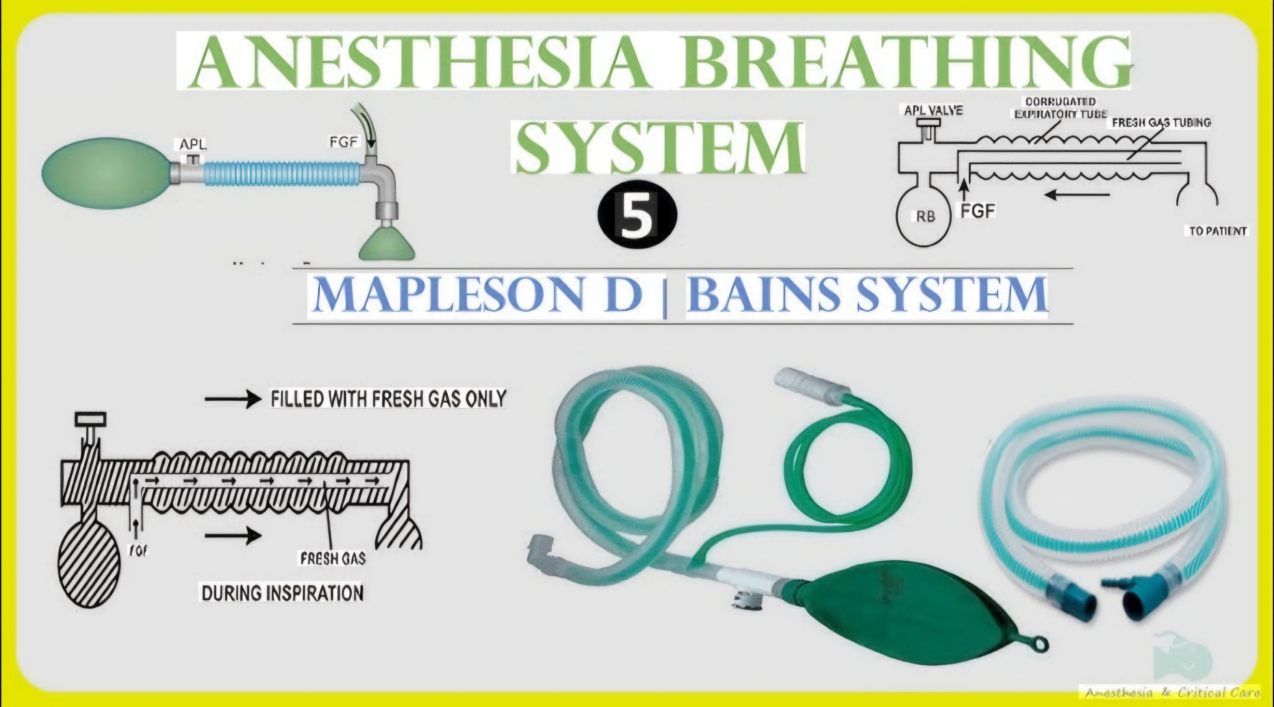
Discuss the Bain circuit in detail.

collections Question Diagrams & Reference Images (5)
Question Reference Diagram
zoom_in View Image
Question Reference Diagram
zoom_in View Image
Question Reference Diagram
zoom_in View Image
Question Reference Diagram
zoom_in View Image
Question Reference Diagram
zoom_in View Image
description Clinical Response
"
⚙ CORE CONCEPT

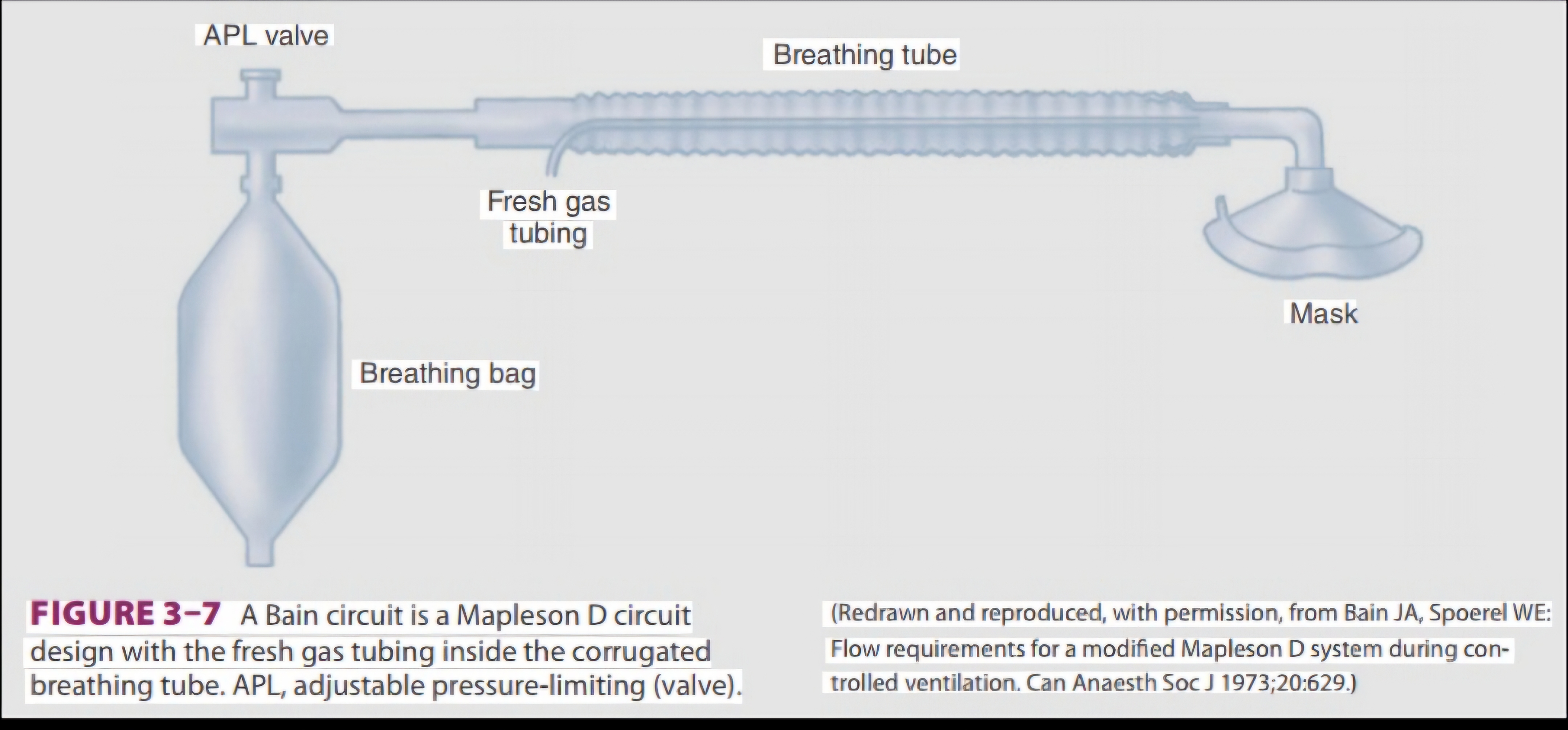
The Bain circuit is a co-axial modification of the MAPLESON D breathing system — the fresh gas flow (FGF) is delivered through an inner tube running INSIDE the outer expiratory tubing, making it compact, lightweight, and particularly efficient for spontaneously breathing patients when used at high FGF. Understanding its classification (Mapleson D), function, FGF requirements, and the critical Pethick test for checking inner tube integrity is essential for every anaesthesiologist.

(Mapleson WW — breathing system classification; Bain JA, Spoerel WE — 1972 original description; Conway CM — FGF requirements for Bain; Willis BA — rebreathing in Mapleson circuits; Spoerel WE — coaxial circuits)
A. Classification and Structure
△ BAIN CIRCUIT — STRUCTURAL DIAGRAM
FRESH GAS INLET: at the MACHINE END (away from patient)
  -> enters the INNER TUBE (narrow, ~7mm diameter)
  -> inner tube runs INSIDE the outer corrugated tube
  -> FGF exits at the PATIENT END (near the patient)

OUTER TUBE: corrugated, 22mm, serves as EXPIRATORY LIMB
  -> patient expires -> gas flows back THROUGH the outer tube
  -> toward the machine end (reservoir bag + APL valve)

  +------- OUTER EXPIRATORY TUBE (22mm corrugated) ---------+
  |  +---- INNER FRESH GAS TUBE (7mm) --------------------+ |
  |  | Fresh gas >>>>>>>>>>>>>>>>>>>>>>>>>>>>> to patient  | |
  |  +-----------------------------------------------------+ |
  |                                                            |
  |  <---- Expired gas flows in outer tube <----              |
  +------------------------------------------------------------+

MACHINE END: APL valve (Pop-off) + Reservoir bag (2L adult)
PATIENT END: Patient connection (mask or ETT connector)
Total length: 180 cm; outer tube 22mm; inner tube ~7mm

MAPLESON CLASSIFICATION: MAPLESON D
(FGF enters near patient; APL valve and reservoir at machine end)
Note: Humphrey ADE circuit is another version of Mapleson A/D/E in one device
B. Functional Analysis and FGF Requirements

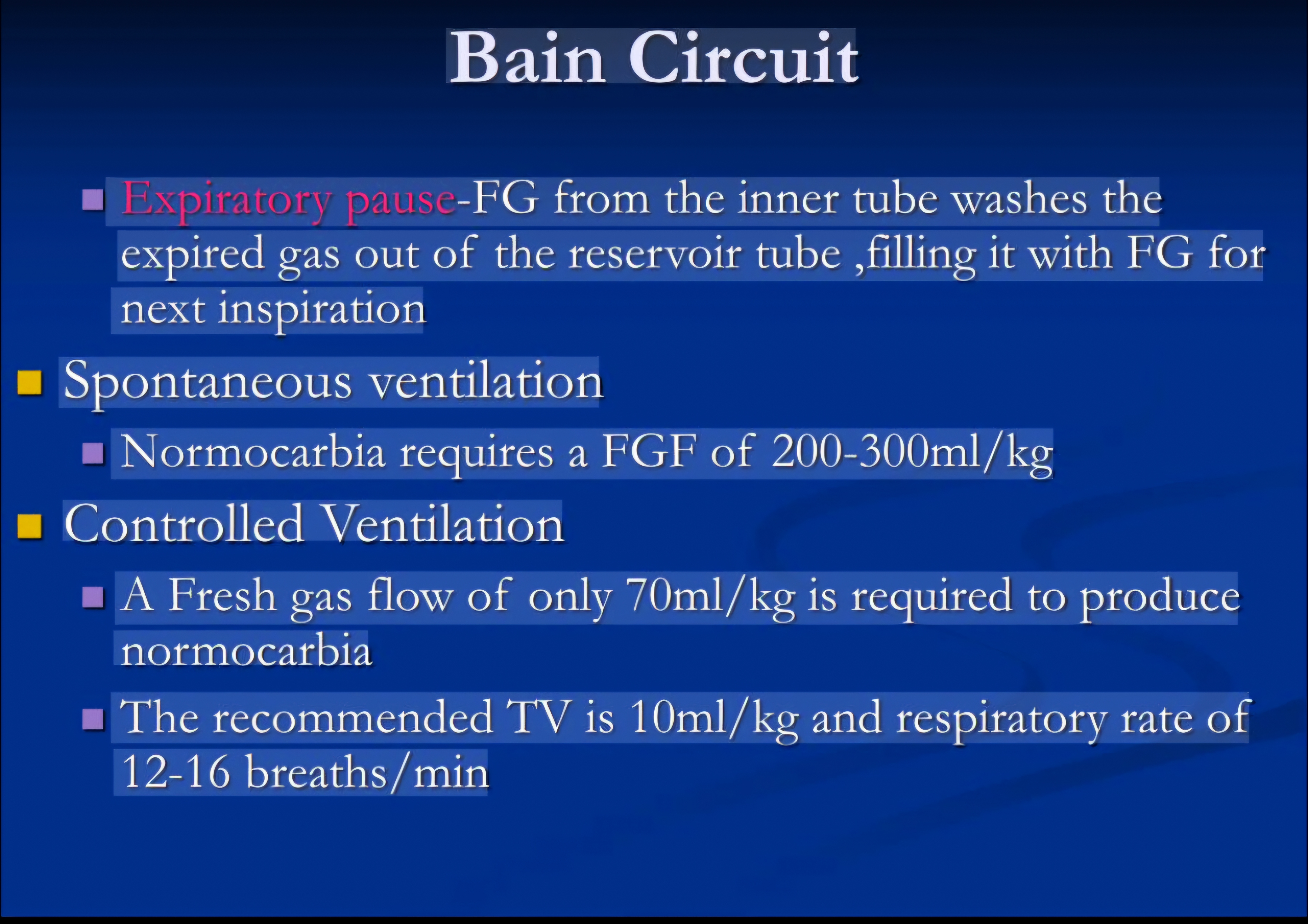
Why FGF enters at the patient end matters: fresh gas exits the inner tube at the patient end → mixes first with ALVEOLAR gas (high CO₂) at the patient end; during EXPIRATION, expired gas travels in the outer tube AWAY from patient toward the bag — this is the KEY to understanding rebreathing in the Bain circuit.

Ventilation ModeFGF Required to Prevent RebreathingMechanism
Controlled (Mechanical) Ventilation70 mL/kg/min (~equal to alveolar minute volume); or FGF = 2.5–3× tidal volume; MOST EFFICIENT use of Bain circuitDuring mechanical inspiration, fresh gas pushes expired gas away from patient toward the bag during expiration; since expired gas was pushed far from the patient end, rebreathing is minimal; BAIN IS MORE EFFICIENT for CONTROLLED ventilation
Spontaneous Ventilation2× minute ventilation = ~200–250 mL/kg/min; or FGF = 2× tidal volume per minute; LESS EFFICIENT than Mapleson AAt low FGF, expired CO₂-laden gas near patient end may be re-inhaled; high FGF required to continuously flush expired gas away before next inspiration
C. Advantages and Disadvantages
AdvantagesDisadvantages
LIGHTWEIGHT and COMPACT (coaxial design); suitable for HEAD AND NECK surgery; easily portable; no valves at patient end (low resistance — important in paediatric use)INNER TUBE DISCONNECTION (most dangerous complication — may not be detected clinically; PETHICK TEST essential); requires HIGH FGF for spontaneous breathing (wasteful); not as efficient as Mapleson A for spontaneous breathing

Comparison of Mapleson circuits for efficiency (least FGF needed to prevent rebreathing):
SPONTANEOUS breathing: A > DFE > CB (Magill attachment = most efficient for spontaneous breathing)
CONTROLLED ventilation: D,E,F > BC > A (Mapleson D = most efficient for controlled ventilation)

D. The Pethick Test — Pre-use Check for Inner Tube Integrity
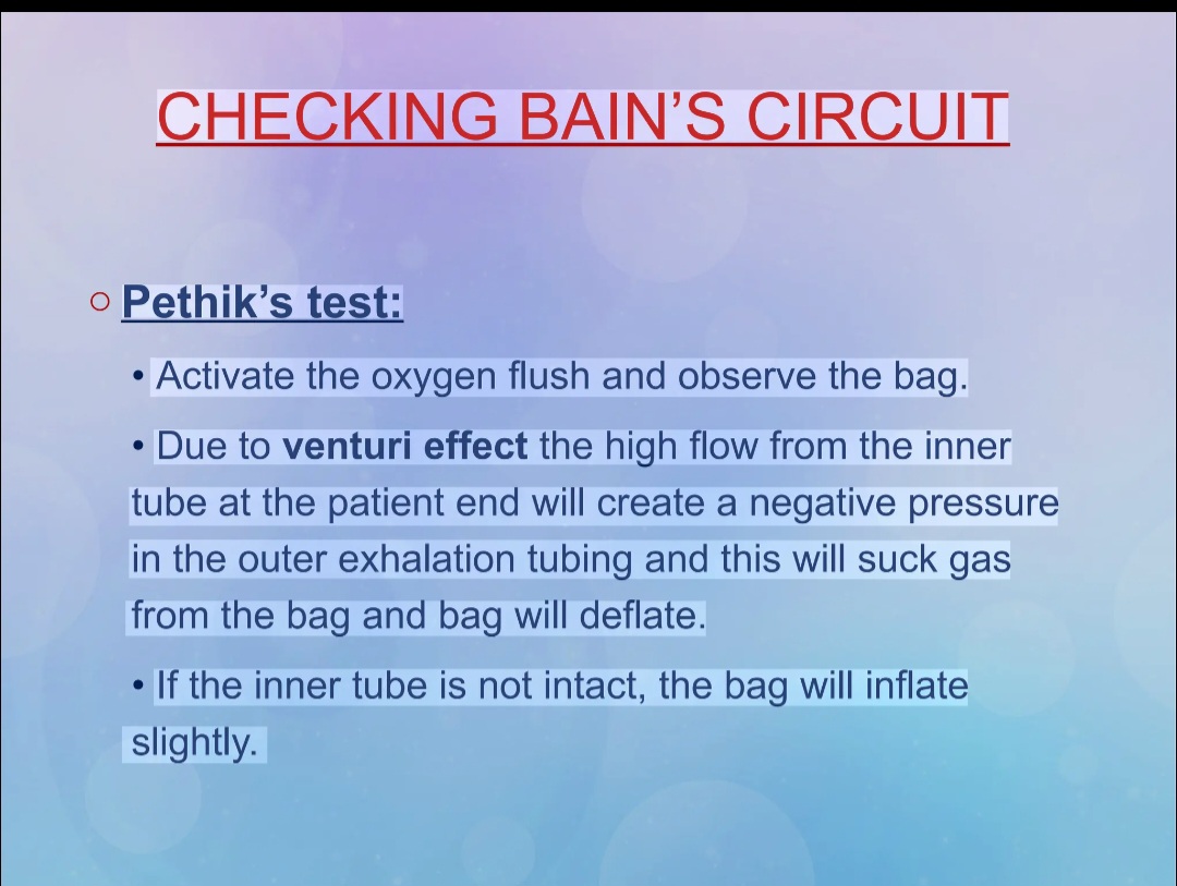
△ PETHICK TEST — STEP BY STEP
PURPOSE: To confirm that the INNER TUBE is intact and patent
(a disconnected inner tube = silent CO2 rebreathing)

STEP 1: OCCLUSION TEST -- CONFIRM INNER TUBE INTEGRITY
- Occlude the PATIENT END of the Bain circuit (thumb over connector)
- Flush O2 at high flow (10 L/min O2 flush button)
- RESULT if inner tube intact:
  -> Pressure builds up in the circuit -> RESERVOIR BAG INFLATES/DISTENDS
  -> The high pressure gas cannot escape (patient end occluded)

STEP 2: VENTURI TEST -- CONFIRM INNER TUBE LUMEN
- Now RELEASE the patient end occlusion
- Activate O2 FLUSH BUTTON (delivers high-flow O2 into inner tube at high velocity)
- RESULT if inner tube is INTACT and lumen is patent:
  -> High-velocity fresh gas from inner tube creates VENTURI EFFECT at patient end
  -> Negative pressure sucks gas from outer tube
  -> RESERVOIR BAG DEFLATES/EMPTIES rapidly

NORMAL (PASS): BAG INFLATES on occlusion -> BAG DEFLATES on flush
ABNORMAL (FAIL): Bag does not inflate (inner tube not transmitting pressure)
  -> inner tube disconnected/kinked/absent -> DO NOT USE THIS CIRCUIT

ALTERNATIVE/QUICK CHECK: Gently breathe into patient end while
occluding inner tube outlet -- no resistance felt = inner tube open and patent
E. Paediatric Considerations — The T-Piece and Jackson-Rees Modification
CircuitClassificationFeaturesFGF
Ayre's T-PieceMapleson ET-shaped connector: fresh gas inlet + patient limb + expiratory limb (open-ended, no reservoir bag, no APL valve); extremely low resistance; no dead space; IDEAL for neonates (<10 kg)2–3× MV for controlled ventilation; no spontaneous breathing capability
Jackson-Rees Modification of T-PieceMapleson F (E + open-tailed bag)Open-tailed reservoir bag added to expiratory limb of Ayre's T-piece; allows visual monitoring of respiration; manual ventilation via bag; most widely used paediatric circuit2–3× MV (controlled); FGF = MV + 30% (spontaneous)
💬 VIVA CORNER

A Bain circuit fails the Pethick test — what does this mean and what do you do?
A failed Pethick test means the INNER TUBE is either disconnected from the machine end, kinked, or otherwise non-functional. With a disconnected inner tube, fresh gas no longer reaches the patient end — instead it enters the outer expiratory tube somewhere in the middle of the circuit. The patient would then be rebreathing their own expired gas (high CO₂, low O₂) without any CO₂-free fresh gas delivery at the patient connection. Clinically this presents as gradually rising EtCO₂ and eventual hypercapnia and hypoxia — without capnography, this could be entirely silent. The action is: DO NOT USE THIS CIRCUIT. Replace the entire Bain circuit with a new one or use an alternative breathing system (standard circle system). Repeat the Pethick test on the replacement circuit. This is precisely why the AAGBI pre-use checklist mandates a Pethick test before using the Bain circuit at the start of every anaesthetic list.
★ EXAMINER'S PEARL

Bain circuit = CO-AXIAL modification of MAPLESON D system; FGF through INNER TUBE (enters at patient end); expired gas returns through OUTER TUBE (back to machine end); reservoir bag + APL valve at machine end. FGF requirements: CONTROLLED VENTILATION = 70 mL/kg/min (most efficient use); SPONTANEOUS BREATHING = 200-250 mL/kg/min (2x MV — inefficient). Mapleson efficiency: Spontaneous = A > DFE > CB; Controlled = D > BC > A. ADVANTAGES: lightweight, compact, no valves at patient end, suitable for head/neck/remote anaesthesia. DISADVANTAGE: inner tube disconnection = silent CO₂ rebreathing (most dangerous failure mode). PETHICK TEST (mandatory pre-use check): Step 1 = Occlude patient end + O₂ flush → BAG INFLATES; Step 2 = Release + O₂ flush → BAG DEFLATES (venturi effect). FAIL = either step fails → replace circuit. Paediatric: Ayre's T-piece (Mapleson E) + Jackson-Rees modification (Mapleson F) — for <10-20 kg.
References: Bain JA, Spoerel WE. Flow requirements for a modified Mapleson D system during controlled ventilation. Can Anaesth Soc J 1973;20:629-636. Willis BA et al. Rebreathing in T-piece. Br J Anaesth 1975. Mapleson WW. The elimination of rebreathing in various breathing systems. Br J Anaesth 1954.
"
lock Protected Clinical Document • Pulse App • Screenshots Strictly Prohibited
QUESTION 266 person Asked by .
bookmark_add

Tests for autonomic dysfunction.

description Clinical Response
⚙ CORE CONCEPT

Autonomic dysfunction — impaired function of the sympathetic or parasympathetic nervous system — significantly increases perioperative risk: severe intraoperative hypotension, cardiovascular instability, aspiration risk (gastroparesis), and impaired thermoregulation. Diabetic autonomic neuropathy (DAN) is the most common cause encountered in anaesthetic practice, affecting 30–50% of long-standing diabetics. A systematic assessment of autonomic function — using simple bedside tests and specialised investigation — identifies at-risk patients and guides perioperative management.

(Ewing DJ — Edinburgh group of autonomic tests; Low PA — clinical autonomic disorders; Boulton AJM — diabetic neuropathy; Vinik AI — autonomic neuropathy in DM; Lahrmann H — autonomic testing)
1. Background — Anatomy and Pathophysiology
SystemAnatomy and FunctionEffect of Dysfunction
SYMPATHETICThoracolumbar outflow (T1–L2); preganglionic (cholinergic); postganglionic (noradrenergic, except sweat glands and adrenal medulla = cholinergic); controls: vasomotor tone, cardiac rate/contractility, pupil dilation, sweating, bladder neck, piloerectionOrthostatic hypotension (loss of vasomotor tone on standing), fixed heart rate (loss of cardiac sympathetic), anhidrosis (no sweating), impaired hypoglycaemia awareness (blunted catecholamine response)
PARASYMPATHETICCraniosacral outflow (CN III, VII, IX, X, S2-S4); cholinergic at both pre- and post-ganglionic; controls: cardiac slowing (SA node — CN X), GI motility, bronchodilation, pupil constriction, bladder detrusor contractionResting tachycardia (loss of vagal tone), gastroparesis (loss of GI motility — aspiration risk), urinary retention, impaired pupillary light reflex
2. Ewing's Battery — The Standard Autonomic Test Protocol

Developed by DJ Ewing at Edinburgh (1985); 5 bedside cardiovascular tests assessing PARASYMPATHETIC (3 tests) and SYMPATHETIC (2 tests) integrity — a classic viva topic.

TestSystem TestedMethodNormal ResponseAbnormal (Dysfunction)
1. Heart Rate Response to DEEP BREATHING (E:I ratio)PARASYMPATHETIC (vagal)Patient breathes deeply at 6 breaths/min (5s in + 5s out); continuous ECG; calculate maximum-minimum HR or E:I RATIO over 3 cyclesMax-min HR difference >15 bpm; E:I ratio >1.21 (younger adults); physiological sinus arrhythmiaMax-min <10 bpm = ABNORMAL; E:I ratio <1.05 = severe autonomic neuropathy; EARLIEST test to become abnormal in DAN
2. Immediate HR Response to STANDING (30:15 ratio)PARASYMPATHETIC (vagal)Patient stands quickly from lying; continuous ECG; 30:15 RATIO = RR interval at beat 30 / RR interval at beat 1530:15 ratio >1.04; initial tachycardia followed by relative bradycardia (vagal rebound)30:15 ratio ≤1.00 = ABNORMAL (parasympathetic failure)
3. HR Response to VALSALVA MANOEUVRE (Valsalva Ratio)PARASYMPATHETIC predominantly; sympathetic contributesForced expiration against closed glottis (40 mmHg) for 15 sec; VALSALVA RATIO = longest RR after / shortest RR duringValsalva ratio >1.21; post-Valsalva overshoot bradycardia (vagal)Ratio <1.21 = impairment; <1.10 = significant; CONTRAINDICATED in proliferative retinopathy, recent MI, aortic stenosis, high ICP
4. BP Response to STANDING (Orthostatic Hypotension)SYMPATHETIC (vasomotor)BP lying then immediately + 2 min standing; active standing preferredSystolic BP fall <20 mmHg (diastolic <10 mmHg) within 3 minOH: systolic fall ≥20 mmHg / diastolic ≥10 mmHg = ABNORMAL; risk of severe intraoperative hypotension
5. BP Response to SUSTAINED HANDGRIPSYMPATHETIC (vasomotor)Sustained handgrip at 30% MVC for 5 min; measure diastolic BP rise at 5 minDiastolic BP rises >16 mmHg (muscle metaboreceptor-mediated pressor response)Diastolic rise <10 mmHg = ABNORMAL ("flat" response); poor prognosis
3. Specialised Autonomic Tests
TestWhat It AssessesMethod and Significance
QSART (Quantitative Sudomotor Axon Reflex Test)Post-ganglionic sympathetic sudomotor fibres; most sensitive test for small-fibre sympathetic neuropathyIontophoresis of acetylcholine into skin → local axon reflex → sweat output measured at 4 sites; reduced/absent = postganglionic fibre loss
Thermoregulatory Sweat Test (TST)Whole-body sudomotor function (pre + post-ganglionic)Controlled heated environment; sweat visualised by indicator powder (alizarin red); anhidrosis pattern maps distribution of failure
Tilt Table Test (Head-Up Tilt)Sympathetic vasomotor and cardiac response; distinguishes vasovagal syncope from POTS and OHPassive 60–70° head-up tilt 30–45 min; POTS: HR ↑>30 bpm without BP fall; Neurally mediated syncope: progressive BP+HR fall to syncope; OH: progressive BP fall without compensatory HR rise
Plasma Norepinephrine Levels (supine vs standing)Sympathetic noradrenergic functionNE measured supine and after 5 min standing; NORMAL: NE doubles on standing; LOW standing NE = postganglionic sympathetic failure
Cardiac MIBG ScintigraphyPostganglionic cardiac sympathetic innervation¹²³I-MIBG (NE analogue) uptake; REDUCED = postganglionic denervation (Parkinson's, Lewy body dementia, pure autonomic failure — distinguishes from MSA)
Skin Conductance / Galvanic Skin ResponseSympathetic cholinergic sudomotor functionElectrodes on palm/sole measure conductance changes; rapid, simple, non-invasive bedside test
Heart Rate Variability (HRV) AnalysisBeat-to-beat RR interval variation; reflects vagal + sympathetic cardiac modulation24-hour Holter; HF power = vagal, LF power = sympathetic+vagal; REDUCED HRV = autonomic dysfunction; predicts post-MI mortality and SCD
4. Perioperative Implications of Autonomic Dysfunction
ImplicationManagement
HYPOTENSION (intraoperative)Most dangerous consequence; prepare vasopressors BEFORE induction (phenylephrine, metaraminol); prefer ETOMIDATE induction; invasive arterial line; volume preloading; AVOID spinal anaesthesia if OH documented
ASPIRATION RISK (gastroparesis)Assume full stomach in long-standing diabetic; RSI; metoclopramide 10 mg IV 30 min before; H2-blocker/PPI prophylaxis
THERMOREGULATORY FAILUREImpaired sweating and vasoconstriction; forced air warming throughout; mandatory temperature monitoring
RESTING TACHYCARDIA + FIXED HRHR does not reliably reflect hypovolaemia; rely on invasive haemodynamic monitoring rather than HR
IMPAIRED HYPOGLYCAEMIA AWARENESSNo catecholamine response to hypoglycaemia — no warning symptoms; hourly glucose monitoring mandatory; avoid tight glucose control
★ EXAMINER'S PEARL

Ewing's 5 tests: PARASYMPATHETIC (3): (1) Deep breathing HR variation (E:I ratio, max-min >15 bpm); (2) Standing 30:15 ratio (>1.04); (3) Valsalva ratio (>1.21) — CONTRAINDICATED in retinopathy, ↑ICP, recent MI. SYMPATHETIC (2): (4) Orthostatic hypotension (systolic fall ≥20 mmHg on standing = abnormal); (5) Sustained handgrip diastolic rise (>16 mmHg = normal). Additional tests: QSART (sudomotor — most sensitive for small-fibre); tilt table (vasovagal vs POTS vs OH); plasma NE supine/standing (preganglionic vs postganglionic); cardiac MIBG (Parkinson's disease postganglionic denervation); HRV (HF power = vagal; LF/HF ratio = sympathovagal balance). Perioperative implications: profound hypotension (have vasopressors ready, avoid spinal), full stomach (RSI in gastroparesis), fixed heart rate (use invasive monitoring), impaired hypoglycaemia awareness (hourly glucose). EARLIEST test to become abnormal in DAN = deep breathing HR variation (E:I ratio).
References: Ewing DJ et al. Autonomic neuropathy: its diagnosis and prognosis. Clin Endocrinol Metab 1986. Low PA (ed). Clinical Autonomic Disorders 3rd Ed. American Autonomic Society and American Academy of Neurology: Consensus on autonomic testing.
lock Protected Clinical Document • Pulse App • Screenshots Strictly Prohibited
QUESTION 267 person Asked by .
bookmark_add

Perioperative hypothermia and thermoregulation.

description Clinical Response
"
⚙ CORE CONCEPT

Inadvertent perioperative hypothermia (IPH) — a core temperature <36°C at any point in the perioperative period — occurs in 50–80% of unwarmed surgical patients and is associated with a cascade of devastating consequences: coagulopathy, increased surgical site infections, increased blood loss and transfusion requirements, prolonged recovery room stays, shivering, and increased cardiovascular events. Despite being almost entirely preventable with active warming, it remains one of the most common and underappreciated anaesthetic complications.

(Sessler DI — thermoregulation NEJM 2016; NICE Guideline CG65 — prevention of inadvertent hypothermia; Frank SM — perioperative hypothermia; Madrid E — Cochrane review active warming; Lenhardt R — mild hypothermia and coagulopathy)
A. Normal Thermoregulation
△ THERMOREGULATION — CONTROL SYSTEM OVERVIEW
TEMPERATURE SENSORS:
- Peripheral (skin): A-delta (cold, 10-40C), C fibres (warm, >30C)
- Core: Hypothalamic neurons (pre-optic area, anterior hypothalamus)
- Deep tissues: spinal cord, abdominal viscera

CENTRAL CONTROL:
HYPOTHALAMUS (pre-optic area) = ""the thermostat""
- Receives afferents from periphery and core sensors
- Compares input to SET POINT (37.0C +/- 0.2C = INTER-THRESHOLD RANGE)
- Within inter-threshold range: NO thermoregulatory responses
- ABOVE upper threshold (37.2C): heat loss responses activated
- BELOW lower threshold (36.8C): heat conservation responses activated

HEAT GAIN MECHANISMS (response to cold):
1. VASOCONSTRICTION (most important, most sensitive): cutaneous vessels ->
   down heat loss to environment; A-V shunts in digits close; shunts blood to core
2. NON-SHIVERING THERMOGENESIS (NST): brown adipose tissue (BAT) activation
   (important in neonates, infants -- minimal in adults); UCP-1 protein uncouples
   oxidative phosphorylation -> heat not ATP
3. SHIVERING: involuntary rhythmic muscle contractions; 3-5x up heat production
   (most powerful heat generation mechanism in adults); mediated by posterior
   hypothalamus; inhibited by: anaesthetic agents, opioids, meperidine (pethidine)
4. Voluntary behaviour (clothing, posture, seeking warmth)

HEAT LOSS MECHANISMS (response to heat):
1. SWEATING: most important; eccrine sweat glands; cholinergic sympathetic
   innervation; evaporation removes 580 kcal/L water evaporated
2. CUTANEOUS VASODILATION: up blood flow to skin -> up radiation and convection
3. RADIATION (60% of heat loss at rest): electromagnetic IR radiation from skin
4. CONVECTION (15-20%): air movement over skin surface
5. CONDUCTION (<5%): direct contact heat transfer
6. EVAPORATION (10-20%): respiratory (25%) + sweat (75%)
B. Effects of Anaesthesia on Thermoregulation
MechanismEffectClinical Consequence
WIDENING OF THE INTER-THRESHOLD RANGEAll GA agents widen the inter-threshold range from 0.4°C to ~2–4°C; vasoconstriction threshold shifts down to ~34.5°C; sweating threshold shifts upPatients can become profoundly hypothermic before vasoconstriction/shivering occurs; agents ranked by impairment: volatiles ≈ propofol >> opioids; neuraxial anaesthesia impairs regional thermoregulation
REDISTRIBUTION HYPOTHERMIA (most important cause — first hour)GA vasoconstriction disappears → warm core blood redistributes to cold peripheral compartment → core temperature falls 1–1.5°C within 30–40 min of inductionAccounts for 80% of core temp fall in first hour; CANNOT be prevented by external warming alone once started; BEST PREVENTION: pre-op skin surface warming for 30 min before induction
ENVIRONMENT AND RADIATION LOSSESOT temperature typically 18–22°C; ongoing heat loss via radiation and convection (laminar flow)Major contributor in prolonged surgery (>2h); SOLUTION: forced air warming blankets, warmed IV fluids, warm irrigation fluids, increased OT temperature
C. The Three Phases of Perioperative Hypothermia
PhaseTimingMechanismRate of Temp Fall
PHASE 1 (Redistribution)First 30–60 min after inductionInternal redistribution of heat from core to peripheral compartment (vasodilation from anaesthetic-induced loss of vasoconstriction)RAPID: 1.0–1.5°C in first hour; THE MOST DRAMATIC PHASE
PHASE 2 (Linear decline)60–210 min (1–3.5h)Heat loss to environment exceeds metabolic heat production; anaesthetic ↓metabolic heat production ~20%GRADUAL: 0.5–1°C per hour
PHASE 3 (Plateau)After 3–4 hoursCore temperature falls to new (impaired) vasoconstriction threshold (~34–35°C) → cutaneous vasoconstriction re-establishes → steady stateSTABLE: temperature no longer falls, but plateau is at a LOW level unless active warming applied
D. Consequences of Perioperative Hypothermia
SystemConsequence of HypothermiaEvidence
COAGULOPATHYPlatelet dysfunction (↓thromboxane A₂); impaired clotting factor enzyme kinetics (↓1–2°C → activity ↓10–15%); standard PT/aPTT (done at 37°C) UNDERESTIMATE in vivo coagulopathy; TEG/ROTEM better reflects true haemostasisWatts DD 1998: 1°C reduction → 16% ↑blood loss; Rajagopalan S 2008: hypothermia → 16% ↑transfusion requirement
SURGICAL SITE INFECTION (SSI)↓Tissue O₂ delivery to wound (vasoconstriction) → impaired neutrophil oxidative killing; ↓T-cell function; ↓macrophage phagocytosisKurz A (NEJM 1996): SSI 6% (normothermic) vs 19% (hypothermic) in colorectal surgery; NNT ~8
CARDIAC EVENTS<35°C → myocardial irritability → ventricular arrhythmias; prolonged QT; ↑sympathetic activity (shivering) → ↑cardiac demandFrank SM (Anesthesiology 1997): hypothermia → 55% ↑adverse cardiac events in high-risk patients; 2.2× ↑perioperative MI in vascular surgery
DRUG PHARMACOKINETICS↓Hepatic/renal drug metabolism → PROLONGED DRUG EFFECT; NMBD duration significantly prolonged; volatile MAC decreases ~5% per °C fallNMBD prolongation → delayed extubation; monitor NMJ quantitatively; confirm TOF ≥0.9 before extubation
SHIVERINGPost-operative shivering — increases O₂ demand 3–5×; problematic in cardiac/pulmonary/vascular disease; increases pain perception; delays recoveryMeperidine (pethidine) 25–50 mg IV = most effective (κ-opioid); clonidine 75 mcg IV; physostigmine; magnesium sulphate
PROLONGED RECOVERYProlonged drug effects + shivering → delayed awakening, impaired ventilator weaning, ↑PACU timeLenhardt R 1997: PACU arrival temperature inversely correlated with PACU discharge time
E. Prevention and Management of Perioperative Hypothermia
StrategyMethodEffectiveness
FORCED AIR WARMING (FAW) — GOLD STANDARDBair Hugger / Hot Dog forced warm air blanket; air 38–43°C; pre-warming 30 min before induction; continued intraoperatively; NICE: mandatory for surgery >30 minMost effective active warming device; prevents redistribution hypothermia when started pre-op; Cochrane review: FAW superior to electric heating mattress, passive insulation, warm IV fluids
WARM IV FLUIDS AND BLOOD PRODUCTSAll IV fluids warmed to 37–40°C via in-line fluid warmers; 1 L cold (4°C) fluid lowers core temp by 0.25°C; blood products warmed (never microwave)Effective adjunct with large volumes; NICE: all IV fluids >500 mL should be warmed
WARM IRRIGATION FLUIDSWarm (37–40°C) irrigation fluid for body cavity irrigation; prevents conductive heat lossSignificant reduction in hypothermia in laparoscopic/open abdominal surgery; simple, inexpensive, often overlooked
PASSIVE INSULATIONCotton blankets, reflective "space blankets," head covering (20–30% of heat loss from head); minimum skin exposureReduces heat loss but CANNOT rewarm; insufficient alone for surgery >30 min; adjunct to active warming
INCREASED OT TEMPERATUREOT temperature 21–23°C (rather than 18–19°C); reduces radiation/convection loss; especially important for neonates (26–28°C)Effective but uncomfortable for surgical team; important compromise for paediatric anaesthesia
HEATED HUMIDIFIER (HME vs active)HME (passive) or active heated humidifier reduces respiratory heat loss (10–20% of total)Modest effect on core temperature; important for prolonged surgery and NICU; active humidifiers more effective than HME
TEMPERATURE MONITORINGContinuous core temperature monitoring mandatory for procedures >30 min (NICE CG65); sites: nasopharynx, oesophagus, tympanic membrane, bladder, pulmonary artery, axilla, foreheadEssential prerequisite for any warming strategy; target 36.5–37.5°C throughout
★ EXAMINER'S PEARL

Normal thermoregulation: hypothalamus pre-optic area = "thermostat"; inter-threshold range 36.8-37.2°C. Anaesthesia WIDENS the inter-threshold range (36.8°C → ~34.5°C for vasoconstriction) — patient does not vasoconstrict until very cold. THREE PHASES: Phase 1 (0-1h) = REDISTRIBUTION (1-1.5°C drop — the most dramatic phase); Phase 2 (1-3.5h) = LINEAR DECLINE (0.5°C/hr); Phase 3 (3-4h+) = PLATEAU at low temperature. CONSEQUENCES: coagulopathy (standard labs done at 37°C UNDERESTIMATE coagulopathy; use TEG at patient's actual temp), SSI ↑ (NEJM Kurz 1996: 6% vs 19% — NNT 8), cardiac events ↑, prolonged drug effects (NMBD prolongation + ↓MAC), shivering (3-5x ↑O₂ demand). PREVENTION: FORCED AIR WARMING (Bair Hugger — gold standard, 30 min pre-op warming most effective), warm IV fluids (>500 mL), warm irrigation fluids, passive insulation, ↑OT temperature. Treatment of shivering: MEPERIDINE (pethidine) 25-50 mg IV (most effective) or clonidine 75 mcg IV. Core temperature monitoring site: NASOPHARYNX (most accurate under GA).
References: Sessler DI. Perioperative thermoregulation. NEJM 2016;374:2053-2063. Kurz A et al. Perioperative normothermia to reduce surgical site infections. NEJM 1996;334:1209-1215. NICE Clinical Guideline 65 — Inadvertent perioperative hypothermia 2008 (updated 2016). Madrid E et al. Warming measures for prevention of inadvertent perioperative hypothermia. Cochrane Database 2016.
"
lock Protected Clinical Document • Pulse App • Screenshots Strictly Prohibited
QUESTION 268 person Asked by .
bookmark_add

Intralipid — discuss in detail.

description Clinical Response
"
⚙ CORE CONCEPT

Intralipid (20% intravenous fat emulsion) has emerged as one of the most important ANTIDOTES in clinical anaesthesia — the treatment of LAST RESORT for life-threatening local anaesthetic systemic toxicity (LAST). Simultaneously, it is the cornerstone of parenteral nutrition. The mechanism of "lipid rescue" — once considered implausible — is now physiologically explained and supported by remarkable clinical success stories. Beyond LAST, Intralipid has applications in toxicology as a systemic antidote for lipophilic drug poisoning.

(Weinberg GL — lipid emulsion therapy NEJM; Fettiplace MR — lipid emulsion mechanistic review; AAGBI Safety Guideline LAST 2010; Cave G — intralipid for poisoning; Grunau BE — Lipid Emulsion for LAST)
A. Composition of Intralipid
Component20% Intralipid30% IntralipidFunction
Soybean oil (vegetable fat)20 g/100 mL (200 g/L)30 g/100 mLEnergy substrate; source of essential fatty acids; lipid "sink" for lipophilic drugs
Glycerol2.25 g/100 mL2.25 g/100 mLOsmolarity adjustment → isotonic with blood; gluconeogenic substrate
Egg lecithin (phospholipid emulsifier)1.2 g/100 mL1.2 g/100 mLEMULSIFIER — stabilises oil-in-water emulsion; keeps fat globules 0.1–0.4 µm; AVOID in egg allergy
Particle size0.1–0.4 µm dropletsSameMimic chylomicrons; metabolised by lipoprotein lipase in adipose and muscle
Caloric density2.0 kcal/mL3.0 kcal/mLHigh energy density; 20% = standard for LAST treatment; higher concentration NOT used for LAST
Osmolarity~350 mOsm/L~310 mOsm/LIsotonic — can be given peripherally without phlebitis
B. Local Anaesthetic Systemic Toxicity (LAST) — Mechanism and Epidemiology
AspectDetail
Incidence~1:1,000–1:10,000 regional anaesthesia procedures; higher with peripheral nerve blocks (1:1,000) than neuraxial (1:10,000); bupivacaine and levobupivacaine carry highest cardiac toxicity risk; ropivacaine less cardiotoxic
Mechanism of toxicityIntravascular injection or systemic absorption → LA distributes to all tissues → CNS toxicity first (Na⁺ channel blockade in inhibitory interneurons → seizures); cardiac toxicity at higher concentrations (Na⁺/K⁺/Ca²⁺ channel blockade, mitochondrial dysfunction); bupivacaine: "fast in, slow out" = cardiotoxic
Clinical presentationBIPHASIC: CNS first (circumoral numbness, metallic taste, tinnitus, agitation → seizures → CNS depression) then CARDIAC (ECG changes: PR/QRS/QT prolongation → arrhythmias → haemodynamic collapse); cardiac arrest is characteristically REFRACTORY to standard resuscitation
C. Mechanisms of Intralipid (Lipid Emulsion Therapy) in LAST
MechanismDetailEvidence
1. LIPID SINK (Primary mechanism)Large intravascular lipid emulsion volume creates an expanded intravascular lipid phase ("lipid sink"); highly lipophilic LAs (bupivacaine) partition out of tissue sites (heart, brain) INTO the lipid phase; drug is carried away from heart/brain to less critical tissues for metabolism — analogous to dynamic dialysisWeinberg GL (1998) — original animal experiments; explains why Intralipid works better for bupivacaine than for less lipophilic mepivacaine
2. DIRECT CARDIAC EFFECT (Secondary mechanism)Fatty acids liberated from Intralipid provide direct energy substrate to the poisoned myocardium; bupivacaine impairs carnitine-mediated fatty acid transport into mitochondria; acute fatty acid load may bypass this block and rescue mitochondrial functionFettiplace MR (2016) — metabolic/mitochondrial mechanisms; Intralipid improves contractility before drug redistribution could account for it
3. POST-CONDITIONING (Cardioprotection)Lipid emulsion activates PKC and PI3K/Akt cardioprotective pathways → reduces ischaemia-reperfusion injuryEmerging evidence from animal models; relevant in post-resuscitation period
D. AAGBI Protocol for Management of LAST (20% Intralipid)
△ AAGBI / ASRA LIPID EMULSION PROTOCOL — LAST MANAGEMENT
AT FIRST SIGN OF TOXICITY (seizures, cardiac arrhythmia, cardiovascular collapse):
- STOP local anaesthetic injection IMMEDIATELY
- CALL FOR HELP (emergency call + anaesthetic colleague)

IF SEIZURES:
  -> Benzodiazepine (midazolam 2-5 mg IV or diazepam 5-10 mg IV)
  -> Thiopentone 100-150 mg IV (if available) -- rapid termination
  -> AVOID propofol in high dose (>3 mg/kg/h if haemodynamically compromised)

IF CARDIAC ARREST: -> START CPR IMMEDIATELY (standard ALS algorithm)

*** INTRALIPID 20% -- THE ANTIDOTE: ***
  STEP 1 -- BOLUS: Intralipid 20% 1.5 mL/kg IV over 1 minute
            = 100 mL for 70 kg adult (give RAPIDLY)
            Repeat bolus x2 if haemodynamic recovery not achieved
            (at 5-min intervals -- total maximum 3 boluses)

  STEP 2 -- INFUSION: 0.25 mL/kg/min (for 70 kg = 17.5 mL/min)
            Continue until haemodynamically stable (typically 30-60 min)
            If instability continues -> DOUBLE RATE to 0.5 mL/kg/min

  MAXIMUM DOSE: 12 mL/kg total (beyond this, lipid emulsion itself
  causes adverse effects)

  FOR 70 KG PATIENT:
    - Bolus: 100 mL over 1 min
    - Infusion: ~17 mL/min (1 L/hr approx)
    - Maximum total: 840 mL

DURING CARDIAC ARREST:
  -> CPR as per ALS algorithm (30:2 or continuous if ETT)
  -> ADRENALINE: reduce dose to 10-100 mcg IV (NOT 1 mg -- catecholamines
     may worsen bupivacaine cardiac toxicity by increasing myocardial O2 demand)
  -> AVOID: lidocaine (adds to toxicity), vasopressin, beta-blockers, CCBs

REFRACTORY CARDIAC ARREST:
  -> CARDIOPULMONARY BYPASS (CPB) or VA-ECMO -- ULTIMATE RESCUE
  -> DO NOT STOP RESUSCITATION EARLY -- bupivacaine cardiac arrest
     may require 60-90 min CPR until drug redistributes

POST-RESUSCITATION:
  -> ICU admission; monitor for recurrence (drug still redistributing)
  -> Avoid hypoxia, hypothermia, acidosis
  -> Serum lipase monitoring (pancreatitis risk)
  -> Report to AAGBI/ASRA toxicity registry
E. Other Clinical Applications of Intralipid
ApplicationIndicationDose/Details
PARENTERAL NUTRITION (TPN)Caloric support when enteral feeding not possible; provides essential fatty acids; prevents EFAD1–1.5 g/kg/day; 30–40% of non-protein calories from lipid; monitor triglycerides (hold if >5 mmol/L)
TOXICOLOGY — Lipophilic Drug PoisoningCCBs (verapamil, diltiazem), TCAs, lipophilic beta-blockers, lamotrigine, sertraline, bupropion, haloperidol (LogP >2)Same protocol as LAST (1.5 mL/kg bolus + 0.25 mL/kg/min infusion); mostly case reports; poison control consultation recommended
PROPOFOL VEHICLEPropofol is poorly water-soluble — dissolved in 10% IntralipidExplains milky appearance, injection pain, and bacterial growth risk (use within 12h of opening)
NUTRITIONAL SUPPORT IN NEONATESPremature neonates on TPN; DHA critical for neuronal membrane development2–3 g/kg/day; start slowly (0.5 g/kg/day, increase over days); monitor triglycerides
F. Adverse Effects and Contraindications
Adverse Effect / ContraindicationDetail
HypertriglyceridaemiaHold Intralipid if TG >5 mmol/L (pancreatitis risk); monitor TG regularly in TPN patients
PancreatitisHypertriglyceridaemia-mediated; monitor amylase/lipase post LAST treatment
Fat overload syndromeExcessive infusion rates → lipoprotein lipase saturation → lipid embolism, hepatic dysfunction, coagulopathy, fever, anaemia
Egg and soy allergyRELATIVE CONTRAINDICATION; in life-threatening LAST, benefit of treatment outweighs allergy risk
Interference with lab testsLipemic serum interferes with triglyceride assays, some glucometers, co-oximetry haemoglobin, pseudohyponatraemia
💬 VIVA CORNER

A patient collapses with refractory VF 10 minutes after an interscalene brachial plexus block using bupivacaine. Three defibrillations have failed. What is your immediate management?
This is LOCAL ANAESTHETIC SYSTEMIC TOXICITY (LAST) with bupivacaine-induced cardiac arrest. (1) Continue CPR at all times — DO NOT STOP (may require 60–90 minutes while drug redistributes). (2) INTRALIPID 20% immediately: 1.5 mL/kg IV bolus (~100 mL for 70 kg) rapidly over 1 minute, then infusion 0.25 mL/kg/min; bolus can be repeated twice at 5-min intervals. (3) Continue defibrillation and ALS — always after Intralipid. (4) REDUCE adrenaline dose to 10–100 mcg (not 1 mg). (5) AVOID high-dose propofol, lidocaine, vasopressin. (6) If refractory → activate VA-ECMO or cardiopulmonary bypass. (7) Admit to ICU post-ROSC and monitor for recurrent arrhythmias. Key messages: Intralipid is the antidote; CPR must continue; expect prolonged resuscitation to succeed; VA-ECMO is the ultimate rescue.
★ EXAMINER'S PEARL

Intralipid 20% = 20g soybean oil + 1.2g egg lecithin (emulsifier) + 2.25g glycerol per 100 mL; 2 kcal/mL. MECHANISMS OF ACTION IN LAST: (1) LIPID SINK (primary — lipophilic LA partitions from cardiac muscle/brain into intravascular lipid phase → carried to liver for metabolism); (2) Direct cardiac energy substrate (fatty acids rescue bupivacaine-poisoned mitochondria); (3) Post-conditioning cardioprotection (PI3K/Akt). LAST PROTOCOL (AAGBI): BOLUS 1.5 mL/kg (=100 mL for 70 kg) over 1 min → REPEAT ×2 at 5 min if needed; INFUSION 0.25 mL/kg/min; double rate if instability; MAXIMUM TOTAL 12 mL/kg. Adrenaline in LAST arrest: REDUCE dose to 10-100 mcg (NOT 1 mg). REFRACTORY: VA-ECMO/CPB as ultimate rescue — CONTINUE CPR (bupivacaine arrest needs 60-90 min). Other applications: TPN (1-1.5 g/kg/day; prevents EFAD); lipophilic drug poisoning (CCBs, TCAs, propranolol); propofol vehicle (10% Intralipid = propofol carrier). Adverse effects: hypertriglyceridaemia (hold if TG >5 mmol/L), pancreatitis (monitor post LAST), fat overload syndrome, egg/soy allergy (relative CI — in LAST, risk of withholding > allergy risk).
References: Weinberg GL et al. Pretreatment or resuscitation with a lipid infusion shifts the dose-response to bupivacaine-induced asystole in rats. Anesthesiology 1998;88:1071-1075. AAGBI Safety Guideline: Management of Severe Local Anaesthetic Toxicity 2010. Fettiplace MR et al. Mechanisms of lipid emulsion treatment of local anaesthetic poisoning. BJA 2016;117(Suppl 1):i46-i58. Cave G, Harvey MG. Intravenous lipid emulsion as antidote. Clin Tox 2009;47:875-885.
"
lock Protected Clinical Document • Pulse App • Screenshots Strictly Prohibited
QUESTION 269
bookmark_add

Anesthetic implications in a case of posterior fossa tumour.

picture_as_pdf Clinical Reference Documents & PDFs (1)
Protected Inline Viewer
description j6hLxTTXEwbswIGO8TgyNOX4jP8kywEZEYO2hZvH.pdf
lock Protected Clinical Document • Pulse App • Screenshots Strictly Prohibited
QUESTION 270
bookmark_add

Zones of Lungs

collections Question Diagrams & Reference Images (2)
Question Reference Diagram
zoom_in View Image
Question Reference Diagram
zoom_in View Image

Showing 261270 of 337 questions

account_tree

Subcategory Tree

Explore Anesthesia subcategories

folder_special Anesthesia
Main
lock

Category Subscription

Subscribe to Anesthesia to unlock this module and all nested subcategories.

  • check_circle Access Anesthesia & all subcategories
  • check_circle Detailed, Peer-Reviewed Answers
  • check_circle High-yield visual aids & imaging
Get Category Subscription arrow_forward
Secure 256-bit SSL Connection

Anatomical Models

Explore high-fidelity 3D visualizations included in premium modules.

Case Reviews

Real-world clinical scenarios narrated by senior consultants.

Pulse App