Anesthesia | Eklavya Medical
vaccines
medical_services Main Specialty Domain lock Subscription Required

Anesthesia

Clinical anesthesiology, local and general anesthesia delivery systems, monitoring, and perioperative care.

verified Verified Medical Faculty menu_book 337 Q&A Modules
lock Protected Clinical Document • Pulse App • Screenshots Strictly Prohibited
QUESTION 241 person Asked by .
bookmark_add

Define autonomic dysreflexia (AD) — pathophysiology, triggers and emergency management [4]. Describe the

description Clinical Response
"haemodynamic management challenges in patients with chronic spinal cord injury [3]. What are the specific considerations for anaesthesia below, at, and above T6 injury level? [3] ⚙ Core Concept Spinal cord injury (SCI) creates a permanent physiological reorganisation — loss of descending supraspinal control results in spastic paralysis, autonomic instability, and loss of normal thermoregulation. Autonomic dysreflexia (AD) is a potentially lethal hypertensive emergency unique to patients with SCI above T6 that can be triggered by any stimulus below the injury level — including surgical incision, urinary catheter, or even a tight clothing waistband. (Karlsson AK — autonomic dysreflexia; Teasell R — SCI management; Miller's Anaesthesia 9th Ed) A. Autonomic Dysreflexia 4 marks Definition and mechanism: AD is an acute, massive, unmodulated sympathetic reflex response to a stimulus below the level of the SCI, occurring in patients with SCI at or above T6; normally, supraspinal centres modulate and limit sympathetic outflow below the injury — in SCI above T6 (above the major splanchnic sympathetic outflow at T6-T10), this modulation is lost; a noxious stimulus below the level → enters the spinal cord below the lesion → triggers sympathetic reflex via the intact spinal cord below the injury → massive sympathetic vasoconstriction below the lesion → acute severe hypertension (SBP may rise to 200– 300 mmHg); the ascending signal cannot reach the brain (blocked by the SCI) → cerebral baroreceptors detect the hypertension → activate parasympathetic response (bradycardia, vasodilatation ABOVE the injury — flushing, nasal congestion, sweating) → but this parasympathetic response cannot reach below the injury → the sympathetic vasoconstriction persists below the lesion; the result: severe hypertension + bradycardia + diaphoresis/flushing ABOVE the injury + piloerection/pallor BELOW Common triggers: bladder distension (most common — 85%) or urinary tract infection; bowel distension (faecal impaction, rectal examination); surgical incision; pressure sores; tight clothing; spasticity; DVT; labour in pregnant SCI patients (AD occurs in virtually all T6 SCI patients during uterine contractions → spinal anaesthesia is strongly recommended) Emergency management of AD episode: Sit the patient upright immediately (orthostatic ↓ BP from lower limb venous pooling — helps ↓ BP while cause is identified) Identify and remove the trigger (catheterise bladder or unblock catheter — most common; loosen tight clothing; remove any pressure; check skin) If BP remains severely elevated (SBP >150 mmHg): sublingual nifedipine 10 mg (rapid onset vasodilation — bite and swallow) OR glyceryl trinitrate (GTN) spray sublingually → rapid vasodilation; IV labetalol (combined α+β) or IV hydralazine if sublingual route inadequate; do NOT use β-blocker alone (unopposed α activation → worse hypertension) Intraoperatively: deepen anaesthesia (most effective intervention — removes surgical stimulus); regional anaesthesia (spinal or epidural) provides complete afferent block → prevents AD even in SCI patients; IV GTN or SNP infusion for refractory hypertension B. Haemodynamic Challenges in Chronic SCI 3 marks Chronic hypotension: loss of supraspinal sympathetic tone → chronic vasodilation → resting BP 90–110/60–70 mmHg (normal for that patient); supine hypertension does not apply; ""hypertension"" threshold effectively lower Orthostatic hypotension: loss of sympathetic venoconstriction → pooling in lower limbs when upright → ↓ venous return → ↓ CO; treat with compression stockings, elevation, fludrocortisone (↑ intravascular volume), midodrine (α₁ agonist) Succinylcholine contraindication: upregulated extrajunctional nAChRs → hyperkalaemia risk (same as burn/denervation patients); contraindicated after 24– 72h following acute SCI; safe only within the first 24h after acute SCI C. Anaesthetic Considerations by SCI Level 3 marks SCI Level Respiratory Impact AD Risk Key Anaesthetic Consideration Above T6 (cervical/high thoracic) Significant — diaphragm ± intercostals paralysed; may be ventilator-dependent HIGH — all surgery below injury → AD risk; spinal or epidural strongly recommended Regional anaesthesia (spinal/epidural) prevents AD afferent stimulus from reaching cord; GA: careful airway management (cervical SCI → cervical spine instability); post-op ventilatory support T6–T10 (thoracic) Intercostal muscles affected; intact diaphragm; can breathe spontaneously but ↓ cough efficacy Variable — may or may not develop AD depending on exact level Regional preferred for procedures below injury; chest physiotherapy post- op; epidural or PVB for thoracic procedures; secretion clearance important Below T10 (lumbar/sacral) Respiratory function usually preserved LOW — sympathetic outflow below T6 not involved Standard anaesthetic approach; succinylcholine contraindicated in chronic SCI regardless of level ★ Examiner's Pearl AD: SCI at/above T6 + stimulus below injury → massive sympathetic reflex → severe hypertension + bradycardia + diaphoresis/flushing above lesion. Most common trigger: bladder distension. Emergency: sit upright → remove trigger → sublingual nifedipine or GTN → IV labetalol. Intraoperative AD: DEEPEN ANAESTHESIA (most effective); regional (spinal/epidural) prevents AD completely. Succinylcholine contraindicated >24h post-SCI (upregulated extrajunctional nAChRs → hyperkalaemia). References: Karlsson AK. Autonomic dysreflexia (Spinal Cord 1999;37:383-391). Colachis SC. Autonomic hyperreflexia with spinal cord injury (J Am Paraplegia Soc 1992;15:171-186). Miller RD et al. Miller's Anaesthesia, 9th Ed."
lock Protected Clinical Document • Pulse App • Screenshots Strictly Prohibited
QUESTION 242 person Asked by .
bookmark_add

Explain ABO blood group genetics and compatibility requirements for red cell transfusion [3]. Classify transfusion

description Clinical Response
"reactions and describe the pathophysiology and management of TRALI and TACO [4]. Summarise the SHOT reporting system and its impact on transfusion safety [3]. ⚙ Core Concept Transfusion is the most common therapeutic intervention in hospital patients — 2.3 million units of blood are transfused annually in the UK. Transfusion errors and reactions are significant causes of morbidity and mortality, yet many are preventable. The SHOT haemovigilance system provides the evidence base for transfusion safety improvements. (SHOT Annual Report 2023; Kleinman S — TRALI; AABB Technical Manual; Miller's Anaesthesia 9th Ed) A. ABO Compatibility 3 marks ABO genetics: ABO locus on chromosome 9 — three alleles: A, B, O (O is recessive); A gene → N-acetylgalactosaminyl transferase → A antigen; B gene → galactosyl transferase → B antigen; O gene → non-functional enzyme → H antigen (no A or B); antibodies: naturally occurring IgM anti-A and/or anti-B formed in the first year of life (stimulated by gut bacteria cross-reactive antigens) Blood groups and compatibility: Group O (universal donor — RBC): neither A nor B antigens; compatible with A, B, AB, and O recipients; however contains anti-A and anti-B antibodies → donated plasma may cause haemolysis in non-O recipients if given in large volumes Group AB (universal plasma donor): has both A and B antigens; no naturally occurring anti-A or anti-B; can receive RBCs from all groups; AB plasma contains no anti-A or anti-B → universal plasma donor Emergency: O-negative RBCs for unknown blood group (O Rh-negative = truly universal donor); AB-positive FFP for unknown group plasma Rhesus (D) system: Rh D antigen — most immunogenic after ABO; Rh D negative patients who receive Rh D positive blood → form anti-D IgG antibodies (immune alloimmunisation) → subsequent exposure or pregnancy → haemolytic disease of the fetus and newborn (HDFN); anti-D immunoglobulin (Rhogam) prevents sensitisation in Rh-negative women B. Transfusion Reactions — TRALI and TACO 4 marks Feature TRALI (Transfusion-Related Acute Lung Injury) TACO (Transfusion-Associated Circulatory Overload) Definition Acute lung injury occurring within 6h of transfusion; ARDS-like presentation; new bilateral pulmonary infiltrates on CXR; PaO₂/FiO₂ <300 mmHg; no evidence of cardiac failure/volume overload Cardiogenic pulmonary oedema occurring during or within 6h of transfusion; new or worsened pulmonary oedema; hypertension; tachycardia; ↑ BNP/NT-proBNP Mechanism Donor leucocyte antibodies (anti-HLA or anti-HNA in donor plasma — usually multiparous female donors) bind to recipient's neutrophils → neutrophil activation → ↑ capillary permeability → non-cardiogenic pulmonary oedema; ""two-hit model"": first hit = patient's pre- existing condition (surgery, sepsis); second hit = transfusion trigger Volume overload — transfusion volume exceeds cardiac capacity to compensate → ↑ LVEDP → ↑ LAP → hydrostatic pulmonary oedema; risk: elderly, cardiac failure, renal failure, low body weight Incidence ~1:10,000 units FFP or platelets (highest); rarer with RBCs; leading cause of transfusion- related mortality in UK (SHOT) Most common transfusion complication reported to SHOT (approximately 1:1,000 transfusions); severely underreported Management STOP transfusion; 100% O₂; mechanical ventilation if severe (lung-protective strategy — TV 6 mL/kg IBW); NO diuretics (non-cardiogenic oedema — diuretics worsen hypovolaemia without benefit); supportive care; report to blood bank and SHOT STOP or slow transfusion; diuretics (furosemide 40–80 mg IV — excellent response to diuresis unlike TRALI); sit upright; O₂; GTN if severe hypertension; haemofiltration if refractory Prevention Male-only or previously untested female plasma donations (removes anti-HLA antibodies); leucodepletion of blood products; pathogen inactivation reduces TRALI risk Single-unit transfusion strategy (transfuse one unit then reassess — do not ""routine"" 2-unit transfuse); restrict transfusion trigger (Hb <7–8 g/dL); use diuretic prophylaxis in high-risk patients (furosemide between units) C. SHOT Reporting System 3 marks SHOT (Serious Hazards of Transfusion): established 1996; confidential, anonymous, voluntary haemovigilance scheme in the UK; collects data on all significant adverse events and reactions from transfusion; annual reports identify trends, learning points, and recommendations; key findings: wrong blood in tube (WBIT) errors — patient identification failures remain the commonest avoidable transfusion error; TACO: consistently the most commonly reported reaction; TRALI: has dramatically declined since male-only plasma policy introduction (2004 UK); recommendations from SHOT have driven: electronic patient identification systems; two-person bedside checks; pre-transfusion sample identity checks Key SHOT categories: ABO incompatible transfusion (most feared — mortality ~1:2,000,000 UK); TRALI; TACO; FNHTR (febrile non-haemolytic transfusion reaction — most common acute reaction — donor leucocyte cytokines → fever/chills — not dangerous; treat with paracetamol; no need to stop transfusion if mild); allergic reactions (urticaria → diphenhydramine; anaphylaxis → see Q33); transfusion-associated graft-vs-host disease (TA-GvHD) — prevented by irradiation of cellular products in immunocompromised recipients ★ Examiner's Pearl O-negative = universal RBC donor (no A, B, D antigens); AB = universal plasma donor (no anti-A or anti-B). TRALI vs TACO: TRALI = non-cardiogenic (do NOT use diuretics); TACO = cardiogenic (diuretics excellent). TRALI mechanism: donor anti-HLA/anti-HNA antibodies → neutrophil activation → capillary leak. SHOT: WBIT = most common preventable error; TACO = most commonly reported reaction; TRALI declined after male-only plasma policy. Key distinction: TRALI → no diuretics; TACO → furosemide. References: SHOT Annual Report 2023. Kleinman S et al. TRALI: ISBT working group update (Transfusion 2019;59:374-382). Bolton-Maggs PHB. SHOT Reports. Miller RD et al. Miller's Anaesthesia, 9th Ed."
lock Protected Clinical Document • Pulse App • Screenshots Strictly Prohibited
QUESTION 243 person Asked by .
bookmark_add

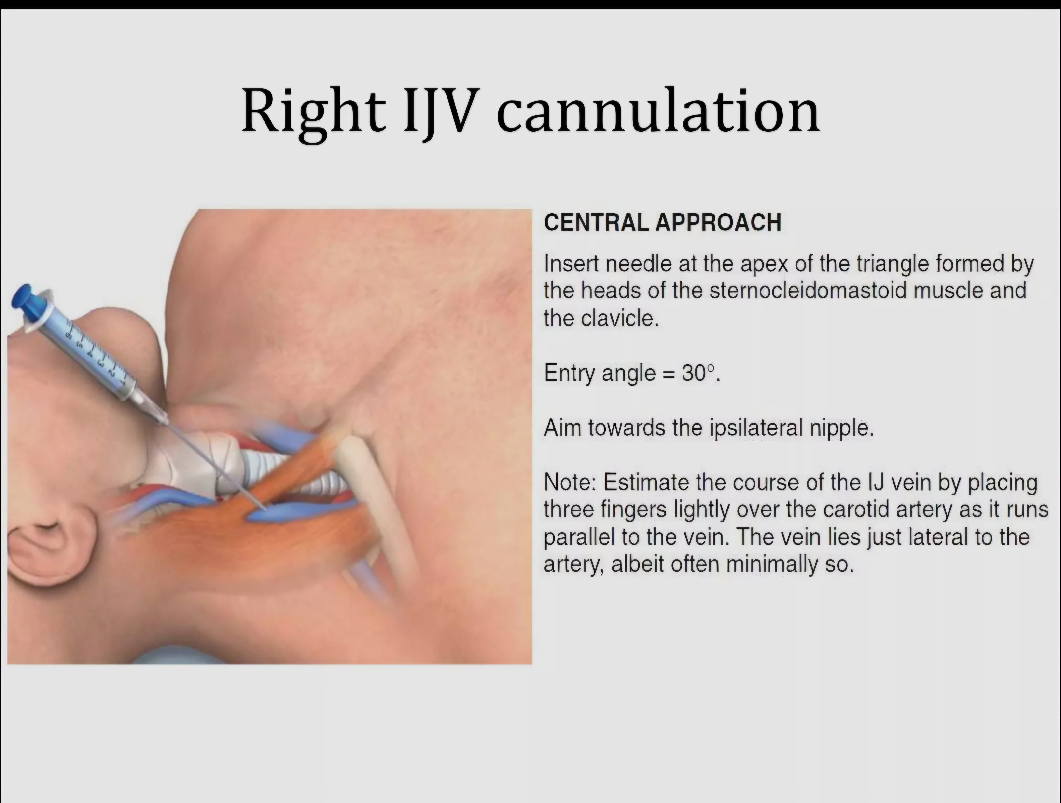
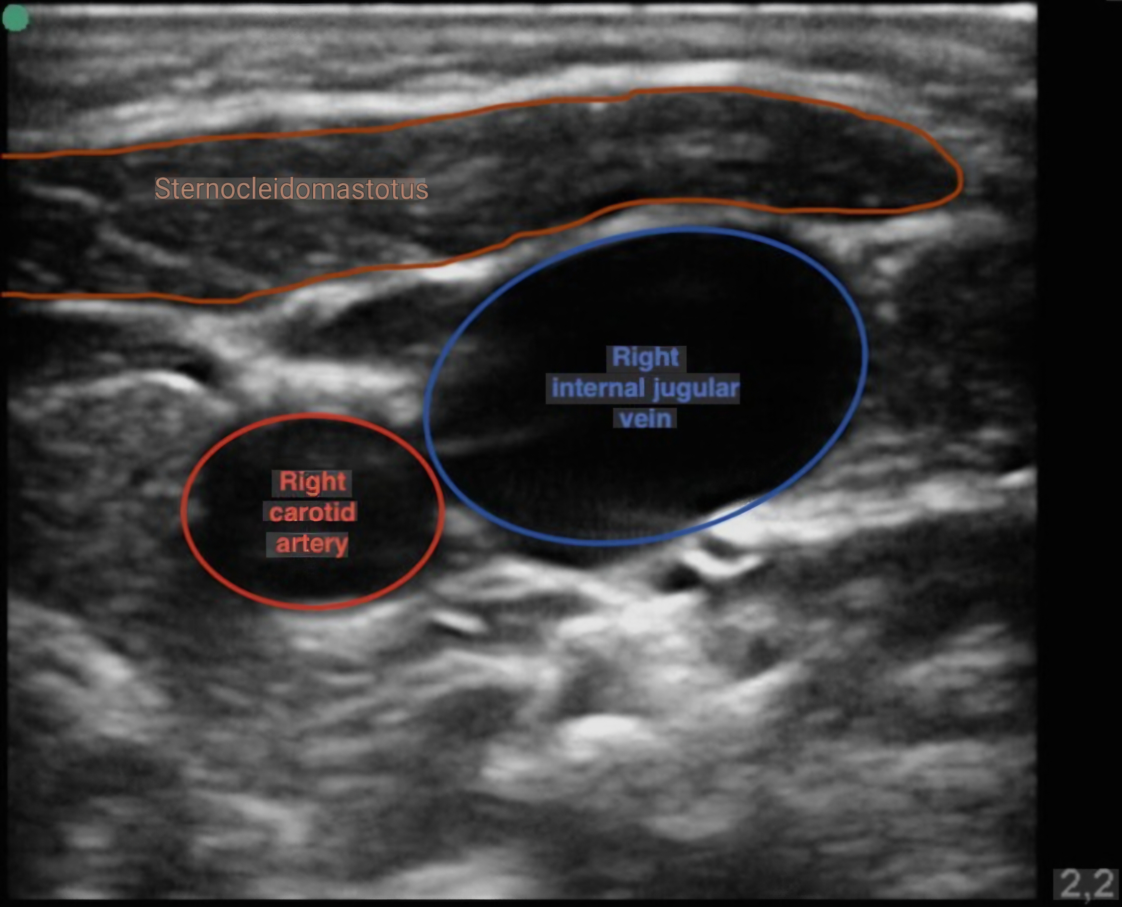
Describe the anatomical landmarks and ultrasound-guided technique for internal jugular CVC insertion [4]. Compare

collections Question Diagrams & Reference Images (3)
Question Reference Diagram
zoom_in View Image
Question Reference Diagram
zoom_in View Image
Question Reference Diagram
zoom_in View Image
description Clinical Response
"internal jugular, subclavian, and femoral approaches — indications, advantages and complications [3]. Outline the immediate and delayed complications of CVC insertion and how to confirm correct placement [3]. ⚙ Core Concept Central venous catheterisation is a core procedural skill in anaesthesia and critical care — used for drug delivery, haemodynamic monitoring, renal replacement therapy, and cardiac pacing. Ultrasound guidance has transformed the safety of CVC insertion, reducing arterial puncture rates from 5–10% (landmark technique) to <1% (USG). (Sznajder JI — CVC complications; McGee DC — evidence for CVC complications; NICE guidance TA49; Miller's Anaesthesia 9th Ed) A. Right Internal Jugular Anatomy and USG Technique 4 marks Anatomy: right IJ runs within the carotid sheath; at the level of the cricoid cartilage (C6): IJ lies anterior and lateral to the carotid artery; at the base of the neck: IJ lies anterolateral to the common carotid → joins subclavian vein to form the brachiocephalic vein; the right IJ is preferred for CVC because: (1) straight path to the SVC (left IJ has an acute angle at the left brachiocephalic junction → more difficult); (2) avoids the thoracic duct (on the left); (3) landmark: apex of the triangle between sternal and clavicular heads of sternocleidomastoid (SCM) Ultrasound-guided technique (NICE TA49 — mandatory for IJ CVC in elective settings): Position: patient supine, head 15° Trendelenburg (↑ IJ diameter + reduces air embolism risk); head turned slightly to the left (exposes right neck); sterile field USG probe: high-frequency linear probe (7–12 MHz); apply sterile sheath + sterile gel; identify IJ in short axis (transverse view): IJ = compressible anechoic (black) circular structure; carotid = pulsatile non-compressible, anterior and medial to IJ; confirm: compression collapses IJ not carotid; Doppler shows venous flow in IJ Needle insertion: under continuous real-time USG guidance; needle visualised as hyperechoic dot entering the IJ in short-axis view; confirm intraluminal position (needle tip seen inside IJ lumen) before advancing guide wire Seldinger technique: aspirate blood (dark, non-pulsatile = venous); confirm with transduction or blood gas (PvO₂ vs PaO₂); thread J-wire through needle; remove needle; nick skin at wire entry point; thread dilator over wire (hold wire at all times — prevent embolisation); remove dilator; thread CVC over wire; remove wire; aspirate and flush all lumens Confirm placement: CXR (tip at the cavoatrial junction — junction of SVC and right atrium — or SVC 2 cm above); exclude pneumothorax; tip position: ideal = SVC/RA junction (not in RA — risk of cardiac perforation or arrhythmia); not in a small vein or contralateral) B. IJ vs Subclavian vs Femoral — Comparison 3 marks Feature Right Internal Jugular Subclavian Femoral Infection risk (CLABSI) Intermediate LOWEST (preferred for long-term lines) HIGHEST (proximity to perineum — avoid if possible; use only in emergency) DVT risk Intermediate Lowest HIGHEST (femoral DVT + PE risk) Pneumothorax risk Low (with USG) HIGHER — needle passes above clavicle under pleura (not compressible with USG — landmark technique has 1–2% pneumothorax) None Arterial puncture risk Low with USG (<1%) Subclavian artery injury — difficult to compress (behind clavicle) Femoral artery puncture — easily compressible Preferred use Standard first choice (USG); emergency; haemodynamic monitoring; Swan-Ganz Long-term (TPN, chemotherapy); coagulopathy (compressible); when IJ not possible Emergency — fastest access in cardiac arrest; coagulopathy (most compressible) C. Complications and Confirmation of Placement 3 marks Immediate complications: arterial puncture (5–10% landmark; <1% USG) → remove needle + apply pressure; if dilator advanced into carotid → DO NOT remove — surgical consultation (removing a dilator from carotid → massive haemorrhage); air embolism (head-down position + cap all ports immediately); cardiac arrhythmias during wire/catheter insertion (usually transient from wire in RV — withdraw slightly); pneumothorax (subclavian approach); haemothorax; thoracic duct injury (left-sided approach) Delayed complications: central line-associated bloodstream infection (CLABSI) — Staphylococcus epidermidis most common; catheter-associated thrombosis; catheter malposition (kinking in subclavian bend, migration to right atrium — arrhythmias, cardiac perforation → tamponade); catheter fracture/embolism; superior vena cava perforation (rare — can cause haemorrhage or chylothorax); fibrin sheath formation → difficult aspiration Confirming correct placement: CXR (tip at SVC-RA junction — level of carina on CXR); transduction (waveform — CVP trace should be biphasic venous waveform, NOT pulsatile arterial); blood gas comparison (central venous PO₂ < arterial PO₂ confirms venous position); contrast CT in doubt; ECG-guided catheter tip positioning (emerging — QRS amplitude changes as tip enters the heart) ★ Examiner's Pearl Right IJ preferred: straight path to SVC, avoids thoracic duct, compressible. NICE TA49: USG mandatory for IJ CVC in elective settings. IJ USG: compressible + non- pulsatile = vein; pulsatile non-compressible = carotid. CLABSI risk order: subclavian (lowest) < IJ < femoral (highest — avoid if possible). DVT risk: femoral highest. Pneumothorax: subclavian highest (non-compressible landmark technique). CXR tip position: SVC/RA junction (level of carina). References: NICE Technology Appraisal 49 (TA49). Ultrasound locating devices for placing central venous catheters (2002). McGee DC, Gould MK. Preventing complications of central venous catheterization (NEJM 2003;348:1123-1133). Miller RD et al. Miller's Anaesthesia, 9th Ed."
lock Protected Clinical Document • Pulse App • Screenshots Strictly Prohibited
QUESTION 244 person Asked by .
bookmark_add

Describe the principles of cardiopulmonary bypass (CPB) — components, physiological consequences and

description Clinical Response
"management [4]. Explain myocardial protection strategies including cardioplegia [3]. Outline the haemodynamic management of protamine administration and its adverse effects [3]. ⚙ Core Concept Cardiopulmonary bypass is the technological foundation of open-heart surgery — temporarily assuming the roles of the heart (pump function) and lungs (gas exchange) while the surgeon operates on a still, bloodless heart. Its physiological consequences are profound and multisystemic. Myocardial protection — preventing ischaemic injury to the arrested heart — is the key technical challenge of cardiac surgery. (Gravlee GP — CPB textbook; Buckberg GD — cardioplegia; Cook DJ; Miller's Anaesthesia 9th Ed) A. Principles of CPB 4 marks CPB components: (1) venous reservoir — collects blood draining by gravity from the right heart via venous cannulae (placed in SVC and IVC, or a single two- stage RA cannula); (2) oxygenator — membrane oxygenator (hollow fibre polypropylene membrane) replaces lung function: O₂ delivered + CO₂ removed via gas flow across the membrane; heat exchanger integrated for temperature control; (3) arterial pump — roller pump (non-occlusive) or centrifugal pump; delivers blood at a set flow rate back into the aorta via the aortic cannula; (4) arterial filter — removes microemboli (air, particles) from the oxygenated blood before it enters the aortic cannula; (5) cardiotomy suction — aspirates blood from the operative field → returns to reservoir (defoamed and filtered); (6) heparinisation — systemic heparin 300–400 U/kg to achieve ACT (activated clotting time) >480 seconds before CPB (prevents clotting in the circuit) Physiological consequences of CPB: non-pulsatile flow (↓ renal perfusion, ↑ RAAS, ↓ microvascular autoregulation — see AKI Q53); haemodilution (prime volume 1–1.5 L crystalloid → ↓ Hb, ↓ colloid oncotic pressure); hypothermia (moderate 28–32°C) — ↓ metabolic rate (protective but → coagulopathy, arrhythmias); SIRS (blood-foreign surface contact → complement activation → cytokine release → capillary leak → post-bypass oedema and organ dysfunction); gaseous and particulate microemboli (→ neurocognitive dysfunction — ""pumphead"" syndrome: subtle cognitive impairment in 30–80% after CABG — reduced with membrane oxygenators vs bubble); coagulopathy (haemodilution + consumption + platelet dysfunction from pump circuit) Management during CPB: MAP 50–80 mmHg (autoregulation range in adults); flow 2.4 L/min/m²; haematocrit target 21–25% (minimum); temperature monitoring (nasopharyngeal + rectal); ACT every 30 min (maintain >480s); glucose control (hyperglycaemia → worse neurological outcomes — target BGL 6– 10 mmol/L) B. Myocardial Protection — Cardioplegia 3 marks Goals of myocardial protection: achieve rapid cardiac arrest (still field for surgery); maintain arrest during the ischaemic period; maximise recovery of cardiac function on reperfusion; prevent ischaemia-reperfusion injury Cardioplegia types: Cold crystalloid cardioplegia (St Thomas' solution): high K ⁺ (20–25 mEq/L → depolarises and arrests the heart in diastole); hypothermia (4°C — ↓ CMRO₂ equivalent ↓ myocardial metabolic demand); repeated doses every 20–30 min to maintain arrest and hypothermia Blood cardioplegia (Buckberg — warm or tepid blood cardioplegia + high K ⁺ ): uses the patient's own blood as the vehicle → delivers O₂ and nutrients even during arrest; superior metabolic protection compared to crystalloid; ""warm induction"" of arrest + cold maintenance + warm reperfusion (""hot shot"") Del Nido solution (currently popular for paediatric and adult cardiac surgery): single dose provides 60–90 min of arrest; higher Mg² ⁺ (membrane stabilisation); mannitol and lidocaine additives; reduces the number of re-dose interruptions to surgery Delivery routes: antegrade (into the aortic root via the aortic root cannula → flows into coronary ostia); retrograde (into coronary sinus → delivers to myocardium even when coronary arteries are severely diseased — useful in CABG); combined: antegrade + retrograde provides most complete distribution C. Protamine Administration and Adverse Effects 3 marks Protamine — heparin reversal: at the end of CPB, heparin must be neutralised to restore coagulation; protamine is a strongly positively-charged protein (from salmon sperm) that forms electrostatic complexes with heparin (strongly negatively charged) → inactive complex excreted by kidneys; dose: 1 mg protamine per 100 U of initial heparin dose (1:1 ratio); given over 10–15 minutes (slow administration reduces adverse reactions); ACT used to confirm heparin reversal (target ACT returns to baseline ± 10%); residual heparin monitored by heparin assay or anti-Xa Adverse effects of protamine: Type I (hypotension): most common — rapid injection → systemic vasodilation + ↓ CO → ↓ MAP; mechanism: direct vasoactive effect; management: slow the infusion rate; phenylephrine/noradrenaline; vasopressin Type II (anaphylactic/anaphylactoid): IgE-mediated or complement-mediated; ↑ risk in: fish allergy (cross-react with salmon protamine); NPH insulin users (protamine-insulin conjugate → sensitisation — 50× higher risk → use regular insulin or analogues perioperatively in cardiac surgery); prior vasectomy (anti-sperm antibodies may cross-react); treatment: adrenaline, antihistamines, steroids; may require going back on CPB Type III (pulmonary hypertension): protamine-heparin complexes activate complement → thromboxane release → massive pulmonary vasoconstriction → acute right heart failure; rare but potentially catastrophic; presentation: sudden ↑ PAP + ↓ CO + ↑ RV dilation on TOE; management: inhaled NO, inhaled prostaglandins, sildenafil; may require ECMO ★ Examiner's Pearl CPB ACT target: >480 seconds (heparinisation). CPB flow: 2.4 L/min/m² (non-pulsatile). SIRS on CPB: blood-foreign surface → complement activation → cytokine SIRS → post-bypass organ dysfunction. Cardioplegia arrest mechanism: high K ⁺ (depolarises + arrests in diastole). Protamine adverse effects: Type I = hypotension (slow infusion); Type II = anaphylaxis (↑ risk: fish allergy, NPH insulin, vasectomy); Type III = pulmonary hypertension (thromboxane complement-mediated → RV failure → inhaled NO). References: Gravlee GP et al. Cardiopulmonary Bypass: Principles and Practice, 3rd Ed. Buckberg GD. A proposed ""solution"" to the cardioplegic controversy (J Thorac Cardiovasc Surg 1979;77:803-815). Miller RD et al. Miller's Anaesthesia, 9th Ed, Chapter 67."
lock Protected Clinical Document • Pulse App • Screenshots Strictly Prohibited
QUESTION 245 person Asked by .
bookmark_add

Define chronic pain and distinguish it from acute pain [2]. Explain the mechanisms of central sensitisation and

description Clinical Response
"neuropathic pain [4]. Outline the pharmacological management of neuropathic pain with mechanisms of action [4]. ⚙ Core Concept Chronic pain is not simply prolonged acute pain — it is a distinct pathophysiological state in which the nervous system itself is altered: central sensitisation, peripheral sensitisation, structural reorganisation, and descending disinhibition create a self-sustaining pain state independent of the original injury. Neuropathic pain (WHO: pain caused by a lesion or disease of the somatosensory nervous system) affects 7–10% of the population and is notoriously refractory to conventional analgesics. (Treede RD — NeuPSIG neuropathic pain definition; Woolf CJ — central sensitisation; IASP Chronic Pain Classification; Finnerup NB — NeuPSIG pharmacological treatment guidelines 2015) A. Definitions — Chronic vs Acute Pain 2 marks Acute pain (IASP): pain of recent onset, usually with an identifiable cause; serves a biological protective function — warns of actual or impending tissue damage; typically resolves as healing occurs; associated with autonomic activation (tachycardia, hypertension, diaphoresis); responds to conventional analgesics (paracetamol, NSAIDs, opioids) Chronic pain (IASP, ICD-11 definition): pain persisting or recurring for more than 3 months; may occur with or without identifiable ongoing tissue damage; the biological protective purpose is lost — chronic pain itself becomes the disease; ICD-11 (2019) introduced ""Chronic Primary Pain"" (e.g., fibromyalgia, chronic widespread pain — pain is the primary disorder) and ""Chronic Secondary Pain"" (chronic pain as a symptom of an identified underlying condition — e.g., chronic cancer pain, chronic post-surgical pain, chronic neuropathic pain); in chronic pain: autonomic activation is often absent; conventional analgesics frequently inadequate Chronic post-surgical pain (CPSP): pain developing after surgical procedure, persisting beyond 3 months, when other causes excluded; incidence after major surgery: thoracotomy 30–50%, mastectomy 20–30%, inguinal hernia repair 10–15%; major risk factors: pre-operative pain, high acute post-operative pain, nerve injury during surgery, psychological vulnerability (catastrophising, anxiety) B. Central Sensitisation & Neuropathic Pain Mechanisms 4 marks Peripheral Sensitisation Tissue injury/inflammation → release of inflammatory mediators (bradykinin, prostaglandins, H ⁺ , NGF, substance P, glutamate) → activation of TRPV1 (transient receptor potential vanilloid 1 — the ""pain receptor"" for heat and capsaicin) and TRPA1 channels on C-fibre nociceptors → ↓ activation threshold (primary hyperalgesia — pain from stimuli at the site of injury that were previously not painful or less painful); also: previously ""silent"" C-nociceptors become active in inflamed tissue → expanded receptive field Central Sensitisation (Woolf CJ, 1983 — described in dorsal horn) Wind-up and NMDA receptor activation: repetitive C-fibre stimulation → repeated release of glutamate + substance P into the dorsal horn → glutamate binds AMPA receptors initially (short EPSPs) → sustained depolarisation displaces Mg² ⁺ blocking the NMDA channel → NMDA receptors now activated (normally blocked by Mg² ⁺ at resting potential) → ↑ Ca² ⁺ influx → activation of protein kinases (PKA, PKC) → phosphorylation of NMDA and AMPA receptors → ↑ receptor sensitivity and expression → ""wind-up"" — each subsequent stimulus produces greater and longer response; this is the cellular basis of central sensitisation Consequences of central sensitisation: (1) Allodynia — pain from normally non-painful stimuli (e.g., light touch via Aβ-fibre activation of sensitised dorsal horn neurons — the spinal cord now responds to innocuous input as if it were noxious); (2) Secondary hyperalgesia — pain hypersensitivity in tissue surrounding (but not at) the site of injury; (3) Expanded receptive fields — spinal neurones respond to stimuli from a wider area; (4) Temporal summation — increasing pain from repeated identical stimuli Descending pain modulation failure: normally, descending inhibitory pathways (locus coeruleus → noradrenaline; raphe nuclei → serotonin; PAG → endorphins) suppress dorsal horn transmission; in chronic pain states: descending inhibition is reduced + descending facilitation (from rostral ventromedial medulla) is enhanced → the spinal cord is no longer appropriately ""gated"" Structural reorganisation: chronic pain → sprouting of Aβ-fibres into superficial dorsal horn laminae (normally occupied only by C and Aδ nociceptors) → touch stimuli now physically access the pain-processing circuitry → structural basis of allodynia that is self-sustaining even after peripheral healing Neuropathic Pain — Specific Mechanisms Mechanism Pathophysiology Clinical Example Ectopic discharge Damaged peripheral nerve → abnormal Na ⁺ channel expression (Nav1.3, Nav1.8) at the site of injury (neuroma) and in the dorsal root ganglion → spontaneous high-frequency firing without peripheral stimulus Burning shooting pain in PHN, diabetic neuropathy; pain at rest; ""electric shock"" character Sympathetically maintained pain α-adrenoceptors expressed on regenerating nociceptors → sympathetic activity directly activates pain fibres; sympatho-afferent coupling at the DRG CRPS Type I/II — sympathetic blocks (stellate ganglion, lumbar sympathetic) provide relief Loss of inhibitory interneurons Nerve injury → death of GABA-ergic and glycinergic inhibitory interneurons in the dorsal horn → disinhibition → spinal cord ""hyperexcitable"" without normal gating Allodynia to light touch after nerve injury; gabapentin/pregabalin partially restores inhibitory tone C. Pharmacological Management of Neuropathic Pain (NeuPSIG Guidelines 2015) 4 marks Drug Class Examples & Doses Mechanism NBS / NNT Gabapentinoids (FIRST LINE) Pregabalin 75–300 mg BD; Gabapentin 300– 1200 mg TDS Bind α₂δ-1 subunit of voltage-gated Ca² ⁺ channels in the dorsal horn → ↓ Ca² ⁺ influx → ↓ release of excitatory neurotransmitters (glutamate, substance P) from central terminals of primary afferents; reduce wind-up and central sensitisation; also have anxiolytic properties (useful in pain- anxiety comorbidity) NNT ~6 for ≥50% pain relief; onset 1–2 weeks; side effects: sedation, dizziness, peripheral oedema, cognitive blunting; abuse potential (pregabalin Schedule 3 CD in UK since 2019) TCAs — tricyclic antidepressants (FIRST LINE) Amitriptyline 10–75 mg nocte; Nortriptyline (preferred in elderly — less anticholinergic) Inhibit reuptake of noradrenaline and serotonin in descending inhibitory pathways (NAd > 5-HT for analgesic effect) → ↑ descending inhibition of dorsal horn; also: Na ⁺ channel block (membrane stabilisation → ↓ ectopic discharge); NMDA antagonism; analgesic effect is independent of antidepressant effect (seen at lower doses, faster onset) NNT ~4; excellent evidence for diabetic neuropathy and PHN; side effects: anticholinergic (dry mouth, urinary retention, constipation), QTc prolongation (ECG before use in cardiac patients), sedation (useful if pain disrupts sleep); contraindicated in recent MI, arrhythmias, glaucoma SNRIs (FIRST LINE) Duloxetine 30–120 mg daily; Venlafaxine 75– 225 mg daily Serotonin and noradrenaline reuptake inhibitor → ↑ descending inhibition (noradrenergic pathway most important for analgesia); fewer side effects than TCAs; duloxetine has strongest evidence for diabetic peripheral neuropathy (approved by FDA/EMA for this indication) and fibromyalgia NNT ~6; side effects: nausea (dose with food), hypertension (venlafaxine — check BP), sexual dysfunction; safer than TCAs in cardiac disease; avoid abrupt withdrawal Topical agents (FIRST LINE for localised neuropathic pain) Lidocaine 5% patch (Versatis); Capsaicin 8% patch (Qutenza — high-concentration, single application clinic procedure) Lidocaine 5%: Na ⁺ channel block in the skin → ↓ ectopic peripheral discharge without systemic absorption; capsaicin 8%: TRPV1 agonist → massive Ca² ⁺ influx → temporary defunctioning of C-fibres (""defunctionalisation"") → reduced nociceptor density in skin for 3–6 months Lidocaine patch NNT ~4 for PHN; capsaicin 8% effective for PHN and HIV neuropathy; minimal systemic side effects — ideal for elderly/polypharmacy; capsaicin 8% requires topical anaesthetic pre-treatment and clinic application Opioids (SECOND LINE) Tramadol 50–100 mg QDS (weak opioid + SNRI); Tapentadol (MOR agonist + NAd reuptake inhibitor); Strong opioids (morphine, oxycodone) — third line Tramadol: μ-opioid receptor (weak — 1/6000th morphine) + serotonin and noradrenaline reuptake inhibition → dual mechanism; tapentadol: μ-opioid + stronger NAd reuptake inhibition (ratio NAd:MOR more favourable than tramadol → better neuropathic efficacy, fewer GI side effects); strong opioids: μ-receptor activation → dorsal horn inhibition, descending pathway activation; evidence for neuropathic pain but addiction risk limits long-term use Tramadol NNT ~5; risk of serotonin syndrome (avoid with SSRIs/MAOIs); tramadol lowers seizure threshold; strong opioids: ↑ risk of addiction, hyperalgesia (OIH), endocrine effects; use only if first/second-line failed; lowest effective dose; regular review ★ Examiner's Pearl Central sensitisation: wind-up → NMDA receptor activation (Mg² ⁺ block removed by sustained depolarisation) → ↑ Ca² ⁺ → PKC phosphorylation → ↑ receptor sensitivity → allodynia + secondary hyperalgesia + expanded receptive fields. NeuPSIG first-line for neuropathic pain: gabapentinoids (α₂δ Ca² ⁺ channel), TCAs (NAd reuptake + Na ⁺ block), SNRIs (duloxetine — DPN), topical lidocaine/capsaicin 8%. Second-line: tramadol (μ + SNRI). Amitriptyline: NNT ~4, best evidence; check ECG. Pregabalin: Schedule 3 CD in UK. NMDA antagonists (ketamine, memantine): adjuncts in refractory cases. References: Finnerup NB et al. Pharmacotherapy for neuropathic pain — NeuPSIG (Lancet Neurol 2015;14:162-173). Woolf CJ. Central sensitisation (Pain 2011;152:S2-S15). Treede RD et al. Neuropathic pain: redefinition (Neurology 2008;70:1630-1635). IASP Chronic Primary Pain ICD-11 Classification 2019. Wall PD, Melzack R. Textbook of Pain, 6th Ed."
lock Protected Clinical Document • Pulse App • Screenshots Strictly Prohibited
QUESTION 246 person Asked by .
bookmark_add

Classify the types and causes of drug errors in anaesthetic practice with examples [3]. Describe the human factors

description Clinical Response
"framework as applied to error prevention in anaesthesia [4]. Explain the WHO Surgical Safety Checklist and the role of critical incident reporting in improving patient safety [3]. ⚙ Core Concept Anaesthesia is intrinsically a high-risk, high-reliability industry — the anaesthetist administers potent drugs with narrow therapeutic indices to unconscious patients who cannot report symptoms. Drug errors are the most common preventable cause of anaesthetic harm — estimated to occur in 1 in 133 anaesthetics (Webster 2001). The human factors approach (from aviation safety science) and structured systems interventions (WHO Checklist, AIMS) have transformed patient safety. (Webster CS — drug error incidence; Reason J — Swiss Cheese Model; WHO Surgical Safety Checklist 2009; NPSA/NRLS; NAP reports; Flin R — human factors in anaesthesia) A. Types and Causes of Drug Errors in Anaesthesia 3 marks Incidence: Webster et al. (2001): drug administration errors in 1 in 133 anaesthetics (0.75%); Orser et al. (2001): 1 in 10 hospital drug orders contain an error; Merry et al.: systematic review suggests drug errors may be even more frequent — many go undetected; approximately 1 in 300 anaesthetic drug errors results in serious patient harm Error Type Definition Anaesthetic Examples Frequency Wrong drug (substitution error) A drug different from the one intended is administered Suxamethonium given instead of vecuronium (both clear solutions, adjacent on the trolley); adrenaline 1:1,000 instead of 1:10,000 (10× overdose); morphine instead of midazolam (similar syringe labelling) Most common drug error class — ~40% of all anaesthetic drug errors Wrong dose Correct drug but incorrect amount Paediatric weight-based dosing errors (e.g., propofol 200 mg instead of 20 mg due to decimal error); neostigmine 5 mg instead of 2.5 mg; 10-fold concentration errors with infusions (e.g., noradrenaline concentration mix-up) ~25% of errors; most dangerous in paediatrics and high-alert drugs Wrong route Drug given by an unintended route Epidural drug given IV (e.g., concentrated KCl intended for epidural, given IV); IV drug given epidurally; oral drug given IV; IV potassium chloride bolus (classic fatal error) Rare but frequently fatal — wrong-route errors have disproportionate harm Wrong patient Drug intended for one patient given to another Allergy-listed drug administered after a handover; blood transfusion ABO mismatch (wrong patient — see SHOT) Rare in anaesthesia; more common in ward drug administration Omission errors Failure to give a required drug Failure to give antibiotic prophylaxis; failure to give thromboprophylaxis; failure to give reversal agent Common and often undetected — particularly antibiotic timing errors Timing errors Drug given at the wrong time Antibiotic given 2h after incision (prophylaxis window missed); anticoagulant given too early after neuraxial block Significant — timing of antibiotics critically affects SSI rates Root Causes of Drug Errors (Reason's Error Taxonomy) Slips and lapses (skill-based errors): errors of execution in a well-known task — attention failure; e.g., picking up the wrong ampoule during induction under cognitive pressure; most drug substitution errors are slips Mistakes (rule/knowledge-based errors): wrong intention even if execution is correct; e.g., choosing an incorrect dose of a drug (knowledge error), or applying a rule incorrectly (rule-based error) Violations: deliberate deviation from rules or protocols; e.g., bypassing the drug check because of time pressure Contributory system factors: look-alike/sound-alike (LASA) drugs on the same shelf; similar ampoule appearance; illegible handwriting; unlabelled syringes; workload and fatigue; interruptions during drug preparation; inadequate monitoring B. Human Factors Framework in Anaesthesia 4 marks ⚙ Core Concept Human factors (also: ergonomics) is the scientific discipline that studies the interaction between humans and the systems they work within — with the goal of optimising human performance and reducing errors. In anaesthesia, human factors analysis recognises that the vast majority of ""human errors"" are precipitated or enabled by system failures, not personal failings. (Reason J — Human Error 1990; Helmreich RL — aviation safety to medicine; Flin R; RCOA human factors in anaesthesia) James Reason's Swiss Cheese Model Every defensive barrier in a system (training, protocols, checklists, monitoring, double-checks) has ""holes"" (weaknesses, failures); normally multiple barriers overlap and holes do not align; an adverse event occurs when all barriers fail simultaneously — holes align to create an uninterrupted pathway from hazard to harm; the key implication: errors are rarely the result of a single person failing — they result from system-wide weaknesses aligning; blame of the ""sharp-end"" individual is counterproductive — the ""blunt-end"" (organisational, management, design) failures must be addressed Human Factors Domain Application to Anaesthetic Drug Error Prevention Situational awareness (SA) The ability to perceive what is happening (Level 1), understand its meaning (Level 2), and project future states (Level 3); maintaining SA requires active scanning — checking monitors, reassessing drug labels, anticipating the next step; SA loss (""fixation error"") is a common precursor to drug errors in crisis situations (e.g., cardiac arrest → fixating on defibrillation → missing a drug dose error) Cognitive aids (checklists, protocols) Working memory is limited — under stress, normal cognitive capacity reduces dramatically; checklists offload memory to the system; WHO Surgical Safety Checklist; Stanford Emergency Manuals (Delphi-developed crisis checklists for MH, AFE, anaphylaxis, LAST); ISBAR handover structure; colour-coded syringe labels (AAGBI/ISO standard: opioids = blue, muscle relaxants = orange/red, hypnotics = yellow) Communication and teamwork Closed-loop communication — the receiver confirms receipt and reads back what was heard; avoids ""errors of commission"" where a drug is given because of a miscommunication; crew resource management (CRM) — aviation-derived: speak up culture, assertive follower (anyone in the team can challenge a decision), formal challenge-and-response for drug preparation; pre-operative briefing + debriefing (part of WHO checklist) Drug labelling and workspace design AAGBI recommendation: ALL syringes labelled before use with drug name, concentration, date/time, preparer; pre-filled labelled syringes reduce preparation errors; separate storage locations for LASA drugs (different shelf, staggered, coloured alerts); use of 10 mL syringes for IV and 20 mL syringes for epidural — prevents wrong-route errors (Luer-lock vs non-Luer connectors for epidural/enteral lines — NPSA guidance: use NRFit connector for neuraxial routes since 2020 — physically impossible to connect to an IV line) Fatigue and workload management Sleep deprivation → ↓ cognitive performance equivalent to alcohol intoxication at 24h awake; European Working Time Directive (max 48h/week) partially addresses this; handover standardisation (ISBAR) critical during fatigue-associated periods (night shifts, long cases) Simulation training Deliberate practice of rare, high-stakes events in a safe environment → builds pattern recognition and practised responses; simulation for malignant hyperthermia, anaphylaxis, LAST, laryngospasm, cannot intubate/cannot oxygenate — converts a novel scenario to a familiar one; reduces errors from ""first time in real life"" unpreparedness C. WHO Surgical Safety Checklist & Critical Incident Reporting 3 marks WHO Surgical Safety Checklist (2009) Background: developed by WHO as part of the Safe Surgery Saves Lives initiative (2008); Haynes et al. (NEJM 2009): implementation of the checklist in 8 hospitals globally → ↓ in-hospital mortality from 1.5% to 0.8% (47% reduction); ↓ complication rate from 11% to 7%; became mandatory in NHS England from February 2010 Three phases of the checklist: Sign In (before induction): patient confirmed identity, site, procedure, consent; site marked if applicable; anaesthesia machine and drugs checked; pulse oximeter applied and working; patient allergy check; difficult airway/aspiration risk assessment; blood loss >500 mL anticipated (≥7 mL/kg in children) — blood/fluids/warming equipment available? Time Out (before skin incision — ENTIRE TEAM pauses): confirmation of patient, procedure, site; surgeon, anaesthetist, nurse introduce themselves by name and role; surgeon states anticipated critical steps, blood loss, specific concerns; anaesthetist states specific patient concerns; nurse confirms sterility, equipment, other concerns; prophylactic antibiotic confirmation (given within last 60 minutes) Sign Out (before patient leaves operating room): nurse verbally confirms: procedure name recorded; instrument/swab/needle counts correct; specimen labelling; equipment problems to address; surgeon, anaesthetist, nurse review key recovery/post-op concerns Critical Incident Reporting Definition (Heinrich, aviation → medicine): a critical incident is any event or situation which did, or could have, led to unintended harm to a patient; includes ""near misses"" (prevented from causing harm by chance or intervention) — near misses are particularly valuable learning resources because they reveal system failures before harm occurs UK reporting systems: NRLS (National Reporting and Learning System) — national NHS incident reporting; AIMS (Anaesthesia Incident Monitoring Study — Australian/NZ; now global); SHOT (haemovigilance); NAP reports (National Audit Projects — RCOA/AAGBI — large-scale prospective audits: NAP3 [neuraxial complications], NAP4 [airway complications], NAP5 [awareness], NAP6 [anaphylaxis], NAP7 [perioperative cardiac arrest]) Principles of effective reporting systems: anonymous + non-punitive (Reason: ""blame-free culture"" — individuals report without fear of repercussion → ↑ reporting → more learning); standardised (structured report format); confidential; timely feedback to reporters (closes the learning loop); systemic analysis (root cause analysis — Ishikawa fishbone diagram identifies: people, methods, machines, materials, measurement, environment contributing to an error); shared learning nationally and internationally Morbidity and Mortality (M&M) meetings: departmental case review of adverse events; culture shift from ""name, blame, shame"" to systems analysis and learning; Safer Anaesthesia From Education (SAFE) — RCOA initiative promoting simulation and human factors training to reduce preventable harm Viva Corner Q. A colleague draws up what they believe is vecuronium but actually draws up suxamethonium — the patient receives it unexpectedly. Analyse this event using a human factors framework and outline what system changes could prevent recurrence. This is a classic wrong-drug (substitution) error — a slip-type error at the sharp end of care, but with multiple blunt-end system vulnerabilities. Analysis: (1) Immediate causation: the two ampoules are likely to be LASA (look-alike, sound-alike) — similar clear solutions, similar vial appearance, stored proximally; under the cognitive load of induction, pattern recognition shortcuts (""this ampoule is the right shape and is in the right location"") replaced careful label reading — a predictable consequence of human cognitive architecture, not personal incompetence. (2) Contributing system factors: ampoules stored together or in similar locations; no double-check protocol for neuromuscular blocking agents (high-alert drugs); no colour-coding or physical differentiation; time pressure at induction; no independent check by another team member. (3) Swiss Cheese analysis: multiple barriers (labelling system, storage separation, double-check, ampoule colour differentiation) all had holes that aligned — any one of these intact would have prevented the error. Recommended system changes: (a) Physical separation — suxamethonium stored in a locked refrigerator separately from other NMBDs; (b) Colour-coded labels — all NMBDs labelled with the AAGBI/ISO standard orange/red neuromuscular blocking agent sticker — applied during preparation; (c) ""Tall Man"" lettering — sUXAMethonium vs VECURONIUM — emphasises distinguishing letters; (d) Pre- drawn labelled syringes — using pharmacy-prepared standardised syringes where possible; (e) Double-check protocol for all high-alert drugs (NMBDs, high- concentration electrolytes, epidural drugs) — a second team member independently confirms drug and dose before administration; (f) Dedicated NMBD labelling — a bright orange warning sticker on all NMBD syringes reading ""NEUROMUSCULAR BLOCKING AGENT — CAUSES PARALYSIS""; (g) Simulation training for drug error recognition and management; (h) Incident report submitted to NRLS — data feeds into national learning and may trigger NPSA alert if a pattern is identified. ★ Examiner's Pearl Drug error incidence: 1 in 133 anaesthetics (Webster 2001); wrong drug = most common (40%). Reason's Swiss Cheese: errors occur when holes in multiple defensive barriers align; blame the system, not the individual. Human factors interventions: syringe labelling (ALL syringes labelled before use); NRFit connector for neuraxial (prevents wrong-route); LASA drug separation; closed-loop communication; CRM simulation training. WHO Checklist: Sign In + Time Out + Sign Out; Haynes NEJM 2009 — 47% ↓ mortality. Critical incident reporting: anonymous + non-punitive = ↑ reporting + learning. NAP reports: the gold standard of UK anaesthetic adverse event surveillance. References: Webster CS et al. The frequency and nature of drug administration error during anaesthesia (Anaesth Intensive Care 2001;29:494-500). Reason J. Human Error (Cambridge University Press 1990). Haynes AB et al. WHO Surgical Safety Checklist (NEJM 2009;360:491-499). Flin R et al. Anaesthesists' attitudes to teamwork and safety (Anaesthesia 2006;61:145- 151). NPSA. NRFit neuraxial connectors guidance 2020. RCOA NAP Reports series."
lock Protected Clinical Document • Pulse App • Screenshots Strictly Prohibited
QUESTION 247 person Asked by .
bookmark_add

Thyroid Storm — Anaesthetic Implications

description Clinical Response
CRITICAL TOPIC · EMERGENCY ANAESTHESIA
Thyroid Storm — Anaesthetic Implications
CRITICAL
1
Thyroid storm (thyrotoxic crisis) is a life-threatening EXAGGERATION of thyrotoxicosis — mortality remains 10-30% even with treatment. Triggers in the perioperative period include surgery on a poorly-controlled/undiagnosed hyperthyroid patient (classically thyroidectomy itself, but ANY surgery/stress can precipitate it), infection, trauma, and abrupt withdrawal of antithyroid medication.
2
Burch-Wartofsky scoring system is used clinically to stratify likelihood — scores thermoregulatory dysfunction (fever), CNS effects (agitation→coma), GI/hepatic dysfunction (vomiting, jaundice), cardiovascular dysfunction (tachycardia, AF, heart failure), and a precipitant history — score ≥45 is highly suggestive of storm.
3
Clinical picture: fever (often >39°C — a key distinguishing feature from simple thyrotoxicosis), severe tachycardia/tachyarrhythmia (often AF, may exceed 140bpm), agitation progressing to delirium/coma, GI symptoms (vomiting, diarrhoea), and high-output cardiac failure in severe/prolonged cases.
4
Intraoperative recognition is critical — UNEXPLAINED tachycardia, hyperthermia, and hypertension under anaesthesia (particularly during/after thyroid surgery manipulation) should immediately raise suspicion of thyroid storm — must be differentiated from malignant hyperthermia (MH typically shows a much more RAPID rise in ETCO₂ as the earliest sign, and occurs in relation to volatile/succinylcholine triggers — thyroid storm's CO₂ rise is less dramatic/immediate and the clinical context — recent thyroid surgery, pre-existing thyrotoxicosis — differs).
5
Five-pronged emergency treatment (must be given in this LOGICAL sequence for maximal effect): (1) Beta- blockade (IV propranolol or esmolol) — controls the severe tachycardia/adrenergic symptoms AND has the added benefit of inhibiting peripheral T4→T3 conversion; (2) Thionamides (propylthiouracil or methimazole, oral/NG) — block NEW thyroid hormone synthesis; PTU is often preferred acutely as it ALSO blocks peripheral T4→T3 conversion; (3) Iodine solution (Lugol's iodine or potassium iodide) given AT LEAST ONE HOUR AFTER the thionamide — blocks the release of PREFORMED hormone from the gland (Wolff-Chaikoff effect) — giving iodine BEFORE the thionamide can paradoxically provide substrate for NEW hormone synthesis, worsening the crisis; (4) Corticosteroids (hydrocortisone) — reduce peripheral T4→T3 conversion and cover possible relative adrenal insufficiency; (5) Supportive care — active cooling (paracetamol, cooling blankets; AVOID aspirin, which displaces thyroid hormone from binding globulin, worsening free hormone levels), aggressive fluid resuscitation, and treatment of any precipitating cause.
6
Anaesthetic management principles for thyroid surgery in a patient with thyrotoxicosis: ensure the patient is RENDERED EUTHYROID preoperatively wherever possible (elective surgery should be deferred until controlled with thionamides ± beta- blockade) — this single step is the most effective storm-PREVENTION measure.
7
Avoid drugs that stimulate the sympathetic nervous system or release histamine — ketamine, pancuronium, and atropine (which causes tachycardia) are relatively avoided/used cautiously; adequate depth of anaesthesia and analgesia is essential to blunt the surgical stress response that can itself precipitate storm.
8
Beta-blockade should be continued through the morning of surgery and into the immediate postoperative period — abrupt withdrawal removes a key protective/preventive mechanism against storm precipitation.
9
Airway considerations specific to thyroid surgery — large goitres may cause tracheal deviation/compression; assess for stridor and obtain thoracic inlet imaging if significant compression suspected; have smaller ETTs and a difficult airway plan available given potential distortion.
10
Differentiating thyroid storm from other perioperative hyperthermia/tachycardia causes is a frequently tested concept: Malignant Hyperthermia (rapid ETCO₂ rise, muscle rigidity, recent volatile/succinylcholine exposure — treat with dantrolene), Neuroleptic Malignant Syndrome (antipsychotic exposure, gradual onset over days, lead-pipe rigidity), Serotonin syndrome (serotonergic drug combination, clonus/hyperreflexia), Sepsis (clinical context, source identification), and Phaeochromocytoma crisis (paroxysmal severe hypertension rather than the more sustained tachycardia/fever pattern of storm) — accurate history (recent thyroid surgery/known thyrotoxicosis) is usually the decisive differentiating clue.
11
Postoperative vigilance — storm can present with delayed onset, classically 6-18 hours POST-thyroidectomy (rather than purely intraoperatively) — postoperative monitoring in a high-dependency setting is prudent for any patient with poorly- controlled preoperative thyrotoxicosis.
lock Protected Clinical Document • Pulse App • Screenshots Strictly Prohibited
QUESTION 248 person Asked by .
bookmark_add

Cardiopulmonary Resuscitation in Pregnancy — Latest Guidelines

collections Question Diagrams & Reference Images (1)
Question Reference Diagram
zoom_in View Image
description Clinical Response
CRITICAL TOPIC · EMERGENCY ANAESTHESIA
Cardiopulmonary Resuscitation in Pregnancy — Latest Guidelines
GUIDELINE
1
Maternal cardiac arrest is rare (approximately 1:12,000 deliveries) but requires SPECIFIC modification of standard ALS algorithms given the unique physiology of pregnancy — the single most important anatomical modification relates to aortocaval compression by the gravid uterus, which can reduce venous return/cardiac output by up to 30% in the supine position from approximately 20 weeks gestation onward.
2
Manual left uterine displacement (LUD) — current resuscitation council guidance (ERC, AHA) recommends CONTINUOUS manual displacement of the uterus to the LEFT, performed by an assistant using a two-handed technique from the LEFT side of the patient, throughout chest compressions — this has REPLACED the older practice of using a wedge/tilt under the patient, because tilting the WHOLE body compromises the ability to deliver effective, high-quality, FLAT chest compressions (compression depth and quality are significantly reduced on a tilted surface) — manual LUD allows the patient to remain SUPINE AND FLAT for optimal compression mechanics while still relieving aortocaval compression.
3
Compression technique — standard rate (100-120/min) and depth (5-6cm) apply, but hand position may need to be slightly HIGHER on the sternum than standard adult CPR, reflecting the cephalad displacement of abdominal/thoracic contents by the gravid uterus.
4
Defibrillation — performed exactly as in the non-pregnant patient, with standard pad positions and energy levels — fetal current exposure during defibrillation is negligible and should NEVER be a reason to delay or withhold defibrillation for a shockable rhythm. Remove any fetal monitoring equipment before shock delivery.
5
Airway management — pregnant patients have a significantly increased risk of difficult intubation (failed intubation rate approximately 1:300, compared to 1:2000 in the general population) AND increased aspiration risk from 16 weeks gestation — early advanced airway placement by the most experienced available operator is recommended, with a SMALLER endotracheal tube (6.0-7.0mm) anticipated given airway oedema. A second-generation supraglottic airway is an acceptable alternative if intubation is unsuccessful, following standard difficult airway algorithm principles.
6
Perimortem Caesarean Section (PMCS) — the single most important pregnancy-specific intervention. If there is NO return of spontaneous circulation within 4 minutes of high-quality CPR in a woman at or beyond approximately 20 weeks gestation (or fundus palpable at/above the umbilicus), PMCS should be COMMENCED at the 4-minute mark, with the aim of delivery by 5 minutes — this is NOT primarily intended to save the fetus (though it may), but to IMPROVE MATERNAL RESUSCITATION by relieving aortocaval compression and improving venous return/cardiac output achievable with ongoing CPR.
7
PMCS should be performed AT THE RESUSCITATION LOCATION — there should be NO attempt to transfer the patient to an operating theatre, as this wastes critical time; CPR continues throughout the procedure; no anaesthesia is required given the patient is in cardiac arrest; a scalpel and basic instruments are the only essential equipment, reflecting the time-critical, simplified nature of this specific emergency procedure.
8
Reversible causes — modified 4H/4T framework for pregnancy includes the standard causes (Hypoxia, Hypovolaemia, Hypo/hyperkalaemia and metabolic, Hypothermia, Tension pneumothorax, Tamponade, Thrombosis, Toxins) PLUS specific obstetric considerations memorably summarised as BEAU-CHOPS: Bleeding/DIC, Embolism (amniotic fluid, pulmonary, venous air), Anaesthetic complications, Uterine atony, Cardiac disease, Hypertension/pre-eclampsia/eclampsia, Other (standard differential), Placental abruption/praevia, Sepsis.
9
Magnesium toxicity must specifically be considered/excluded in any pregnant patient on magnesium sulphate therapy (pre- eclampsia/eclampsia) who arrests — give IV calcium gluconate empirically if this is a plausible cause given the rapid, low-risk reversal it offers if magnesium toxicity is contributory.
10
Local anaesthetic systemic toxicity (LAST) should be considered if the arrest follows a regional anaesthetic procedure — Intralipid 20% should be available wherever neuraxial/regional blocks are performed in obstetric units, with the standard LAST resuscitation protocol applied in addition to modified obstetric CPR principles.
11
Post-ROSC care follows standard principles (targeted oxygenation avoiding hyperoxia, normocapnia, treating the underlying cause) with the additional consideration that if PMCS has been performed and the fetus delivered, ongoing maternal care should proceed as standard post-cardiac-arrest care, while the neonate requires simultaneous, separate resuscitation by a paediatric team.
12
Simulation-based training for maternal cardiac arrest (specifically including PMCS technique and timing) is now widely recommended for ALL obstetric units, given the rarity of the event in individual clinical practice but the catastrophic consequence of delayed/incorrect management — 'thinking about it before it happens' through regular drills is considered essential preparation, similar in principle to other rare-but-critical emergency drills (failed intubation, major haemorrhage) emphasised throughout obstetric anaesthesia practice.
lock Protected Clinical Document • Pulse App • Screenshots Strictly Prohibited
QUESTION 249 person Asked by .
bookmark_add

Magnesium Sulphate — Pharmacology and Clinical Applications

collections Question Diagrams & Reference Images (1)
Question Reference Diagram
zoom_in View Image
description Clinical Response
CRITICAL TOPIC · EMERGENCY ANAESTHESIA
Magnesium Sulphate — Pharmacology and Clinical Applications
PHARMACOLOGY
1
Magnesium is the SECOND most abundant intracellular cation (after potassium) — acts as a cofactor for over 300 enzymatic reactions and has multiple, clinically important pharmacological actions: it is a physiological calcium channel antagonist (competitively blocks calcium entry at voltage-gated channels), an NMDA receptor antagonist (blocking the magnesium- dependent channel pore at resting membrane potential), and reduces acetylcholine release at the neuromuscular junction while also REDUCING the sensitivity of the post-junctional membrane to acetylcholine.
2
Pre-eclampsia/eclampsia — seizure prophylaxis and treatment. The landmark Magpie Trial (Lancet 2002) demonstrated magnesium sulphate HALVES the risk of eclampsia in women with pre-eclampsia (compared to placebo) and significantly reduces the risk of RECURRENT seizures in established eclampsia (superior to both diazepam and phenytoin in the Collaborative Eclampsia Trial) — now the universally established first-line agent for both PREVENTION (in severe pre-eclampsia) and TREATMENT (in established eclampsia) of eclamptic seizures.
3
Standard eclampsia/pre-eclampsia dosing regimen: Loading dose 4g IV over 5-15 minutes (or 4-6g per some protocols), followed by a maintenance infusion of 1-2g/hour, typically continued for 24 hours after delivery or after the last seizure, whichever is later. If a seizure recurs despite maintenance therapy, a further bolus of 2g IV can be given.
4
Mechanism of anticonvulsant action in eclampsia is NOT primarily a generalised CNS depressant effect (unlike benzodiazepines/phenytoin) — magnesium is thought to act principally as a CEREBRAL VASODILATOR (reversing the cerebral vasospasm that underlies the pathophysiology of eclamptic seizures and the related condition PRES — Posterior Reversible Encephalopathy Syndrome) and via NMDA receptor antagonism reducing excitotoxic neuronal injury — this distinct mechanism explains its specific superiority over standard anticonvulsants in this particular condition.
5
Fetal/neonatal neuroprotection — a SEPARATE, distinct indication from the pre-eclampsia use above: magnesium sulphate given to mothers at risk of VERY PRETERM delivery (typically <30-32 weeks gestation, per various guidelines) significantly REDUCES the risk of cerebral palsy and major motor dysfunction in the surviving infant (evidence from the BEAM, MagNET, and ACT trials, and subsequent meta-analyses) — administered as a similar loading/maintenance regimen but for FETAL neuroprotective benefit rather than primarily maternal seizure prevention, representing an important distinct clinical application worth differentiating from the eclampsia indication in examination contexts.
6
Tocolysis — magnesium has historically been used as a tocolytic agent for preterm labour (via its calcium-channel-blocking effect on uterine smooth muscle), though contemporary evidence shows it is LESS EFFECTIVE than nifedipine or atosiban for this specific indication, and its tocolytic use has therefore declined in favour of these more effective agents — magnesium's PRIMARY contemporary obstetric roles are eclampsia management and fetal neuroprotection rather than tocolysis.
7
Magnesium toxicity — dose-dependent clinical progression is a critical, frequently examined safety concept: Therapeutic range 2-3.5 mmol/L (or 4-7 mg/dL depending on units used) → Loss of deep tendon reflexes occurs first, typically around 3.5-5 mmol/L, representing the EARLIEST and most clinically useful warning sign of impending toxicity → Respiratory depression/paralysis occurs around 5-6.5 mmol/L → Cardiac conduction abnormalities (widened QRS, prolonged PR) and eventually cardiac arrest (asystole) occur at very high levels (>7.5-10 mmol/L).
8
Clinical monitoring during magnesium infusion is therefore based on REGULAR clinical assessment rather than routine serum level checking in most straightforward cases: hourly assessment of deep tendon reflexes (patellar reflex — absence is the key early warning sign), respiratory rate (should remain >12-14/min), and urine output (magnesium is renally excreted — reduced urine output risks accumulation and toxicity, particularly relevant given pre-eclampsia's frequent association with renal impairment) — serum magnesium levels are checked if toxicity is clinically suspected, if renal impairment is present, or per specific local protocol.
9
Antidote for magnesium toxicity: Calcium gluconate (typically 1g/10mL of 10% solution, given IV over several minutes) — calcium directly antagonises magnesium's effects at the neuromuscular junction and cardiac conduction system, providing rapid (though not necessarily prolonged) reversal of toxic effects — should be IMMEDIATELY available wherever magnesium infusions are administered, analogous to other critical antidote-availability principles throughout anaesthetic/obstetric practice.
10
Drug interactions of major anaesthetic relevance: Magnesium SIGNIFICANTLY POTENTIATES both depolarising (succinylcholine — prolonged action) AND non-depolarising neuromuscular blocking agents (REDUCED dose requirements, prolonged duration of block) — patients on magnesium therapy require REDUCED NMBA doses with mandatory quantitative neuromuscular monitoring, and may show unexpectedly profound/prolonged block at standard doses if this interaction is not anticipated. This is a frequently tested clinical pharmacology point directly relevant to anaesthetising any pre- eclamptic/eclamptic patient.
11
Magnesium also potentiates the effects of other CNS depressants (opioids, benzodiazepines, volatile anaesthetic agents) to some degree, and combined with calcium-channel-blocking antihypertensives (particularly nifedipine, commonly co- administered in pre-eclampsia management) can produce SIGNIFICANT, sometimes severe, additive hypotension and neuromuscular weakness — careful, conservative dosing and vigilant monitoring is required when these agents are used concurrently.
12
Non-obstetric uses of magnesium relevant to anaesthesia/critical care: torsades de pointes (first-line treatment, 2g IV bolus, regardless of serum magnesium level), severe asthma/refractory bronchospasm (bronchodilator effect via smooth muscle calcium channel blockade), perioperative analgesic adjunct (reduces postoperative opioid consumption via NMDA antagonism, similar conceptual mechanism to ketamine though generally a less potent effect), and atrial fibrillation rate/rhythm control adjunct therapy.
13
Renal impairment is the key risk factor for toxicity given magnesium's near-exclusive renal excretion — extreme caution, REDUCED maintenance infusion rates, and more frequent monitoring (including formal serum level checks) are required in any patient with significant renal impairment, and this risk should be specifically anticipated and planned for in any pre-eclamptic patient who also has renal involvement from their disease (a common overlap given pre-eclampsia's frequent renal manifestations).
lock Protected Clinical Document • Pulse App • Screenshots Strictly Prohibited
QUESTION 250 person Asked by .
bookmark_add

Amniotic Fluid Embolism

description Clinical Response
CRITICAL TOPIC · EMERGENCY ANAESTHESIA
Amniotic Fluid Embolism
CRITICAL
1
Amniotic fluid embolism (AFE) is a rare (estimated 1-12 per 100,000 deliveries) but CATASTROPHIC obstetric emergency with mortality historically reported as high as 20-60% (improving in more recent series with aggressive, early resuscitation, but remaining a leading cause of direct maternal death in developed countries) — it is now understood NOT to be a simple 'mechanical embolism' in the traditional sense, but rather an ANAPHYLACTOID/IMMUNE-MEDIATED response triggered by exposure of the maternal circulation to fetal antigens/amniotic fluid components.
2
Revised pathophysiological understanding — the older term 'amniotic fluid embolism' is increasingly considered a misnomer; the contemporary understanding frames AFE as an anaphylactoid syndrome of pregnancy — amniotic fluid/fetal cells entering the maternal circulation (typically via uterine/cervical vein disruption during labour, delivery, or uterine trauma) trigger a complement-mediated, mast-cell/inflammatory cascade analogous in many respects to anaphylaxis, rather than purely a mechanical obstruction from particulate debris.
3
Classic triad of clinical presentation: (1) Acute hypoxaemia/respiratory failure — sudden dyspnoea, cyanosis; (2) Cardiovascular collapse — sudden, severe hypotension, often progressing rapidly to cardiac arrest; (3) Coagulopathy (DIC) — often presenting as sudden, severe, otherwise unexplained haemorrhage (classically from the uterus/placental site, or generalised oozing from venepuncture sites) — the COMBINATION and SUDDEN onset of these three features, typically during labour, immediately postpartum, or during Caesarean section, is highly suggestive of AFE.
4
Two-phase haemodynamic pattern is increasingly recognised: an INITIAL phase of severe pulmonary vasoconstriction/pulmonary hypertension causing acute RIGHT ventricular failure and profound hypoxaemia, FOLLOWED (in patients surviving the initial phase) by a SECONDARY phase of LEFT ventricular failure/dysfunction (mechanism debated — possibly related to the inflammatory mediator cascade, myocardial depression from hypoxia/acidosis during the initial phase, or a direct cardiodepressant effect of the triggering substances) — understanding this biphasic pattern is relevant to interpreting echocardiographic findings and tailoring resuscitation/inotropic support appropriately to the specific phase.
5
Diagnosis is CLINICAL — AFE remains a diagnosis of EXCLUSION made on the characteristic clinical picture (sudden collapse with the triad above in the appropriate obstetric context) — there is NO definitive, rapid confirmatory laboratory test available for acute clinical decision-making (historical tests looking for fetal squames/debris in maternal pulmonary circulation samples are neither sensitive nor specific and are not used to guide acute management) — management must proceed on clinical suspicion WITHOUT awaiting any specific confirmatory test.
6
Immediate management is SUPPORTIVE AND RESUSCITATIVE, following a structured 'call for help' approach activating the full multidisciplinary emergency team (obstetrics, anaesthesia, haematology, ICU, neonatology) simultaneously: secure the airway and provide 100% oxygen/early intubation given the severity of hypoxaemia typically present; initiate CPR immediately if cardiac arrest occurs, following standard modified obstetric CPR principles (manual left uterine displacement, and PERIMORTEM CAESAREAN SECTION at 4 minutes if no ROSC — AFE is one of the classic precipitants specifically associated with maternal cardiac arrest requiring this intervention).
7
Cardiovascular support — given the right-heart-predominant initial pathophysiology, vasopressor support (noradrenaline first-line) is typically required for the profound hypotension; INOTROPIC support (e.g., dobutamine, or milrinone if pulmonary hypertension/RV failure is prominent) may be added based on the specific haemodynamic picture and any available echocardiographic assessment; AVOID excessive fluid administration in the context of acute RV failure/pulmonary hypertension, as this can worsen RV strain — careful, echo-guided fluid management is preferable to empirical large-volume resuscitation in this specific context.
8
Massive transfusion/coagulopathy management — DIC is a near-universal, severe component of AFE, often manifesting with dramatic, life-threatening haemorrhage — the MASSIVE TRANSFUSION PROTOCOL should be activated EARLY and AGGRESSIVELY (covered in detail in the dedicated MTP pearl), with balanced ratios of red cells, FFP, platelets, and early cryoprecipitate/fibrinogen concentrate given the severe hypofibrinogenaemia characteristic of AFE-associated DIC — point-of- care viscoelastic testing (ROTEM/TEG) is particularly valuable for guiding targeted component therapy in this rapidly evolving coagulopathy.
9
Uterine atony commonly accompanies/follows AFE (related both to the consumptive coagulopathy impairing normal haemostasis at the placental bed, and potentially to the cardiovascular instability itself) — the standard uterotonic cascade (oxytocin, ergometrine, carboprost, misoprostol — see PPH-related content elsewhere) should be employed alongside the broader resuscitative/haemostatic management, with low threshold for surgical haemostatic measures (balloon tamponade, compression sutures, or hysterectomy) if atony contributes significantly to ongoing haemorrhage.
10
ECMO (Extracorporeal Membrane Oxygenation) is increasingly utilised as a RESCUE therapy in specialist centres for AFE patients with refractory cardiopulmonary failure not responding to conventional resuscitation — represents an important, evolving component of the most severe AFE cases where conventional management alone is insufficient, reflecting improving survival outcomes reported in more recent case series from centres with this capability.
11
Differential diagnosis of sudden peripartum collapse with which AFE must be distinguished includes: massive pulmonary embolism (thromboembolic, typically without the same degree of early coagulopathy), eclampsia (typically preceded by hypertension/proteinuria, seizure-predominant presentation), local anaesthetic systemic toxicity (clear temporal relationship to recent regional block, characteristic CNS-then-cardiovascular progression), high/total spinal (clear temporal relationship to neuraxial injection, typically without the coagulopathy component), peripartum cardiomyopathy (more gradual onset, typically without the coagulopathy/DIC component), and septic shock (typically with a more gradual onset and clear infective source/fever) — the SUDDEN, often dramatic onset combined with the specific triad of hypoxia/cardiovascular collapse/coagulopathy in the appropriate obstetric timing context remains the most useful clinical differentiator favouring AFE.
12
Multidisciplinary debrief and psychological support for the clinical team AND the family is an important component of post-event management given the catastrophic, often rapidly fatal or near-fatal nature of this condition — AFE registries (e.g., the UK Obstetric Surveillance System, UKOSS) collect data on cases to improve collective understanding of this rare, poorly- understood condition, and reporting/contributing to such registries is encouraged where available, given the relative rarity limiting any single centre's accumulated experience and the resulting ongoing evolution in understanding and management of this condition.

Showing 241250 of 337 questions

account_tree

Subcategory Tree

Explore Anesthesia subcategories

folder_special Anesthesia
Main
lock

Category Subscription

Subscribe to Anesthesia to unlock this module and all nested subcategories.

  • check_circle Access Anesthesia & all subcategories
  • check_circle Detailed, Peer-Reviewed Answers
  • check_circle High-yield visual aids & imaging
Get Category Subscription arrow_forward
Secure 256-bit SSL Connection

Anatomical Models

Explore high-fidelity 3D visualizations included in premium modules.

Case Reviews

Real-world clinical scenarios narrated by senior consultants.

Pulse App