
Anesthesia | Eklavya Medical
vaccines
medical_services Main Specialty Domain lock Subscription Required

Anesthesia

Clinical anesthesiology, local and general anesthesia delivery systems, monitoring, and perioperative care.

verified Verified Medical Faculty menu_book 337 Q&A Modules
lock Protected Clinical Document • Pulse App • Screenshots Strictly Prohibited
QUESTION 251 person Asked by .
bookmark_add

TURP Syndrome

description Clinical Response
CRITICAL TOPIC · EMERGENCY ANAESTHESIA
TURP Syndrome
CRITICAL
1
TURP (Transurethral Resection of the Prostate) syndrome is a potentially life-threatening complication arising from SYSTEMIC ABSORPTION of the irrigating fluid used during the procedure through the venous sinuses exposed at the resection bed (the prostatic venous plexus is extensively exposed and opened during resection, providing direct vascular access for fluid absorption) — represents a classic, frequently examined topic combining fluid/electrolyte physiology with a specific surgical context.
2
Mechanism — why GLYCINE/non-electrolyte irrigation fluid is traditionally used: standard monopolar electrocautery (used in conventional TURP) requires a NON-CONDUCTIVE, electrolyte-FREE irrigation fluid (since normal saline would conduct/dissipate the electrosurgical current, preventing effective cutting/coagulation) — historically 1.5% glycine has been the most widely used irrigant, with sorbitol/mannitol mixtures and plain water as historical/alternative options, each carrying SPECIFIC additional toxicity profiles beyond the shared hyponatraemia/volume overload risk.
3
TURP syndrome pathophysiology is MULTIFACTORIAL, comprising several SIMULTANEOUS pathological processes from absorption of large volumes of hypotonic, electrolyte-free fluid: (1) Dilutional hyponatraemia — the dominant, defining feature; (2) Fluid overload/volume excess — pulmonary oedema, cardiac failure, particularly in patients with limited cardiac reserve; (3) Hypoosmolality — causing cerebral oedema as water moves into the relatively hypertonic intracellular/cerebral compartment; (4) Specific irrigant toxicity — glycine specifically can cause a TRANSIENT, REVERSIBLE visual disturbance ('glycine encephalopathy', including transient blindness) thought related to glycine's role as an inhibitory neurotransmitter in the retina, and is also METABOLISED to ammonia, which can cause/contribute to encephalopathy, particularly with very large absorbed volumes.
4
Clinical presentation is highly variable in timing and severity depending on the volume absorbed and rate of absorption — classically develops INTRAOPERATIVELY or in the IMMEDIATE postoperative period, presenting with: CNS symptoms (confusion, agitation, restlessness progressing to seizures/coma — particularly prominent and an EARLY sign in the AWAKE patient under spinal/regional anaesthesia, which is actually ADVANTAGEOUS for early detection — see below), cardiovascular changes (initial hypertension/bradycardia from the acute volume expansion, potentially progressing to hypotension and cardiovascular collapse as the syndrome progresses/cardiac decompensation develops), respiratory symptoms (dyspnoea, hypoxia from pulmonary oedema), nausea/vomiting, and VISUAL DISTURBANCE (specifically associated with glycine toxicity — described as 'seeing white' or transient blindness, a relatively specific clinical clue pointing toward glycine as the causative irrigant).
5
Risk factors for significant fluid absorption include: PROLONGED resection time (the single most important factor — absorption risk rises significantly with procedures exceeding approximately 60 minutes), LARGE prostate size (more extensive resection bed, more venous sinuses exposed), HEIGHT of the irrigation fluid bag above the patient (greater hydrostatic pressure driving absorption — should be kept as low as effectively possible, typically no more than 60cm above the patient), and the NUMBER/extent of venous sinuses opened during resection (related to surgical technique and the depth/extent of resection, particularly if the prostatic capsule is breached).
6
Why REGIONAL (spinal/epidural) anaesthesia is PREFERRED over general anaesthesia for TURP — this represents one of the clearest, most specific examples in anaesthetic practice where regional technique offers a DIRECT, mechanism-specific safety advantage: the AWAKE patient under spinal anaesthesia can be CONTINUOUSLY, DIRECTLY assessed for the EARLY neurological symptoms of TURP syndrome (confusion, restlessness, visual disturbance) — these are the EARLIEST, most sensitive clinical indicators of developing TURP syndrome, and would be COMPLETELY MASKED by general anaesthesia, where the syndrome might not be recognised until much later, more severe cardiovascular/respiratory manifestations develop. A spinal block to approximately T10 level also provides excellent surgical conditions for this procedure.
7
Monitoring strategies to detect fluid absorption EARLY (beyond clinical observation in the awake regional-anaesthesia patient) include: monitoring the DEFICIT between irrigation fluid INSTILLED versus fluid RECOVERED in the collection bag (a significant, growing deficit suggests ongoing systemic absorption — though this requires meticulous fluid balance tracking by theatre staff and is not always precisely reliable), and some centres use ETHANOL as a marker added to the irrigation fluid with breath alcohol monitoring of the patient (a rise in measured breath ethanol indicates systemic absorption of the irrigant, since the alcohol marker is absorbed proportionally with the fluid itself) — though this technique is less universally used in contemporary practice.
8
Management of established TURP syndrome: (1) Immediately notify the surgeon and request the procedure be expedited/completed as rapidly as safely possible to stop further fluid absorption; (2) Stop/restrict further IV fluid administration; (3) Oxygen therapy and respiratory support as needed; (4) Diuretics (furosemide) to promote excretion of the absorbed fluid and treat volume overload; (5) Correct hyponatraemia CAREFULLY — severe, symptomatic hyponatraemia (seizures, significantly depressed conscious level) may require hypertonic (3%) saline, but correction must be CONTROLLED and GRADUAL (general principle: no faster than 8-10 mmol/L per 24 hours, with even more caution in the acute, dramatic dilutional hyponatraemia of TURP syndrome) — RAPID overcorrection risks osmotic demyelination syndrome (central pontine myelinolysis), a devastating and potentially irreversible complication; (6) Treat seizures with standard anticonvulsant therapy (benzodiazepines first-line) if they occur; (7) Cardiovascular support as needed based on the specific haemodynamic picture (which can range from volume-overload hypertension to cardiovascular collapse depending on the stage/severity).
9
Bipolar resection technology represents an important MODERN PREVENTIVE advance — bipolar TURP systems allow the use of NORMAL SALINE (isotonic, electrolyte-containing) as the irrigant, since the electrical circuit is completed locally between the two poles of the resecting instrument rather than requiring a non-conductive fluid medium — this LARGELY ELIMINATES the hyponatraemia/hypoosmolality components of TURP syndrome (though significant fluid OVERLOAD/volume excess from large- volume isotonic fluid absorption remains theoretically possible with very prolonged procedures, representing a persisting though much less severe and less specifically 'TURP syndrome'-characteristic risk) — bipolar technology has become increasingly standard in contemporary urological practice specifically because of this safety advantage.
10
Laser prostatectomy techniques (e.g., Holmium laser enucleation, HoLEP) represent a further evolution that typically uses normal saline irrigation throughout, similarly avoiding the classic hyponatraemic TURP syndrome, and increasingly represent the preferred technique in many contemporary urological centres for larger prostates particularly, partly motivated by this safety consideration alongside other surgical advantages (reduced bleeding, shorter catheterisation time).
11
TUR-related syndromes are NOT EXCLUSIVE to prostate resection — an analogous syndrome (sometimes termed 'TURBT syndrome' or simply 'irrigation fluid absorption syndrome') can occur with TRANSURETHRAL RESECTION OF BLADDER TUMOURS (TURBT) using similar monopolar technique/glycine irrigation, particularly with larger tumours, more extensive resection, or perforation of the bladder wall allowing more direct fluid absorption — the same underlying principles of risk, recognition, and management apply, representing an important extension of TURP syndrome knowledge to this related urological procedure context.
12
Postoperative monitoring should continue with appropriate vigilance even after uneventful TURP, given that significant fluid absorption can sometimes manifest with DELAYED onset of symptoms in the early postoperative recovery period — checking serum sodium postoperatively is reasonable practice particularly after prolonged resections, large prostates, or any intraoperative concern regarding fluid absorption, even in the absence of overt intraoperative symptoms.
lock Protected Clinical Document • Pulse App • Screenshots Strictly Prohibited
QUESTION 252 person Asked by .
bookmark_add

Massive Transfusion Protocol

description Clinical Response
CRITICAL TOPIC · EMERGENCY ANAESTHESIA
Massive Transfusion Protocol
GUIDELINE
1
Massive transfusion is variably defined but commonly described as: transfusion of ≥10 units of red blood cells within 24 hours, OR ≥4 units within 1 hour with anticipated ongoing need, OR replacement of more than one total blood volume within 24 hours — Massive Transfusion Protocols (MTPs) are standardised, PRE-PLANNED institutional pathways designed to deliver blood components RAPIDLY and in BALANCED ratios, removing the delay and inconsistency of ad-hoc, sequential component ordering during a genuine emergency.
2
Core rationale — haemostatic resuscitation — the fundamental philosophy underlying modern MTPs is a shift AWAY from historical practice (large-volume crystalloid resuscitation followed by red cells, with FFP/platelets given reactively, late, based on laboratory results) TOWARD EARLY, PROACTIVE, BALANCED component therapy — recognising that large-volume crystalloid/red- cell-only resuscitation DILUTES clotting factors and platelets, WORSENING the 'lethal triad' of coagulopathy, acidosis, and hypothermia that drives a self-perpetuating cycle of ongoing haemorrhage.
3
PROPPR trial (Holcomb JB et al., JAMA 2015) — the landmark RCT comparing FFP:Platelets:RBC ratios of 1:1:1 versus 1:1:2 in trauma patients with major haemorrhage — found the 1:1:1 ratio achieved superior HAEMOSTASIS (more patients achieved haemostatic control, fewer deaths from exsanguination within the first 24 hours) WITHOUT a statistically significant difference in OVERALL 24-hour or 30-day mortality — this evidence underpins the widespread adoption of approximately 1:1:1 ratio protocols as standard contemporary practice for major haemorrhage of any cause, not only trauma.
4
Activation criteria — most institutional MTPs use a SCORING SYSTEM or simple clinical triggers to activate the protocol RAPIDLY without requiring extensive deliberation — the Assessment of Blood Consumption (ABC) score (penetrating mechanism, SBP ≤90mmHg, HR ≥120bpm, positive FAST scan — score ≥2 predicts need for massive transfusion in trauma) is one validated tool; many units simply activate based on clinical gestalt of major, ongoing, life-threatening haemorrhage with haemodynamic instability, recognising that DELAY in activation while awaiting formal scoring is itself harmful — 'activate early, de-escalate later if not needed' is the generally favoured operational philosophy.
5
Typical MTP pack composition (institution-specific but broadly standardised in concept) — Pack 1 typically released IMMEDIATELY on activation often as O-negative (or O-positive in males/post-menopausal females per local policy) UNCROSSMATCHED red cells plus thawed AB (universal donor) plasma, given the time required for formal crossmatching; subsequent packs delivered at fixed intervals (e.g., every 30 minutes, or triggered by ongoing need) containing balanced RBC:FFP:Platelet ratios approximately 1:1:1, with CRYOPRECIPITATE/fibrinogen concentrate incorporated at a defined point (often pack 2 or based on fibrinogen result) given fibrinogen's particular importance and tendency to fall to critically low levels EARLY in major haemorrhage.
6
Fibrinogen — the 'first factor to fail' — fibrinogen is typically the FIRST coagulation factor to fall to critically low levels during major haemorrhage (due to a combination of consumption, dilution, and increased fibrinolysis) — target fibrinogen level in major haemorrhage is generally ≥1.5-2.0 g/L (higher target, around 2.0g/L, specifically recommended in OBSTETRIC haemorrhage given evidence that lower fibrinogen levels are particularly predictive of progression to severe PPH) — replaced via cryoprecipitate (typically 2 pools, each pool containing multiple donor units) or fibrinogen concentrate where available, guided by laboratory or point-of-care testing.
7
Point-of-care viscoelastic testing (ROTEM/TEG) is increasingly integrated into modern MTPs to allow TARGETED, individualised component therapy rather than purely empirical fixed-ratio administration once the initial emergency packs have been given — provides rapid (15-20 minute) assessment of clot formation kinetics, clot strength, and fibrinolysis, allowing specific identification of WHICH component deficiency is driving ongoing coagulopathy (e.g., prolonged clotting time suggesting factor deficiency → FFP; low clot amplitude/MCF suggesting fibrinogen or platelet deficiency → cryoprecipitate/platelets specifically; evidence of hyperfibrinolysis → confirms/reinforces tranexamic acid indication) — represents a more SOPHISTICATED, second-phase approach following the initial empirical balanced-ratio resuscitation.
8
Tranexamic acid (TXA) — antifibrinolytic, given EARLY (within 3 hours of bleeding onset, per the CRASH-2 trauma trial and the analogous WOMAN trial in obstetric haemorrhage) at a typical dose of 1g IV bolus followed by a further 1g infusion over 8 hours if bleeding continues — reduces death from bleeding by 15-30% depending on the specific trial/context when given within this early time window; GIVEN LATE (beyond 3 hours) shows NO benefit and may be associated with harm — TXA should be considered a standard, near-universal EARLY component of major haemorrhage management across trauma, obstetric, and surgical bleeding contexts, administered alongside (not instead of) the balanced component therapy discussed above.
9
Calcium management — citrate (the anticoagulant present in stored blood products) CHELATES ionised calcium — large- volume transfusion can cause significant, clinically important HYPOCALCAEMIA, which itself WORSENS coagulopathy (calcium is an essential cofactor in the clotting cascade) and can cause cardiac depression/arrhythmia — REGULAR calcium monitoring (ionised calcium, not total calcium) and PROACTIVE calcium replacement (typically 10mL of 10% calcium chloride or calcium gluconate, calcium chloride providing more readily bioavailable elemental calcium) is an ESSENTIAL, often under-emphasised component of massive transfusion management, particularly important to administer alongside rapid component administration rather than only treating reactively once hypocalcaemia is confirmed.
10
Hypothermia prevention — completes the 'lethal triad' alongside acidosis and coagulopathy discussed throughout this pearl set — ALL transfused blood products and other IV fluids should be ACTIVELY WARMED via a rapid infusion/blood warming device, combined with active patient warming measures (forced-air warming, warmed theatre environment) — hypothermia independently worsens coagulopathy (reduces enzymatic clotting factor activity and platelet function) and should be considered as critical a target as the haemostatic component therapy itself, not a secondary consideration.
11
Permissive hypotension (targeting a lower-than-normal blood pressure, e.g., systolic 80-90mmHg or palpable radial pulse, until surgical/definitive haemostatic control is achieved) is used in SPECIFIC contexts — particularly penetrating trauma without associated traumatic brain injury — to avoid 'popping the clot' that might be forming at the bleeding site with aggressive, normalising fluid resuscitation/blood pressure targets — this principle does NOT apply uniformly to all massive haemorrhage contexts (e.g., it is NOT appropriate in the presence of significant traumatic brain injury, where adequate cerebral perfusion pressure must be maintained, or necessarily in obstetric haemorrhage, where uteroplacental perfusion and the absence of a single, surgically-controllable bleeding point change the risk-benefit calculation) — representing an important, context-specific rather than universal MTP principle.
12
Definitive haemostatic control remains the ultimate priority alongside haematological resuscitation — MTP/component therapy SUPPORTS the patient's physiology while the underlying source of bleeding is identified and definitively controlled (surgically, via interventional radiology embolisation, or via specific obstetric measures such as uterotonics/balloon tamponade/surgical haemostasis as detailed in the dedicated PPH pearl) — blood product administration alone, without progress toward definitive source control, will not resolve ongoing massive haemorrhage, and the MTP should always run in PARALLEL with, not as a substitute for, active efforts at definitive haemorrhage source control.
13
MTP de-activation and laboratory-guided ongoing management — once bleeding is controlled and the patient stabilises, the MTP should be FORMALLY STOOD DOWN (preventing unnecessary continued release of blood products) with transition to laboratory/viscoelastic-testing-guided, individualised ongoing component therapy as needed — clear, defined criteria and communication for de-activation (as well as activation) is an important, sometimes underemphasised component of a well- functioning institutional MTP, alongside regular post-event multidisciplinary debrief to identify learning points for future major haemorrhage events, an established component of high-quality, safety-focused massive transfusion practice.
lock Protected Clinical Document • Pulse App • Screenshots Strictly Prohibited
QUESTION 253 person Asked by .
bookmark_add

Define FRC and discuss its physiological significance during general anaesthesia. Enumerate the factors that decrease FRC perioperatively. Explain the mechanism and clinical utility of PEEP in restoring FRC.

description Clinical Response
"

Paper I — Applied Basic Sciences [10 Marks] — Long Answer

Functional Residual Capacity — Definition, Perioperative Significance, PEEP Restoration

Define FRC and discuss its physiological significance during general anaesthesia. Enumerate the factors that decrease FRC perioperatively. Explain the mechanism and clinical utility of PEEP in restoring FRC. [3+3+4]

⚙ Core Concept

Functional Residual Capacity (FRC) is the lung volume at end of a passive expiration — the equilibrium between the outward recoil of the chest wall and the inward elastic recoil of the lungs. It is the most anaesthesia-relevant lung volume because it determines the O₂ reserve during apnoea, governs small airway patency, and sets the position of the lung on its compliance curve. General anaesthesia consistently and predictably reduces FRC by 15–20% — the single most important perioperative respiratory change. (Nunn's Applied Respiratory Physiology 8th Ed; Hedenstierna G — BJA 2003; Miller's Anaesthesia 9th Ed)

A. Definition and Composition of FRC 1 mark

  • Definition: FRC is the volume of gas remaining in the lungs at the end of a quiet, passive expiration — when the respiratory muscles are at rest and no active inspiratory or expiratory effort is occurring; it is the resting equilibrium volume of the respiratory system
  • Composition: FRC = Expiratory Reserve Volume (ERV) + Residual Volume (RV); normal adult FRC ≈ 2.5–3.0 L (supine); ≈ 3.0–3.5 L (upright)
  • Determinants: FRC is set by the balance between: outward recoil of the chest wall (which tends to spring outward) and inward elastic recoil of the lungs (which tends to collapse); when these two forces balance → FRC is established; any factor that reduces chest wall outward recoil or increases inward lung recoil → ↓ FRC

B. Physiological Significance of FRC During General Anaesthesia 2 marks

Role of FRCClinical Significance
O₂ reservoir during apnoeaFRC provides the alveolar O₂ store that maintains PaO₂ during periods of apnoea (e.g., laryngoscopy, intubation); normal FRC (2.5 L at FiO₂ 1.0 after pre-oxygenation) allows safe apnoea for 5–8 minutes; reduced FRC (obese, pregnant) → rapid desaturation within 1–2 min; this is why pre-oxygenation and HFNO matter
Prevents small airway closureAt FRC, the lung volume is above closing capacity (CC) in normal adults — small airways remain open throughout tidal breathing; if FRC falls below CC → small airways close during normal tidal expiration → atelectasis → V/Q mismatch → hypoxaemia; this is the mechanism of GA-induced hypoxaemia
Positions lung on optimal compliance curveAt FRC, the lung is on the steep, linear part of the pressure-volume curve — small pressure changes produce large volume changes (high compliance); at very low or high volumes, compliance is reduced; loss of FRC → reduced lung compliance → ↑ work of breathing
Buffers alveolar gas tensionsThe large FRC volume buffers moment-to-moment changes in alveolar PO₂ and PCO₂ between breaths; a reduced FRC means smaller buffer → more oscillation in alveolar gas tensions between breaths

C. Factors Decreasing FRC Perioperatively 3 marks

  • General anaesthesia itself (most important): GA reduces FRC by 15–20% (400–500 mL) within minutes of induction regardless of the agent used; mechanism: loss of tonic inspiratory muscle activity (diaphragm and intercostals relax under GA) → chest wall loses its outward elastic recoil contribution → equilibrium shifts inward → FRC falls; most of this reduction is due to cephalad diaphragm displacement
  • Supine position: gravity causes the abdominal viscera to push the diaphragm cephalad → ↓ FRC by ~500 mL from upright to supine in the awake patient; this positional reduction is ADDITIVE to the GA-induced reduction
  • Muscle paralysis: NMBs eliminate residual tonic diaphragm activity → further cephalad diaphragm shift → additional FRC reduction beyond GA alone
  • Obesity: excess abdominal fat → ↑ intra-abdominal pressure → further diaphragm elevation → FRC may be critically reduced; morbidly obese patients (BMI >40) may have FRC approaching or below CC even when awake and sitting → closing capacity may exceed FRC → airway closure during normal breathing
  • Pregnancy: gravid uterus elevates the diaphragm → FRC reduced by 20–25% at term; combined with increased O₂ consumption (20–30% above normal) → extremely rapid desaturation during apnoea
  • Pulmonary oedema, pleural effusion: fluid within or around the lungs → compression of alveoli → ↓ FRC
  • ARDS/diffuse lung disease: extensive alveolar collapse and consolidation → markedly reduced FRC (the “baby lung” concept)

D. Mechanism and Clinical Utility of PEEP in Restoring FRC 4 marks

  • Mechanism — how PEEP restores FRC: PEEP (Positive End-Expiratory Pressure) maintains airway pressure above atmospheric at end-expiration; this prevents the complete deflation of alveoli that would otherwise occur in anaesthetised, supine, relaxed patients; the maintained positive pressure at end-expiration: (1) props open alveoli that would have collapsed (recruits atelectatic units); (2) stabilises already-open alveoli (prevents end-expiratory derecruitment); (3) shifts the end-expiratory lung volume back toward the pre-GA FRC or above it → the effective FRC is increased by approximately the amount of volume recruited at each PEEP level
  • PEEP and closing capacity: by raising the end-expiratory lung volume above closing capacity, PEEP prevents small airway closure during tidal breathing → ↓ V/Q mismatch → ↑ PaO₂; the goal is to select a PEEP level that keeps the lung above CC throughout the tidal cycle
  • Clinical utility: Routine intraoperative PEEP 5–8 cmH₂O (the “lung-protective ventilation” standard for general anaesthesia): prevents atelectasis, maintains oxygenation, reduces post-operative pulmonary complications; LAS3 trial and PROVE Network support PEEP 6–8 cmH₂O for routine use; PEEP in ARDS: higher PEEP (10–18 cmH₂O) with 6 mL/kg IBW tidal volumes (ARDSNet protocol) → reduces mortality; PEEP in obese patients requires higher PEEP (10–12 cmH₂O)
  • Adverse effects of PEEP: ↑ intrathoracic pressure → ↓ venous return → ↓ CO (particularly at PEEP >10–12 cmH₂O or in hypovolaemic patients); ↑ dead space (overdistension of already-open alveoli → wasted ventilation); ↑ risk of barotrauma at very high levels; risk of worsening V/Q in non-recruitable regions (increases West Zone 1 in non-dependent zones)

💬 Viva Corner

Q. How does closing capacity relate to FRC and what happens when CC exceeds FRC?

Closing capacity (CC) is the lung volume at which small airways in the dependent lung zones begin to close during expiration. In young healthy adults, CC is well below FRC (approximately equal to Residual Volume) — airways remain open throughout the entire tidal breathing range, and V/Q matching is excellent. As age increases, CC rises and FRC falls → in adults over 65 years upright, and over 44 years supine, CC begins to approach or exceed FRC. When CC exceeds FRC: small airways close during tidal expiration → trapped gas behind closed airways → ventilation ceases to dependent alveoli while perfusion continues → true intrapulmonary shunt → hypoxaemia. Under GA, FRC falls by 400–500 mL while CC is relatively unchanged → this dramatically worsens the CC-FRC relationship → even patients who had CC below FRC when awake may develop CC above FRC under GA — this is the mechanism of GA-induced atelectasis. PEEP raises the end-expiratory lung volume above CC → reopens the small airways → restores V/Q matching → improves oxygenation.

★ Examiner's Pearl

The three-part answer must address: definition (equilibrium volume — balance of chest wall outward recoil vs lung elastic recoil); factors reducing FRC (GA = 400 mL, supine = 500 mL, obesity, pregnancy, paralysis — all additive); and PEEP mechanism (maintains end-expiratory pressure above CC → recruits alveoli → prevents airway closure). The FRC = ERV + RV decomposition and the normal values (2.5–3.0 L supine; ≈3.5 L upright) are specific numbers frequently asked.

References: Nunn's Applied Respiratory Physiology, 8th Ed. Hedenstierna G, Edmark L. The effects of anesthesia and muscle paralysis on the respiratory system (Intensive Care Med 2005;31:1327-1335). PROVE Network — Intraoperative Protective Ventilation (NEJM 2014;370:1987-1997). Miller RD et al. Miller's Anaesthesia, 9th Ed.
"
lock Protected Clinical Document • Pulse App • Screenshots Strictly Prohibited
QUESTION 254 person Asked by .
bookmark_add

Write short notes on: (a) Zones of West in the lung and their significance under mechanical ventilation [5] (b) Hypoxic Pulmonary Vasoconstriction (HPV) and factors blunting it [5]

description Clinical Response
"

Paper I — Applied Basic Sciences [10 Marks] — Short Notes

West Zones of the Lung & Hypoxic Pulmonary Vasoconstriction — Mechanical Ventilation Effects

Write short notes on: (a) Zones of West in the lung and their significance under mechanical ventilation [5] (b) Hypoxic Pulmonary Vasoconstriction (HPV) and factors blunting it [5]

A. Zones of West 5 marks

⚙ Core Concept

West's three-zone model (published 1964) describes the gravitational distribution of pulmonary blood flow relative to alveolar pressure — explaining why the lung is not uniformly perfused and why V/Q ratios vary from apex to base. Under mechanical ventilation, positive pressure changes the zone distribution significantly, with important clinical consequences for oxygenation and haemodynamics.

  • Zone 1 (apex): Palv > Part > Pven; alveolar pressure exceeds arterial pressure → alveoli collapse the capillaries → no blood flow → pure dead space; minimal in normal spontaneous breathing; Zone 1 EXPANDS under IPPV + PEEP (raises alveolar pressure above pulmonary arterial pressure) → ↑ dead space → ↑ minute ventilation required to maintain normocapnia
  • Zone 2 (middle): Part > Palv > Pven; blood flow determined by the arterial-alveolar pressure difference — the “waterfall” or “Starling resistor” effect; blood flow is intermittent; largest zone in the upright lung during normal breathing
  • Zone 3 (base): Part > Pven > Palv; both arterial and venous pressures exceed alveolar pressure → continuous blood flow determined by the normal arterio-venous pressure gradient; dependent lung base receives the greatest blood flow; most physiological gas exchange occurs here
  • Zone 4 (very base — extra-alveolar vessels): at very low lung volumes (below FRC), extra-alveolar vessels are compressed by the lung parenchyma → paradoxically reduced blood flow despite high hydrostatic pressure; occurs when FRC is critically low (ARDS, obesity under GA)
ZonePressure RelationshipBlood FlowV/Q Ratio
Zone 1 (apex)Palv > Part > PvenZero (dead space)Infinity (wasted ventilation)
Zone 2 (middle)Part > Palv > PvenIntermittent (waterfall)High (>1) — V relative excess
Zone 3 (base)Part > Pven > PalvContinuous maximumLow (<1) — Q relative excess
Zone 4 (extreme base)Part > Pven > Palv but extra-alveolar compressionReducedVariable ↑ (low flow)

Significance during mechanical ventilation: IPPV raises mean intrathoracic pressure → Zone 1 expands (apex becomes dead space) → physiological dead space increases; clinically: ETCO₂ underestimates PaCO₂ (the PaCO₂-ETCO₂ gradient widens from normal 2–5 mmHg to >10 mmHg in patients with large Zone 1); during OLV (one-lung ventilation), the non-dependent lung is collapsed → Zone 3 predominates in the collapsed lung → V/Q = 0 (shunt); total shunt fraction from the non-ventilated lung determines the degree of hypoxaemia during OLV

B. Hypoxic Pulmonary Vasoconstriction 5 marks

⚙ Core Concept

HPV is the PROTECTIVE, auto-regulatory mechanism by which the pulmonary vasculature constricts in response to alveolar hypoxia — diverting blood away from poorly ventilated (hypoxic) alveoli to better-ventilated ones, thereby maintaining V/Q matching. It is the OPPOSITE of the systemic circulation's response to hypoxia. HPV is the primary mechanism limiting the V/Q mismatch from atelectasis and one-lung ventilation.

  • Stimulus: primarily ALVEOLAR O₂ tension (PAO₂) — not arterial PaO₂; PAO₂ threshold for HPV ≈ 60–70 mmHg; response is biphasic: initial fast phase (minutes) followed by sustained late phase (hours); mixed venous PO₂ also contributes
  • Mechanism: hypoxia → O₂-sensitive K⁺ channels in pulmonary artery smooth muscle cells inhibited → membrane depolarisation → voltage-gated Ca²⁺ channels open → Ca²⁺ influx → smooth muscle contraction → ↑ pulmonary vascular resistance in the hypoxic region → ↓ blood flow to that region; endothelin-1 amplifies HPV; NO production simultaneously reduced
  • Clinical importance: HPV limits shunt during OLV and atelectasis; the non-dependent (operated, collapsed) lung during OLV undergoes HPV → blood is diverted to the ventilated lung → limits hypoxaemia; HPV can reduce the shunt fraction in the collapsed lung from 100% (no HPV) to ~50% of the collapsed lung's blood flow
CategoryFactorMechanism
Anaesthetic agentsVolatile agents (dose-dependent) — halothane > isoflurane > sevoflurane > desflurane; all blunt HPV; IV propofol does NOT blunt HPVVolatile agents relax pulmonary vascular smooth muscle → blunt HPV-mediated vasoconstriction → ↑ shunt during OLV; propofol has no effect on HPV at clinical concentrations
VasodilatorsSodium nitroprusside, nitroglycerin, calcium channel blockers, sildenafil, inhaled NODirect pulmonary vasodilation → vasodilate hypoxic AND normoxic regions → abolish the selective regional vasoconstriction of HPV
High or low FiO₂Very high FiO₂ (1.0) in the ventilated lung may blunt HPV in the non-ventilated lungHigh PvO₂ reaching the non-ventilated lung may blunt HPV; some use FiO₂ 0.7–0.8 to maintain HPV stimulus while preventing hypoxaemia
AlkalosisRespiratory or metabolic alkalosis (hypocapnia)Alkalosis directly relaxes pulmonary vascular smooth muscle → blunts HPV; permissive hypercapnia may enhance HPV
Increased pulmonary vascular pressureHyperdynamic states (fever, sepsis), volume overloadHigh perfusion pressure through the hypoxic region overcomes HPV-mediated resistance

💬 Viva Corner

Q. Why is TIVA preferred over volatile anaesthesia during one-lung ventilation?

During OLV, the non-ventilated lung represents a complete shunt. HPV is the only physiological mechanism limiting this shunt, diverting up to 50% of that lung's blood flow to the ventilated lung. Volatile agents dose-dependently blunt HPV by relaxing pulmonary artery smooth muscle → greater shunt → worse hypoxaemia. Studies confirm PaO₂ during OLV is consistently 15–25 mmHg higher with propofol TIVA than with equi-MAC volatile anaesthesia. TIVA also reduces PONV and has lower environmental impact, making propofol-remifentanil the preferred technique for OLV in thoracic surgery.

★ Examiner's Pearl

Zone pressure relationships must be reproduced exactly in order (Zone 1: Palv > Part; Zone 2: Part > Palv > Pven; Zone 3: Part > Pven > Palv). HPV blunting by volatile agents (dose-dependent; propofol does NOT blunt HPV) is the highest-yield clinical application. Zone 1 expansion by IPPV (↑ dead space → ↑ PaCO₂-ETCO₂ gradient) is the most tested clinical consequence.

References: West JB et al. Distribution of blood flow in isolated lung (J Appl Physiol 1964;19:713-724). Marshall BE, Wyche MQ. Hypoxic pulmonary vasoconstriction (Anesthesiology 1972;37:588-609). Benumof JL. Anesthesia for Thoracic Surgery, 2nd Ed. Nunn's Applied Respiratory Physiology, 8th Ed.

"
lock Protected Clinical Document • Pulse App • Screenshots Strictly Prohibited
QUESTION 255 person Asked by .
bookmark_add

Detail the boundaries, contents, and ligaments traversed during epidural puncture [5]. Explain the physiological factors governing the spread of local anaesthetics within the epidural space [5].

description Clinical Response
"

Paper I — Applied Basic Sciences [10 Marks] — Long Answer

Epidural Space Anatomy — Boundaries, Contents, Ligaments & LA Spread Factors

Detail the boundaries, contents, and ligaments traversed during epidural puncture [5]. Explain the physiological factors governing the spread of local anaesthetics within the epidural space [5].

⚙ Core Concept

The epidural space is the potential space surrounding the dural sac within the vertebral canal — understanding its three-dimensional anatomy is the foundation of safe and effective epidural anaesthesia. The spread of local anaesthetic within this space is determined by multiple interacting factors, explaining why two patients given identical doses may achieve dramatically different block heights. (Hadzic A — Textbook of Regional Anesthesia; Macintosh RR; Miller's Anaesthesia 9th Ed)

A. Anatomy of the Epidural Space 5 marks

Boundaries

  • Superior: foramen magnum (dura fuses with skull periosteum → closed space superiorly)
  • Inferior: sacrococcygeal membrane (sacral hiatus — entry point for caudal epidural anaesthesia)
  • Anterior: posterior longitudinal ligament (PLL) covering the vertebral bodies and discs
  • Posterior: ligamentum flavum (LF) and anterior surface of the laminae (the firm resistance felt during loss-of-resistance technique)
  • Lateral: pedicles and intervertebral foramina (route by which epidurally-injected LA reaches nerve roots)

Contents of the Epidural Space

StructureSignificance
Epidural fat (adipose tissue)Fills the posterior epidural space; acts as a depot for lipid-soluble LAs; reduced in the elderly (spreads further → higher block for same dose); more fat in obese patients
Epidural venous plexus (Batson's plexus)Valveless network communicating with abdominal/pelvic veins; engorged in pregnancy (IVC compression → ↑ venous pressure → reduced space volume → higher block); inadvertent intravascular injection → LAST
Spinal nerve roots (dural sleeves)Primary target for epidural anaesthesia; LA must penetrate the dural sleeve to block the nerve roots
LymphaticsMinor role in clearance of epidural drugs
Spinal (radicular) arteriesSupply the spinal cord; accidental injection → spinal cord infarction; rare but catastrophic

Ligaments Traversed (Posterior Approach — Midline)

  • Skin and subcutaneous tissue
  • Supraspinous ligament: connects tips of spinous processes; dense fibrous cord; tougher in elderly and lumbar region
  • Interspinous ligament: connects adjacent spinous processes; less dense; “softer” feel
  • Ligamentum flavum (LF): the KEY ligament — 70–80% yellow elastin fibres; produces the characteristic “loss of resistance”; thickest in lumbar region (3–5 mm); thinnest thoracic (2–3 mm); may be discontinuous midline → false loss of resistance
  • Epidural space (destination — negative pressure encountered on entry)

⇗ Depth from Skin to Epidural Space

Average 4–6 cm in adults; range 3–8 cm depending on BMI, level of puncture, patient habitus. Greater at thoracic levels, in obese patients, and in lateral decubitus.

B. Factors Governing LA Spread in the Epidural Space 5 marks

FactorEffect on SpreadMechanism
Volume of LA injected (most important)↑ Volume → ↑ spread; 1–2 dermatomes per mLHigher volume increases longitudinal spread; volume is the PRIMARY determinant for epidural (unlike spinal where baricity/position dominate)
AgeElderly → higher block for same doseReduced epidural fat and smaller foramina → less LA escapes paravertebrally → more spreads longitudinally; older nerve fibres more susceptible
PregnancyHigher block for same volumeEngorged Batson's plexus → ↓ effective epidural space volume → same volume spreads further; ↑ nerve sensitivity (progesterone effect)
Speed of injectionFaster → unpredictably wider spreadRapid injection creates turbulence/pressure wave driving LA cephalad; slow injection = controlled spread
Posture/positioningHead-up → caudal spread; Trendelenburg → cephalad spreadGravity influences initial distribution; effect diminishes as LA spreads through fat
Level of injectionThoracic insertion → smaller volume neededThoracic epidural space is narrower, less fat → same volume gives greater spread
Concentration/pH/pKaAlkalinisation (NaHCO₃ 8.4% 0.1 mEq/mL) speeds onsetNon-ionised base form penetrates nerve membranes; alkalinisation shifts more drug to non-ionised form
Vasoconstrictors (adrenaline)1:200,000 prolongs duration by 30–50%Vasoconstriction slows systemic absorption → more LA remains at site of action → also ↓ LAST risk

💬 Viva Corner

Q. A 70-year-old woman receives an epidural top-up of 15 mL bupivacaine 0.5% and develops a block to T2 — far higher than expected. What factors contributed?

Multiple age-related factors converge: reduced epidural fat (spreads further), sclerosis/calcification of intervertebral foramina (less lateral escape, forces more cephalad spread), reduced neuronal sensitivity threshold (lower concentrations achieve equivalent block), and reduced total epidural space volume from osteoarthritic changes. The combination means 15 mL in a 70-year-old may achieve the spread that 20–22 mL would in a 30-year-old. Management: reduce epidural dose by 20–30% in the elderly; inject incrementally with neurological assessment; have vasopressors, atropine, and airway equipment available; never inject full intended doses rapidly in elderly patients.

★ Examiner's Pearl

Ligaments traversed in order (supraspinous → interspinous → ligamentum flavum → epidural space) must be reproduced with characteristics of each. Ligamentum flavum: 70–80% elastin, produces “loss-of-resistance”, thickest lumbar (3–5 mm). Volume is the PRIMARY determinant of spread in epidural (unlike spinal where baricity + position dominate). Pregnancy → ↑ block height from engorged Batson's plexus is the highest-yield pregnancy-specific fact.

References: Hadzic A. Textbook of Regional Anaesthesia and Acute Pain Management, 2nd Ed. Netter FH. Atlas of Human Anatomy. Miller RD et al. Miller's Anaesthesia, 9th Ed, Chapters 49 and 56.

"
lock Protected Clinical Document • Pulse App • Screenshots Strictly Prohibited
QUESTION 256 person Asked by .
bookmark_add

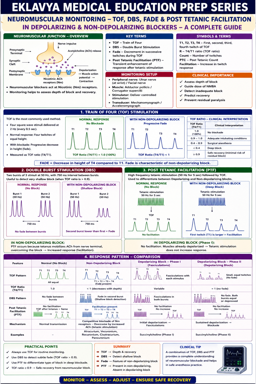
Describe the principles of Train-of-Four (TOF) and Double Burst Stimulation (DBS) [4]. Explain the phenomenon of 'fade' and 'post-tetanic facilitation' in depolarising vs. non-depolarising blocks [6].

collections Question Diagrams & Reference Images (1)
Question Reference Diagram
zoom_in View Image
description Clinical Response
"

Paper I — Applied Basic Sciences [10 Marks] — Long Answer

Neuromuscular Blockade Monitoring — TOF, DBS, Fade & Post-Tetanic Facilitation

Describe the principles of Train-of-Four (TOF) and Double Burst Stimulation (DBS) [4]. Explain the phenomenon of 'fade' and 'post-tetanic facilitation' in depolarising vs. non-depolarising blocks [6].

⚙ Core Concept

Neuromuscular monitoring is the gold-standard tool for guiding NMB dosing, timing reversal, and confirming adequate recovery before extubation. The specific patterns of fade and post-tetanic facilitation distinguish depolarising from non-depolarising block at a fundamental mechanistic level. (Murphy GS; Naguib M — Anesthesiology; Miller's Anaesthesia 9th Ed)

A. Train-of-Four (TOF) Monitoring 2 marks

  • Principle: four supramaximal electrical stimuli (0.2 ms pulse width) at 2 Hz to the ulnar nerve at the wrist; four twitches (T1–T4) measured by acceleromyography
  • TOF Count: number of detectable twitches (0–4); T1 returns first → count 1/4; T4 returns last → count 4/4
  • TOF Ratio (TOFR): T4/T1 amplitude ratio; TOFR ≥0.9 = adequate recovery (safe extubation); 0.7–0.9 = pharyngeal dysfunction/aspiration risk; <0.4 = obvious weakness
  • Monitoring sites: adductor pollicis (ulnar — standard); corrugator supercilii (facial — surrogate for laryngeal muscles); orbicularis oculi

B. Double Burst Stimulation (DBS) 2 marks

  • Principle: two short bursts of 50 Hz tetanic stimulation (each = 3 × 0.2 ms pulses), separated by 750 ms; most common variant DBS₃,₃
  • Clinical advantage: fade between the two DBS responses is more detectable by tactile/visual assessment than TOF fade; the human finger CANNOT reliably detect TOF fade between 0.4–0.7 TOFR but can detect fade in DBS
  • Limitation: DBS does NOT eliminate the need for quantitative accelerometry — cannot reliably detect TOFR >0.7 by tactile assessment

C. Fade and Its Mechanism 3 marks

  • Definition: progressive decrease in twitch amplitude during repetitive stimulation — T4 smaller than T1 in non-depolarising block; TOFR = T4/T1 < 1.0
  • Mechanism: presynaptic nicotinic receptors normally facilitate ACh mobilisation during repetitive firing; non-depolarising NMBs block BOTH postsynaptic AND presynaptic nicotinic receptors → impaired ACh mobilisation → progressively less ACh available → successive end-plate potentials decrease → fade
  • Non-depolarising block: FADE IS PRESENT during TOF and tetanic stimulation — the hallmark diagnostic feature

D. Post-Tetanic Facilitation (PTF) 3 marks

  • Phenomenon: after brief tetanic stimulation (50 Hz for 5 seconds), subsequent single-twitch/TOF responses are temporarily ENHANCED beyond pre-tetanic level
  • Mechanism: tetanic stimulation → massive presynaptic Ca²⁺ influx → very large ACh release overwhelms postsynaptic competitive block; terminal Ca²⁺ load also temporarily increases ACh mobilisation for 1–3 minutes
  • Post-Tetanic Count (PTC): twitches detectable in 5 seconds following tetanic stimulus; used during PROFOUND non-depolarising block (TOF count = 0); PTC 1–5 = very deep block; PTC >10 = recovery approaching

E. Comparison: Depolarising vs Non-Depolarising Block Patterns

FeatureDepolarising Block (Succinylcholine)Non-Depolarising Block (Rocuronium, Vecuronium)
TOF fadeNO FADE — all four twitches decrease equallyFADE PRESENT — T4 < T1 (presynaptic ACh mobilisation impaired)
Post-tetanic facilitationNOT PRESENTPRESENT — post-tetanic twitches enhanced
Sustained tetanusYes — sustained contractionFade during tetanus — cannot sustain tetanic contraction
Reversal possibleNo reversal agents; spontaneous recovery (plasma cholinesterase)Neostigmine or sugammadex (for steroidal NMBs)

💬 Viva Corner

Q. Why does fade occur with non-depolarising NMBs but not with succinylcholine?

Non-depolarising NMBs block BOTH postsynaptic and presynaptic nicotinic receptors. The presynaptic auto-receptor normally acts as positive feedback signalling “release more ACh” during rapid firing; blocking it impairs replenishment of the readily-releasable ACh pool → each successive end-plate potential is smaller → T4 < T1 = FADE. Succinylcholine causes depolarisation as a postsynaptic agonist without significantly impairing presynaptic ACh mobilisation → the feedback loop remains intact → all four twitches decrease equally → NO FADE. Similarly, tetanic stimulation enhances ACh release with succinylcholine present, but the postsynaptic receptor is already depolarised-and-blocked, so the extra ACh cannot overcome the block → no post-tetanic facilitation.

★ Examiner's Pearl

The four-part comparison must be reproduced: Depolarising: no fade + no PTF + sustained tetanus. Non-depolarising: FADE present + PTF present + unsustained tetanus. The mechanism of fade (presynaptic nicotinic receptor blockade impairs ACh mobilisation) is the most tested mechanistic explanation. TOFR ≥0.9 for safe extubation (not head-lift ≥5 seconds which only requires TOFR ≥0.6) must be cited.

References: Murphy GS et al. Residual neuromuscular blockade (Anesth Analg 2008;107:130-137). Naguib M et al. Advances in neurobiology of the NMJ (Anesthesiology 2002;96:202-231). Miller RD et al. Miller's Anaesthesia, 9th Ed, Chapter 34.

"
lock Protected Clinical Document • Pulse App • Screenshots Strictly Prohibited
QUESTION 257 person Asked by .
bookmark_add

Describe the pharmacology of dexmedetomidine: (a) Mechanism of action and receptor selectivity profile [3] (b) Clinical applications in modern anaesthesia and ICU [4] (c) Adverse effects and contraindications [3].

description Clinical Response
"

Paper I — Applied Basic Sciences [10 Marks] — Long Answer

Dexmedetomidine — Mechanism, Receptor Selectivity, Clinical Applications & Adverse Effects

Describe the pharmacology of dexmedetomidine: (a) Mechanism of action and receptor selectivity profile [3] (b) Clinical applications in modern anaesthesia and ICU [4] (c) Adverse effects and contraindications [3].

⚙ Core Concept

Dexmedetomidine (Precedex) is a highly selective α₂-adrenoceptor agonist with a selectivity ratio of 1600:1 (α₂:α₁) — 8-fold more selective than clonidine (200:1). Its unique property of producing sedation through the locus coeruleus (mimicking natural sleep) without respiratory depression makes it the only sedative suitable for awake procedures requiring patient cooperation and for ICU sedation where respiratory drive must be preserved. (Kamibayashi T, Mazez M — Anesthesiology 2000; SEDCOM trial JAMA 2009; MENDS trial JAMA 2007; Miller's Anaesthesia 9th Ed)

A. Mechanism of Action & Receptor Profile 3 marks

  • Receptor: α₂-adrenoceptor agonist; subtypes α₂A (predominant — CNS sedation, analgesia, sympatholysis), α₂B (peripheral vasoconstriction, anti-shivering), α₂C (CNS modulation, adrenal medulla)
  • Selectivity: α₂:α₁ ratio = 1600:1 (vs clonidine 200:1)
  • Central mechanism — sedation/analgesia: α₂A activation in the locus coeruleus → hyperpolarisation via Gᵢ protein → ↓ noradrenaline release → ↓ arousal → sedation; EEG resembles natural NREM sleep, unlike GABA-mediated agents which suppress both sleep and arousal pathways — producing unique “rousable cooperative sedation”
  • Analgesia mechanism: α₂ receptors in the dorsal horn → presynaptic inhibition of substance P release; modest as sole analgesic — primarily an adjuvant
  • Sympatholysis: α₂ agonism → ↓ noradrenaline release → ↓ sympathetic tone → ↓ HR, ↓ BP

B. Clinical Applications 4 marks

ApplicationDose/ProtocolRationale
ICU sedation (primary indication)0.2–1.4 mcg/kg/hr IV; loading 1 mcg/kg over 10 min in stable patients; target RASS 0 to −2SEDCOM trial: vs midazolam — less delirium, fewer ventilator days; MENDS trial: vs lorazepam — more delirium-free days; PADIS 2018 recommends over benzodiazepines
Awake fiberoptic intubation (AFOI)Loading 1 mcg/kg over 10 min; maintenance 0.4–0.7 mcg/kg/hrRousable cooperative sedation without respiratory depression; anxiolytic; blunts haemodynamic response to instrumentation
Awake craniotomyTitrated to conscious sedation; stopped during mappingMaintains cooperation for neurological testing without respiratory compromise
Procedural sedation (MRI, bronchoscopy)0.5–1 mcg/kg loading then 0.2–0.7 mcg/kg/hrAvoids respiratory depression where airway support is limited; preserves airway reflexes
Paediatric premedicationIntranasal 1–2 mcg/kg (off-label); oral 1–2 mcg/kgNon-IV route; calm cooperative sedation; avoids paradoxical agitation seen with midazolam
Adjunct for regional anaesthesiaPerineural 50–100 mcg; intrathecal 3–5 mcgα₂ receptors on peripheral nerve/spinal cord prolong analgesia; motor-sparing
Blunting haemodynamic responseIV bolus 0.5–1 mcg/kg before laryngoscopyAttenuates sympathetic surge; useful in hypertensive patients, IHD, phaeochromocytoma

C. Adverse Effects and Contraindications 3 marks

  • Bradycardia: most common significant adverse effect; from central sympatholysis + possible direct SA node effect; treatment: atropine 0.5 mg IV, glycopyrrolate 0.2 mg IV, dose reduction, avoid rapid boluses
  • Biphasic blood pressure response: initial transient hypertension (peripheral α₂B vasoconstriction with rapid bolus) followed by sustained hypotension (central sympatholysis); give loading dose slowly over 10 minutes
  • Respiratory effects: minimal at clinical doses — does NOT cause clinically significant respiratory depression (key safety advantage over opioids/benzodiazepines)
  • Paradoxical agitation: rarely seen in paediatric patients
  • Contraindications: advanced heart block without pacemaker; severe hepatic failure (exclusive hepatic metabolism); sick sinus syndrome without pacemaker; caution in acute cerebrovascular events

💬 Viva Corner

Q. Why is dexmedetomidine preferred over midazolam for ICU sedation according to current guidelines?

PADIS 2018 recommends dexmedetomidine over benzodiazepines for non-deeply sedated ventilated ICU patients, based on SEDCOM (fewer delirium days, ~1.9 days shorter time to extubation) and MENDS (more delirium-free/coma-free days) trials. Mechanistically, dexmedetomidine sedates via the locus coeruleus, producing a physiological NREM sleep-like EEG that preserves sleep architecture, whereas benzodiazepines suppress the cortex via GABA-A and disrupt normal sleep, contributing to delirium via circadian disruption and inflammatory amplification. Dexmedetomidine also lacks respiratory depression and accumulates less than long-acting benzodiazepines in organ failure.

★ Examiner's Pearl

α₂:α₁ selectivity = 1600:1 (dexmedetomidine) vs 200:1 (clonidine). Sedation via locus coeruleus → NREM sleep-like EEG → “rousable cooperative sedation” (unique property). NO clinically significant respiratory depression at clinical doses. SEDCOM trial is the landmark ICU evidence. Bradycardia and biphasic BP response are the most common adverse effects; avoid rapid loading boluses.

References: Kamibayashi T, Maze M. Clinical uses of α₂-adrenergic agonists (Anesthesiology 2000;93:1345-1349). Riker RR et al. SEDCOM trial (JAMA 2009;301:489-499). Devlin JW et al. PADIS Guidelines 2018 (Crit Care Med 2018;46:e825-e873). Miller RD et al. Miller's Anaesthesia, 9th Ed.
"
lock Protected Clinical Document • Pulse App • Screenshots Strictly Prohibited
QUESTION 258 person Asked by .
bookmark_add

Explain the physical principle of infrared absorption spectroscopy [3]. Draw and analyse a normal capnograph waveform, labelling its distinct phases [4]. Describe capnograph variations in bronchospasm, cardiac arrest, and rebreathing [3].

description Clinical Response
"

Paper I — Applied Basic Sciences [10 Marks] — Long Answer

Capnography — Infrared Spectroscopy, Normal Waveform Phases & Pathological Variations

Explain the physical principle of infrared absorption spectroscopy [3]. Draw and analyse a normal capnograph waveform, labelling its distinct phases [4]. Describe capnograph variations in bronchospasm, cardiac arrest, and rebreathing [3].

⚙ Core Concept

Capnography is a mandatory monitor for all intubated patients under general anaesthesia — it is the most reliable and immediate confirmation of tracheal intubation, provides continuous ventilation monitoring, and acts as a window into cardiovascular physiology. (Bhavani-Shankar K — Anesth Analg; AAGBI Monitoring Standards 2021; Miller's Anaesthesia 9th Ed)

A. Physical Principle — Infrared Absorption Spectroscopy 3 marks

  • Principle: CO₂ molecules absorb infrared (IR) radiation at specific wavelengths characteristic of molecular bond vibrations; CO₂ absorbs IR maximally at 4.28 µm — the wavelength used in clinical capnographs
  • Beer-Lambert Law applied: IR absorbed is proportional to CO₂ concentration and path length; capnograph contains an IR source (4.28 µm), a sample cell, and an IR detector — more CO₂ = more absorption = less signal reaching detector
  • Mainstream vs sidestream: Mainstream — in-line, no sampling delay, heavier, can cause burns; Sidestream — aspirates 50–150 mL/min, slight delay (2–3 s), more common clinically

B. Normal Capnograph Waveform — Four Phases 4 marks

⇗ Waveform Description

PhaseCO₂ LevelGas OriginClinical Significance
Phase I — Inspiratory baseline0 mmHg (flat, at zero)Anatomical dead space (no CO₂); inspired fresh gasElevated baseline = REBREATHING: exhausted soda lime, incompetent expiratory valve, insufficient FGF in Mapleson D
Phase II — Expiratory upstrokeRapidly rising from 0 to plateauMixing of dead space gas with alveolar gasProlonged, gradual slope = V/Q inhomogeneity; seen in bronchospasm
Phase III — Alveolar plateauNear-flat plateau at ETCO₂ (typically 35–45 mmHg)Predominantly alveolar gasNormal: slight upward slope (<2 mmHg); STEEP (“shark fin” >5 mmHg) = bronchospasm/COPD
Phase 0/IV — Inspiratory downstrokeRapid fall to zeroFresh inspiratory gas washing out CO₂ETCO₂ is read at the peak just before this downstroke

Normal ETCO₂: 35–45 mmHg (4.5–6.0 kPa); PaCO₂-ETCO₂ gradient normally 2–5 mmHg; widens with increased dead space (IPPV, Zone 1 expansion, PE, ↓ cardiac output)

C. Pathological Capnograph Variations 3 marks

ConditionCapnograph PatternMechanism
Bronchospasm“Shark fin”/reverse sawtooth: prolonged gradual Phase II upstroke; steep upward Phase III slopeInhomogeneous airway resistance; obstructed alveoli empty slowly with higher CO₂ reaching detector later
Cardiac arrest / severe ↓COSudden sustained fall in ETCO₂ to near-zero despite continued ventilationCO₂ delivery to alveoli requires pulmonary blood flow; no flow → no delivery; ETCO₂ ≥10 mmHg during CPR = adequate compressions
RebreathingElevated baseline (Phase I not at zero)Exhausted soda lime; incompetent expiratory valve; FGF too low in non-rebreathing system
Oesophageal intubationAbsent or rapidly diminishing waveform — flat within 4–6 breathsNo sustained CO₂ production from stomach; confirms need to remove tube
Pulmonary embolismSudden fall in ETCO₂; widened PaCO₂-ETCO₂ gradientObstructed pulmonary vessels → ↑ dead space → CO₂ from unperfused regions never delivered to alveoli

💬 Viva Corner

Q. During CPR in theatre, ETCO₂ reads 5 mmHg. What does this tell you and how should it guide your actions?

ETCO₂ 5 mmHg indicates critically inadequate pulmonary blood flow from compressions (benchmark: ≥10 mmHg = adequate). Immediate actions: switch compressor (fatigue is the commonest cause), confirm hand position, depth ≥5 cm, rate 100–120/min, full recoil, minimise pauses. Consider reversible causes (4Hs/4Ts) if compressions are already optimal. Give adrenaline if due. If ETCO₂ stays <10 mmHg after 20 minutes of optimal CPR despite addressing reversible causes, this is associated with failure of ROSC. Conversely, a sudden rise to ≥35 mmHg is the earliest sign of ROSC — stop compressions and check for a pulse.

★ Examiner's Pearl

The waveform must be reproduced as a labelled drawing description with four phases and normal ETCO₂ (35–45 mmHg). The PaCO₂-ETCO₂ gradient (normally 2–5 mmHg; widens with ↑ dead space) is a consistently tested integration question. Shark fin in bronchospasm and the cardiac arrest pattern (sudden fall of ETCO₂ to near-zero) are the two most commonly asked pathological patterns.

References: Bhavani-Shankar K et al. Capnometry and anaesthesia (Can J Anaesth 1992;39:617-632). AAGBI Monitoring Standards 2021. Miller RD et al. Miller's Anaesthesia, 9th Ed.

"
lock Protected Clinical Document • Pulse App • Screenshots Strictly Prohibited
QUESTION 259 person Asked by .
bookmark_add

Write short notes on: (a) Sugammadex: Mechanism of action, dosing criteria, and limitations [5] (b) Hofmann elimination and ester hydrolysis with reference to Cisatracurium and Remifentanil [5].

description Clinical Response
"

Paper I — Applied Basic Sciences [10 Marks] — Short Notes

Sugammadex & Hofmann Elimination — Mechanism, Dosing, Limitations & Cisatracurium/Remifentanil

Write short notes on: (a) Sugammadex: Mechanism of action, dosing criteria, and limitations [5] (b) Hofmann elimination and ester hydrolysis with reference to Cisatracurium and Remifentanil [5].

A. Sugammadex 5 marks

⚙ Core Concept

Sugammadex (Bridion) is a modified γ-cyclodextrin — a toroidal molecule that forms a tight, 1:1 encapsulation complex with steroidal NMBs (rocuronium > vecuronium > pancuronium), effectively removing free drug from the neuromuscular junction. It is the first truly mechanism-specific NMB reversal agent — unlike neostigmine, which increases ACh at all cholinergic synapses.

  • Mechanism: hydrophobic interior cavity encapsulates the lipophilic steroid nucleus of rocuronium/vecuronium; Kd for rocuronium ≈ 0.1 µM (extremely tight); complex is pharmacologically INACTIVE and renally excreted intact; as free rocuronium falls, more is drawn from the NMJ down its concentration gradient → reversal
  • Dosing criteria: 2 mg/kg IV — routine reversal at TOF count ≥2/4 (reversal to TOFR ≥0.9 within 3 min); 4 mg/kg IV — deep block (TOF 0–1, PTC ≥1–2); 16 mg/kg IV — immediate reversal of RSI rocuronium dose (1.2 mg/kg) within 3 minutes
  • Limitations: does NOT reverse succinylcholine or benzylisoquinolinium NMBs (atracurium, cisatracurium, mivacurium); recurarisation risk if rocuronium re-dosed within 24h; reduces efficacy of progesterone contraceptives (advise additional contraception for 7 days); accumulates in renal failure; expensive; rare anaphylaxis (~1:3500–1:6000)

B. Hofmann Elimination & Ester Hydrolysis 5 marks

⚙ Core Concept

Hofmann elimination and ester hydrolysis are organ-independent chemical degradation pathways — the drug degrades spontaneously in plasma without requiring hepatic metabolism or renal excretion, making these agents safe in organ failure. Cisatracurium and remifentanil are the prime examples.

Hofmann Elimination — Cisatracurium

  • pH- and temperature-dependent non-enzymatic breakdown at physiological pH (7.4) and 37°C → laudanosine (active CNS stimulant, sub-clinical at normal doses) + monoquaternary acrylate
  • Cisatracurium is the 1R-cis,1R'-cis isomer of atracurium — 3× more potent (less laudanosine produced); onset 3–5 min; duration 45–60 min
  • Hepatic/renal failure does NOT prolong its action — ideal for ICU infusions (ARDS, status asthmaticus, ICP management)

Ester Hydrolysis — Remifentanil

  • Ester linkage hydrolysed by non-specific tissue and blood esterases → remifentanil acid (essentially inactive)
  • NOT metabolised by plasma pseudocholinesterase — patients with dibucaine-positive pseudocholinesterase deficiency have NORMAL remifentanil metabolism
  • Context-sensitive half-time (CSHT) = 3 minutes, CONSTANT regardless of infusion duration — because hydrolysis occurs everywhere simultaneously, with no peripheral reservoir

💬 Viva Corner

Q. Why does remifentanil have a constant CSHT of 3 minutes while fentanyl's CSHT rises steeply with infusion duration?

Fentanyl is highly lipophilic and redistributes into peripheral fat during infusion; when stopped, drug slowly returns from fat and requires hepatic metabolism — the longer the infusion, the more accumulation, so CSHT rises steeply (60 min after 1h to 300+ min after 8h). Remifentanil undergoes ester hydrolysis ubiquitously throughout the body, including peripheral compartments — there is no reservoir slowly releasing drug back into plasma. The drug disappears from all compartments simultaneously, so CSHT stays constant at 3 minutes regardless of duration. Clinically, long-acting analgesia must be established before stopping remifentanil, as its analgesic effect disappears within 5–10 minutes.

★ Examiner's Pearl

Sugammadex doses must be exact: 2 mg/kg (TOF ≥2); 4 mg/kg (PTC ≥1, TOF 0); 16 mg/kg (immediate reversal post-RSI). Does NOT work for succinylcholine or atracurium — steroidal NMBs ONLY. Remifentanil ester hydrolysis by NON-SPECIFIC esterases (NOT pseudocholinesterase) is the specific frequently-tested distinction.

References: Naguib M. Sugammadex (Anesthesiology 2007;106:1267-1273). Papazian L et al. Neuromuscular blockers in early ARDS (NEJM 2010;363:1107-1116). Minto CF et al. Remifentanil pharmacokinetics (Anesthesiology 1997;86:10-23). Miller RD et al. Miller's Anaesthesia, 9th Ed.

"
lock Protected Clinical Document • Pulse App • Screenshots Strictly Prohibited
QUESTION 260 person Asked by .
bookmark_add

Describe the anatomy of the NMJ. Discuss normal physiology. Describe depolarising and non-depolarising muscle relaxants. Discuss tests for neuromuscular block monitoring.

description Clinical Response
"
⚙ CORE CONCEPT

The Neuromuscular Junction (NMJ) is the specialised synapse between the motor nerve terminal and skeletal muscle fibre — a site of remarkable pharmacological specificity where anaesthesiologists exert precise control over muscle relaxation. Every muscle relaxant, reversal agent, and monitoring technique is designed around this junction's anatomy and physiology. Understanding the NMJ is the foundation of safe paralysis in anaesthesia practice.

(Naguib M — NMJ review NEJM; Bowman WC — pharmacology of NMB; Donati F — neuromuscular pharmacology; Fuchs-Buder T — monitoring NMB)
A. Anatomy of the Neuromuscular Junction
△ NMJ STRUCTURE — DETAILED DIAGRAM
MOTOR NEURON AXON
  | (myelinated)
  |
+----v----------------------------------------------+
| MOTOR NERVE TERMINAL (bouton)                      |
| +-------------------------------------------------+ |
| | ACh VESICLES: ~10,000 vesicles per terminal      | |
| | Each vesicle: ~5,000-10,000 ACh molecules (1 quantum)| |
| | ACTIVE ZONES: docked vesicles ready for release  | |
| | Voltage-gated Ca2+ channels (P/Q-type) at active zones| |
| | Mitochondria: ATP for ACh synthesis              | |
| +-------------------------------------------------+ |
+---------------------+-------------------------------+
                       | SYNAPTIC CLEFT (~50 nm)
                       | Acetylcholinesterase (AChE)
                       | Collagen fibrils
                       | Agrin protein (organises postjxn)
+----------------------v-----------------------------+
| POSTJUNCTIONAL MEMBRANE (Motor End Plate)           |
| JUNCTIONAL FOLDS: deep infoldings ^ surface area ~10x|
| nAChRs densely packed at CRESTS of junctional folds |
| AChE located at DEPTHS of folds                     |
| nAChR density: ~10,000-20,000 per um2 at the crests |
|                                                       |
| nAChR STRUCTURE (pentameric):                        |
| a1 (x2) + b1 + d + e (adult) or g (fetal/extrajxnal) |
| ACh binding site on EACH a1 subunit                  |
| BOTH a1 subunits must be occupied for channel opening|
+-------------------------------------------------------+

PREJUNCTIONAL RECEPTORS: a3b2 nAChRs (autoreceptors)
-> POSITIVE FEEDBACK: ACh released -> binds prejxnal receptors
-> Facilitates further ACh release (mobilisation during tetanus)
Non-depolarising NMBDs block BOTH pre- and postjunctional receptors
-> Explains ""fade"" on sustained/tetanic stimulation
ComponentDetails
ACh Synthesis and StorageSynthesised in the nerve terminal from CHOLINE (taken up by Na⁺-dependent high-affinity choline transporter) + ACETYL-CoA (from mitochondrial pyruvate metabolism); enzyme: CHOLINE ACETYLTRANSFERASE (ChAT); stored in synaptic vesicles (~10,000 molecules = 1 quantum); SPONTANEOUS release of individual quanta = miniature end-plate potentials (MEPPs) — basis of the "safety factor"
Presynaptic voltage-gated Ca²⁺ channelsP/Q-type (CaV2.1) calcium channels at active zones; action potential → membrane depolarisation → Ca²⁺ influx → triggers vesicle fusion (SNARE protein complex: synaptobrevin, SNAP-25, syntaxin) → EXOCYTOSIS of ACh quanta; ~60–100 quanta released per action potential (quantum content); Lambert-Eaton myasthenic syndrome targets these channels
Acetylcholinesterase (AChE)Located at the DEPTHS of junctional folds; collagen-tailed tetramers (A₁₂ form); hydrolyses ACh → CHOLINE + ACETATE within 1 millisecond; remarkably efficient — one enzyme molecule can hydrolyse 25,000 ACh molecules per second; choline is recycled back into the nerve terminal; inhibited by: neostigmine, pyridostigmine (reversible carbamate inhibitors), organophosphates (irreversible)
nAChR Structure and FunctionNICOTINIC ACETYLCHOLINE RECEPTOR: pentameric ligand-gated ion channel (2α₁ + β₁ + δ + ε in adult; γ replaces ε in fetal/denervated/extrajunctional); ion channel is non-selective cation channel (Na⁺ in, K⁺ out); BOTH α₁ subunits must be occupied by ACh (or agonist) for channel opening; opens for ~1 ms → ion flow → END-PLATE POTENTIAL (EPP); EPP must reach threshold → action potential propagates along muscle fibre → contraction; EXTRAJUNCTIONAL RECEPTORS (γ-subunit): appear with denervation, immobility, burns → increased K⁺ release with suxamethonium → hyperkalaemia risk
B. Normal Physiology and Functions of the NMJ
Physiological ProcessDetail
Normal Neuromuscular Transmission (Step-by-Step)(1) Action potential propagates down motor axon to nerve terminal; (2) Depolarisation → P/Q-type Ca²⁺ channels open → Ca²⁺ influx; (3) Ca²⁺ triggers SNARE-mediated exocytosis of ~60–100 ACh quanta; (4) ACh diffuses across 50 nm synaptic cleft; (5) ACh binds BOTH α₁ subunits of nAChR → ion channel opens; (6) Na⁺ influx (+ K⁺ efflux) → END-PLATE POTENTIAL (EPP) +25 to +40 mV; (7) EPP spreads to adjacent muscle membrane → exceeds threshold → action potential in muscle fibre; (8) Muscle action potential propagates along T-tubules → Ca²⁺ release from SR → actin-myosin crossbridge cycling → CONTRACTION; (9) AChE hydrolyses ACh → choline recycled → termination of signal
Safety Factor of NMJThe EPP generated by normal neuromuscular transmission is MUCH LARGER than required to trigger muscle action potential — a "margin of safety" approximately 3–4×; this safety factor means that even with partial nAChR blockade (~75–80%), sufficient receptors remain to generate adequate EPP for contraction; clinical weakness (force <25% of normal) is not detectable by clinical examination until 70–80% of receptors are blocked; critically: PORC (post-operative residual curarisation) = TOF ratio <0.9 — patient can fail clinical tests of strength but still be unable to maintain airway at TOF ratio 0.7–0.9
Tetanic Stimulation and Post-Tetanic PotentiationHIGH-FREQUENCY stimulation (≥50 Hz = tetanus): initially ↑ quantal ACh release (facilitation — mobilisation from reserve pool); maintained tetanus → depletion of immediately available quanta → FADE (↓ force over time); POST-TETANIC POTENTIATION (PTP/PTC): after tetanus ends, residual Ca²⁺ in terminal → ↑ baseline ACh release for 2–4 minutes → allows transmission at some blocked NMJs → post-tetanic count (PTC) reveals response even when TOF count is 0
Fade — the basis of NMJ monitoringNON-DEPOLARISING NMBDs block PREJUNCTIONAL α₃β₂ receptors → prevents ACh mobilisation during high-frequency stimulation → FADE (ratio of 4th to 1st twitch = TOF ratio <1.0); DEPOLARISING NMBDs do NOT cause fade (equal depression of all twitches — a "Phase 1 block"); fade detection = basis of TOF and tetanic stimulation monitoring
C. Non-Depolarising Muscle Relaxants (NDNMBDs)
DrugOnsetDurationDose (intubation)Metabolism/ExcretionKey Features
Atracurium (Benzylisoquinolinium)Intermediate onset (2–3 min)Intermediate (20–35 min)0.5 mg/kgHOFMANN ELIMINATION (pH + temp dependent, organ-independent) + ester hydrolysis; metabolite: laudanosine (CNS stimulant — seizures at high plasma levels; clinically insignificant at usual doses)IDEAL for renal/hepatic failure; HISTAMINE RELEASE (intrinsic, dose-dependent); inject slowly; no cardiovascular effect at clinical doses
Cisatracurium (Benzylisoquinolinium)Intermediate onset (2–3 min)Intermediate (40–60 min)0.15 mg/kgHOFMANN ELIMINATION only (no ester hydrolysis); laudanosine produced in smaller amountsCLEANER than atracurium — NO histamine release; organ-independent elimination; preferred in ICU; more expensive
Mivacurium (Benzylisoquinolinium)Short onset (2–3 min)SHORT (12–20 min)0.2 mg/kgPLASMA CHOLINESTERASE (pseudocholinesterase) — same enzyme as suxamethonium; organ-independentSHORTEST acting NDNMBD; prolonged in pseudocholinesterase deficiency; histamine release
Rocuronium (Steroidal)RAPID onset (60–90s at 0.6 mg/kg; 30s at 1.2 mg/kg)Intermediate (30–60 min)0.6 mg/kg (routine); 1.2 mg/kg (RSI)HEPATIC metabolism + biliary excretion; RENAL (10–25%); prolonged in liver diseaseFASTEST onset of NDNMBDs; ONLY NDNMBD suitable for RSI; REVERSIBLE with SUGAMMADEX (16 mg/kg — CICO rescue)
Vecuronium (Steroidal)Intermediate onset (2–3 min)Intermediate (25–40 min)0.1 mg/kgHEPATIC (80%), renal (20%); active metabolite 3-OH vecuronium accumulates in renal failureNO histamine; NO cardiovascular effects (no vagolytic); AVOID in hepatic failure; reversed by sugammadex or neostigmine
Pancuronium (Steroidal)Slow onset (3–5 min)LONG (60–120 min)0.1 mg/kgRENAL (80%); hepatic (20%); AVOID in renal failureVAGOLYTIC (tachycardia, ↑BP); used for ICU paralysis and cardiac surgery; reversed by neostigmine
D. Depolarising Muscle Relaxants
FeatureDetail
Drug: SUXAMETHONIUM (Succinylcholine)The ONLY clinically used depolarising NMBD; two acetylcholine molecules joined at their acetyl groups (bis-quaternary ammonium compound); dose: 1–1.5 mg/kg IV (RSI); onset: 30–45 seconds; duration: 8–12 min (shortest-acting NMBD); metabolised by PSEUDOCHOLINESTERASE in plasma and liver → succinylmonocholine → succinic acid + choline
Mechanism — Phase 1 (Depolarising) BlockSch acts as an ACh AGONIST — binds BOTH α₁ subunits of nAChR → opens ion channel → SUSTAINED depolarisation; NOT hydrolysed by AChE → remains bound → end plate stays depolarised → adjacent Na⁺ channels inactivated → FLACCID PARALYSIS; initial fasciculations then flaccid block; no fade on TOF or tetanus; PTC normal; abolished by pseudocholinesterase
Phase 2 (Desensitisation) BlockWith LARGE or REPEATED doses of Sch: nAChRs become DESENSITISED; block now shows FADE on TOF (mimics NDNMBD); prolonged and potentially irreversible; occurs with total Sch dose >5–6 mg/kg or prolonged infusion
ADVERSE EFFECTSHYPERKALAEMIA: plasma K⁺ rises 0.5–1.0 mEq/L normally; DANGEROUS in burns (>24h), denervation, prolonged immobility, crush injury, rhabdomyolysis, muscular dystrophies — K⁺ rise 5–10 mEq/L → fatal cardiac arrest.
BRADYCARDIA: muscarinic (M2) stimulation, especially second dose or in children.
MALIGNANT HYPERTHERMIA TRIGGER: treat with dantrolene 2.5 mg/kg IV.
MYALGIA: prevented by defasciculation (vecuronium 0.01 mg/kg or rocuronium 0.06 mg/kg 90 sec before Sch).
RAISED ICP, IOP, INTRAGASTRIC PRESSURE.
PROLONGED BLOCK (Suxamethonium apnoea): pseudocholinesterase deficiency (dibucaine number <30 = homozygous atypical).
INDICATIONSRAPID SEQUENCE INTUBATION (RSI) — fastest onset + shortest duration; LARYNGOSPASM treatment (0.5 mg/kg IV or 4 mg/kg IM)

Mechanism of NDNMBDs: COMPETITIVE ANTAGONISM at BOTH α₁ subunits of postjunctional nAChR — they BIND the receptor without activating the ion channel; ACh is DISPLACED or prevented from occupying both sites; ALSO block PREJUNCTIONAL α₃β₂ receptors → prevent ACh mobilisation → FADE; the blockade is REVERSIBLE. REVERSAL: neostigmine (+ glycopyrrolate) inhibits AChE; SUGAMMADEX encapsulates rocuronium/vecuronium directly.

Factors affecting NDNMBD action: potentiation by volatile anaesthetics, hypokalaemia, hypermagnesaemia, hypothermia, aminoglycosides, respiratory acidosis; RESISTANCE with chronic phenytoin/carbamazepine, denervation.

E. Tests for Neuromuscular Block Monitoring
△ NMJ MONITORING — SITES AND PATTERNS
STIMULATION SITES (peripheral nerve stimulation):
- ULNAR NERVE at wrist (most common) -> adductor pollicis (thumb adduction)
- FACIAL NERVE (orbicularis oculi/corrugator) -- poor indicator of diaphragm/laryngeal recovery
- Posterior tibial nerve -> flexor hallucis brevis
- Common peroneal nerve -> extensor hallucis longus

RESPONSE MEASUREMENT:
- CLINICAL observation (least accurate)
- MECHANOMYOGRAPHY (MMG) -- gold standard but cumbersome
- ACCELEROMYOGRAPHY (AMG) -- most common clinical method (TOF-Watch SX)
  AMG TOF ratio >0.9 = adequate recovery (may overestimate)
- ELECTROMYOGRAPHY (EMG) -- measures muscle electrical activity directly
- KINEMYOGRAPHY (KMG) -- measures movement
Monitoring TestStimulation PatternInterpretationDepth of Block Detected
Single Twitch (ST)Single supramaximal stimulus (0.1–0.2 ms); 0.1 HzHeight of twitch as % of control; 75–80% receptor blockade before visible twitch depressionGROSS measure only; insensitive to early block; not useful clinically without control value
Train-of-Four (TOF)4 supramaximal stimuli at 2 Hz; every 10–12 sec; TOF RATIO = T4/T1TOF COUNT: 0 = profound; 1 = deep; 2 = moderate; 3–4 = light block; TOF RATIO ≥0.9 = adequate recoveryMOST USEFUL single monitoring test; TOF ratio <0.9 = PORC risk; FADE distinguishes NDNMBD from depolarising block
Tetanic Stimulation50 Hz for 5 sec; 100 Hz if very deep block suspectedSUSTAINED = adequate function; FADE = residual NDNMBD blockVery sensitive for RESIDUAL block; PAINFUL — not for routine intraoperative use
Post-Tetanic Count (PTC)50 Hz tetanic x 5 sec → 3 sec pause → single 1 Hz twitches countedPTC 1–2 = very deep; PTC 5–6 = TOF response in ~10 min; PTC >10 = TOF response imminentDetects PROFOUND BLOCK when TOF count = 0
Double Burst Stimulation (DBS)2 short tetanic bursts (3 stimuli at 50 Hz, 750 ms apart): DBS3,3Second burst smaller = FADE = residual block; equal = adequate recoveryDetects RESIDUAL BLOCK when TOF ratio cannot be quantified
F. PORC — Post-Operative Residual Curarisation
AspectKey Points
DefinitionTOF ratio <0.9 at tracheal extubation or in PACU; incidence up to 40–50% when reversal not used; associated with airway obstruction, hypoxaemia, aspiration pneumonia, muscle weakness, impaired hypoxic ventilatory response
Clinical tests (unreliable for TOF <0.9)5-second head lift: can be performed at TOF ratio 0.5 (UNRELIABLE); sustained hand grip; tongue depressor test; vital capacity <15 mL/kg; sustained tetanus (more sensitive but painful); QUANTITATIVE monitoring is the only reliable method
PreventionQUANTITATIVE NMJ monitoring (AMG/EMG) MANDATORY for every case using NDNMBD; SUGAMMADEX for rocuronium/vecuronium — most reliable reversal; neostigmine (ceiling effect); avoid extubation until TOF ratio ≥0.9 confirmed quantitatively
💬 VIVA CORNER

Why do we use TOF ratio >0.9 and not 1.0 as the target for extubation?
A TOF ratio of 1.0 is the physiological ideal, but TOF ratio ≥0.9 correlates with clinically adequate neuromuscular recovery — patients can protect their airway, maintain oxygen saturation, and perform reliable clinical tests. Below 0.9, significant impairment of pharyngeal and laryngeal muscle function occurs. Routine CLINICAL TESTS can be performed successfully at TOF ratio as low as 0.5 — they are UNRELIABLE. Only QUANTITATIVE monitoring can reliably confirm TOF ≥0.9, which is why NAP5 and current guidelines mandate it.

Why is suxamethonium absolutely contraindicated in burns patients but only relatively contraindicated in the first 24 hours?
In the FIRST 24 HOURS after burn injury, extrajunctional acetylcholine receptor proliferation has not yet occurred (takes 24–72h). So in the first 24h, suxamethonium causes only the normal 0.5–1.0 mEq/L rise in plasma K⁺. AFTER 24 HOURS, millions of extrajunctional receptors are expressed — depolarising all simultaneously causes massive K⁺ efflux (5–10 mEq/L) → VF → cardiac arrest. This risk persists indefinitely; the same applies to denervation, prolonged immobility, and severe sepsis.
★ EXAMINER'S PEARL

NMJ ANATOMY: Motor nerve terminal (vesicles, P/Q Ca²⁺ channels, active zones) → 50nm synaptic cleft (AChE at fold depths) → postjunctional membrane (junctional folds, nAChRs at crests: α₁₂β₁δε pentamer). PHYSIOLOGY: AP → Ca²⁺ → 60-100 quanta released → ACh binds BOTH α₁ → ion channel opens → EPP → muscle AP → contraction. SAFETY FACTOR: EPP 3-4x threshold needed; 75-80% receptors must be blocked before twitch depresses. NDNMBDs: COMPETITIVE ANTAGONIST at both α₁ (postjunctional) + prejunctional block → FADE on TOF. Rocuronium (RSI at 1.2 mg/kg, 30 sec); atracurium/cisatracurium (Hofmann — renal/hepatic failure). Suxamethonium: agonist → sustained depolarisation → flaccid block; AVOID: burns >24h, denervation, MH susceptibility, hyperkalaemia, open globe; PHASE 2 block with large doses (shows fade). MONITORING: TOF most important (count + ratio); PTC for profound block (count 0); DBS most sensitive clinical fade detection; QUANTITATIVE monitoring (AMG/EMG) mandatory → TOF ≥0.9 for extubation. PORC: TOF <0.9 — incidence 40-50% without monitoring; head lift unreliable (positive at TOF 0.5).
References: Naguib M et al. Advances in NMJ pharmacology. Anesthesiology 2002;96:202-231. Bowman WC. Neuromuscular block. Br J Pharmacol 2006;147:S277-286. Murphy GS et al. Residual NMB. Anesth Analg 2015;121:1329-1335.
"

Showing 251260 of 337 questions

account_tree

Subcategory Tree

Explore Anesthesia subcategories

folder_special Anesthesia
Main
lock

Category Subscription

Subscribe to Anesthesia to unlock this module and all nested subcategories.

  • check_circle Access Anesthesia & all subcategories
  • check_circle Detailed, Peer-Reviewed Answers
  • check_circle High-yield visual aids & imaging
Get Category Subscription arrow_forward
Secure 256-bit SSL Connection

Anatomical Models

Explore high-fidelity 3D visualizations included in premium modules.

Case Reviews

Real-world clinical scenarios narrated by senior consultants.

Pulse App