Anesthesia | Eklavya Medical
vaccines
medical_services Main Specialty Domain lock Subscription Required

Anesthesia

Clinical anesthesiology, local and general anesthesia delivery systems, monitoring, and perioperative care.

verified Verified Medical Faculty menu_book 337 Q&A Modules
lock Protected Clinical Document • Pulse App • Screenshots Strictly Prohibited
QUESTION 111 person Asked by .
bookmark_add

Describe the perioperative management of patients on warfarin, NOACs (apixaban, rivaroxaban, dabigatran), LMWH, and aspirin/clopidogrel. State the timing windows for stopping and restarting each drug. Outline the safe neuraxial anaesthesia (spinal/epidural) timing relative to each anticoagulant class.

collections Question Diagrams & Reference Images (1)
Question Reference Diagram
zoom_in View Image
description Clinical Response
⚙ Core Concept
Anticoagulant perioperative management requires balancing two competing risks: thromboembolism (stroke in AF, mechanical valve thrombosis, DVT) from withholding anticoagulation, versus haemorrhage (surgical bleeding, epidural haematoma after neuraxial anaesthesia) from continuing it. The ASRA neuraxial guidelines (2018) provide evidence-based minimum time windows between anticoagulant dose and neuraxial procedure — epidural haematoma has a 30-minute window for surgical decompression to prevent paraplegia. (Douketis JD — BRIDGE trial; ASRA Neuraxial Guidelines 2018; Narouze S; Miller's Anaesthesia 9th Ed)
A. Warfarin Perioperative Management2 marks

Stop: 5 days before elective surgery (allows INR to normalise to <1.5 in most patients); check INR on day of surgery

BRIDGE therapy: LMWH bridging was previously recommended for high-risk patients (mechanical heart valves, recent VTE, CHADS₂-VASc ≥4 in AF); BRIDGE trial (Douketis JD, NEJM 2015; n=1884 AF patients): no-bridging was non-inferior to bridging for stroke prevention AND significantly reduced major bleeding; current consensus: bridging NOT routinely indicated for most AF patients; still indicated for: mechanical mitral valves; recent (<3 months) VTE; very high thromboembolic risk conditions

Emergency reversal: vitamin K 5–10 mg IV (effective in 12–24 hours); 4-factor PCC (Beriplex/Octaplex) 25–50 IU/kg IV — immediate reversal for urgent surgery; FFP 10–15 mL/kg if PCC unavailable; target INR <1.5 for surgery

Restart: warfarin the evening of surgery or next day when haemostasis is secure; takes 5–7 days to re-achieve therapeutic INR; LMWH bridge may be needed if rapid anticoagulation is required

B. NOACs (Direct Oral Anticoagulants)3 marks

Drug Mechanism t½ Stop Before Surgery Emergency Reversal Dabigatran Direct 12–17h; renally cleared 80% 48h before low-risk; 96h before Idarucizumab (Praxbind) 5 g IV — monoclonal antibody (Pradaxa) thrombin → SIGNIFICANTLY prolonged high-risk; 4–5 days if eGFR <50 fragment; complete reversal in <5 min; approved for emergency (Factor IIa) in renal failure mL/min surgery/life-threatening bleeding inhibitor Rivaroxaban Direct Factor 5–9h (younger) to 11–13h 24h before low-risk; 48h before Andexanet alfa (Ondexxya) — Factor Xa decoy protein; rapid (Xarelto) Xa inhibitor (elderly) high-risk surgery; can be 24h reversal; expensive; alternative: 4-factor PCC 50 IU/kg (partial due to shorter t½ reversal) Apixaban Direct Factor 12h 48h before high-risk; 24h before Andexanet alfa (same as rivaroxaban); 4-factor PCC as (Eliquis) Xa inhibitor low-risk surgery alternative Edoxaban Direct Factor 10–14h 24–48h depending on renal Andexanet alfa; 4-factor PCC (Lixiana) Xa inhibitor function and bleeding risk There is NO reliable routine coagulation test to measure NOAC anticoagulant effect; anti-Xa levels (for Factor Xa inhibitors) or thrombin time (for dabigatran) can detect drug presence; NOT routinely available; in emergencies, absence of drug effect can be assumed if the drug was last taken >48 hours ago in a patient with normal renal function

C. LMWH and UFH2 marks

Drug Stop Before Surgery Restart LMWH (prophylactic dose — e.g., 12 hours before neuraxial procedure and surgery Resumption: 12 hours after surgery (prophylactic dose); 24 enoxaparin 40 mg OD) hours after high-bleeding-risk surgery LMWH (therapeutic dose — e.g., 24 hours before neuraxial procedure; 24–48h before high-risk 48–72 hours after surgery to ensure haemostasis before enoxaparin 1 mg/kg BD) surgery therapeutic anticoagulation UFH (intravenous, continuous) Stop 4–6 hours before; check APTT — must be normal; APTT Can restart 1 hour after uncomplicated surgery if haemostasis normalisation confirms drug clearance confirmed; IV UFH allows precise titration

D. ASRA 2018 Neuraxial Anaesthesia Windows3 marks
⚠ Epidural Haematoma Risk — These Minimum Intervals Are Patient Safety Minimums, Not Recommendations
Anticoagulant Minimum Interval: LAST DOSE → Neuraxial Minimum Interval: Neuraxial → NEXT DOSE Prophylactic LMWH 12 hours 4–6 hours post-procedure Therapeutic LMWH 24 hours 24 hours post-procedure (for epidural catheter management) UFH prophylactic (SC) 4–6 hours (and normal APTT) 1 hour post-procedure UFH therapeutic (IV) 4–6 hours (and normal APTT) 1 hour post-procedure Warfarin INR ≤1.4 (check on day of procedure) Resume warfarin after catheter removed; timing based on INR target Rivaroxaban/Apixaban 72 hours (or 48h if eGFR ≥50 and low-risk) 6 hours after neuraxial single shot; 6h after catheter removal for indwelling catheter Dabigatran 120 hours (5 days) if eGFR <50; 72 hours if eGFR ≥50 6 hours post-procedure Aspirin alone (75–150 No additional interval required (ASRA 2018: aspirin alone does NOT Can continue aspirin through neuraxial procedure mg) contraindicate neuraxial anaesthesia) Clopidogrel 7 days (irreversible platelet inhibition; new platelets must form) 24 hours post-procedure
🎤 Viva Corner
Q. A patient on rivaroxaban 20 mg OD for AF took his last dose 36 hours ago and requires emergency appendicectomy. Can you perform a spinal anaesthetic?
No — 36 hours is insufficient time after the last rivaroxaban dose for safe neuraxial anaesthesia under ASRA 2018 guidelines. The ASRA guideline for Factor Xa inhibitors (rivaroxaban, apixaban) is a minimum interval of 72 hours from the last dose to neuraxial procedure in standard-risk patients (those with eGFR ≥50 and for whom the procedure is not categorised as lower-risk). At 36 hours, rivaroxaban may still be present at clinically significant anticoagulant concentrations: with a halflife of 5–9 hours in younger patients (up to 11–13 hours in elderly), 36 hours represents approximately 2.5–7 half-lives depending on age and renal function, meaning 1–20% of the original plasma concentration may still be present; this residual concentration may be sufficient to impair haemostasis and increase the risk of epidural haematoma from neuraxial needle insertion — a catastrophic complication that can cause permanent paralysis unless surgically decompressed within 8 hours of symptom onset. For this emergency appendicectomy: general anaesthesia with RSI is the appropriate technique — it avoids the neuraxial bleeding risk entirely while providing appropriate anaesthesia for an emergency case; the appendicectomy can proceed safely under GA without waiting for rivaroxaban clearance. If the patient has normal renal function (eGFR ≥50) and the surgery were truly low-risk/non-major, some ASRA guidelines acknowledge 48 hours may be acceptable — but 36 hours is not sufficient even under these exceptions. The surgical bleeding risk from general anaesthesia in the context of rivaroxaban at 36 hours is relatively low (platelet function is preserved; only the Factor Xa-dependent clotting cascade is impaired at this level); the surgeon should be informed of the residual anticoagulation and be prepared for slightly increased surgical bleeding. There is no specific reversal agent available in most centres in real-time for emergency surgery for rivaroxaban (andexanet alfa is expensive and not universally available); 4-factor PCC 50 IU/kg can provide partial reversal of Factor Xa inhibitors if major bleeding occurs during surgery.
★ Examiner's Pearl
ASRA 2018 neuraxial timing windows must be memorised as a table: LMWH prophylactic = 12h; LMWH therapeutic = 24h; rivaroxaban/apixaban = 72h; dabigatran eGFR ≥50 = 72h; clopidogrel = 7 days; aspirin alone = NO additional interval. The BRIDGE trial (Douketis NEJM 2015 — no-bridging non-inferior for AF; bridging increases bleeding) is the landmark trial that changed warfarin bridging practice. Idarucizumab (Praxbind) for dabigatran reversal is the specific NOAC-specific antidote most tested.
Douketis JD et al. BRIDGE trial — perioperative anticoagulation (NEJM 2015;373:823-833). Narouze S et al. ASRA Practice Advisory on interventional pain management (Reg Anesth Pain Med 2018;43:225-262). Horlocker TT et al. ASRA Neuraxial Anesthesia and Anticoagulation guidelines 2018. Miller RD et al. Miller's Anaesthesia, 9th Ed.
lock Protected Clinical Document • Pulse App • Screenshots Strictly Prohibited
QUESTION 112 person Asked by .
bookmark_add

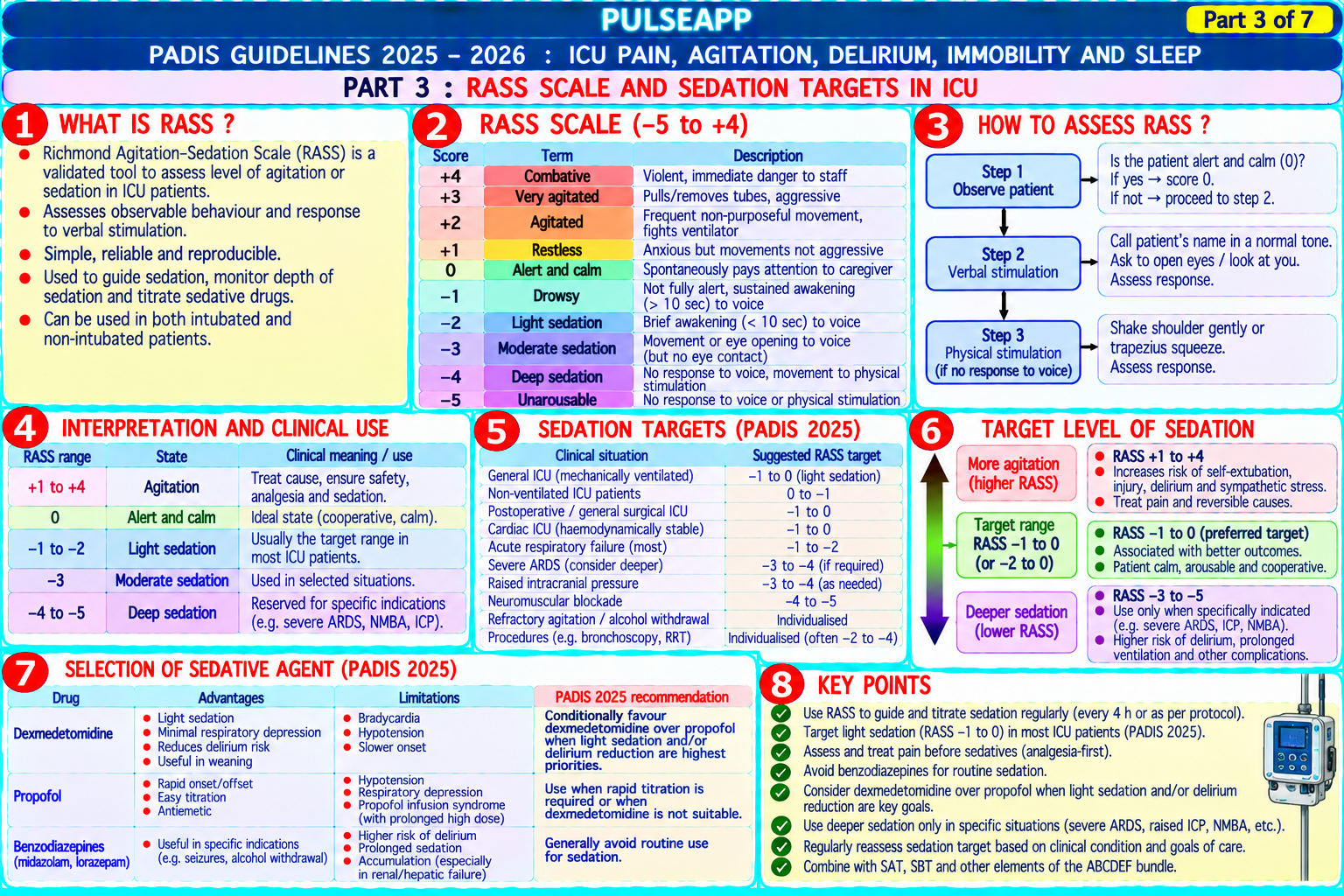
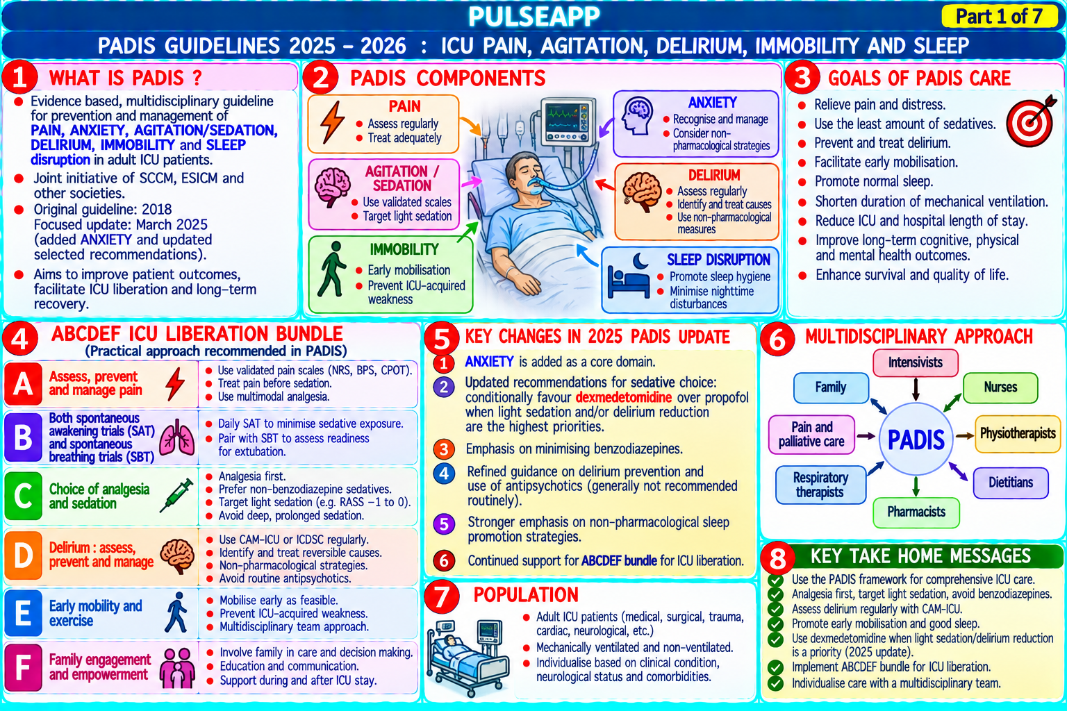
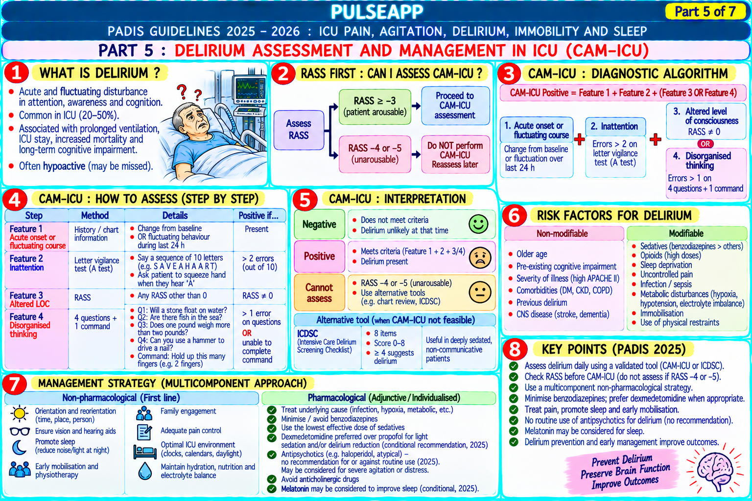
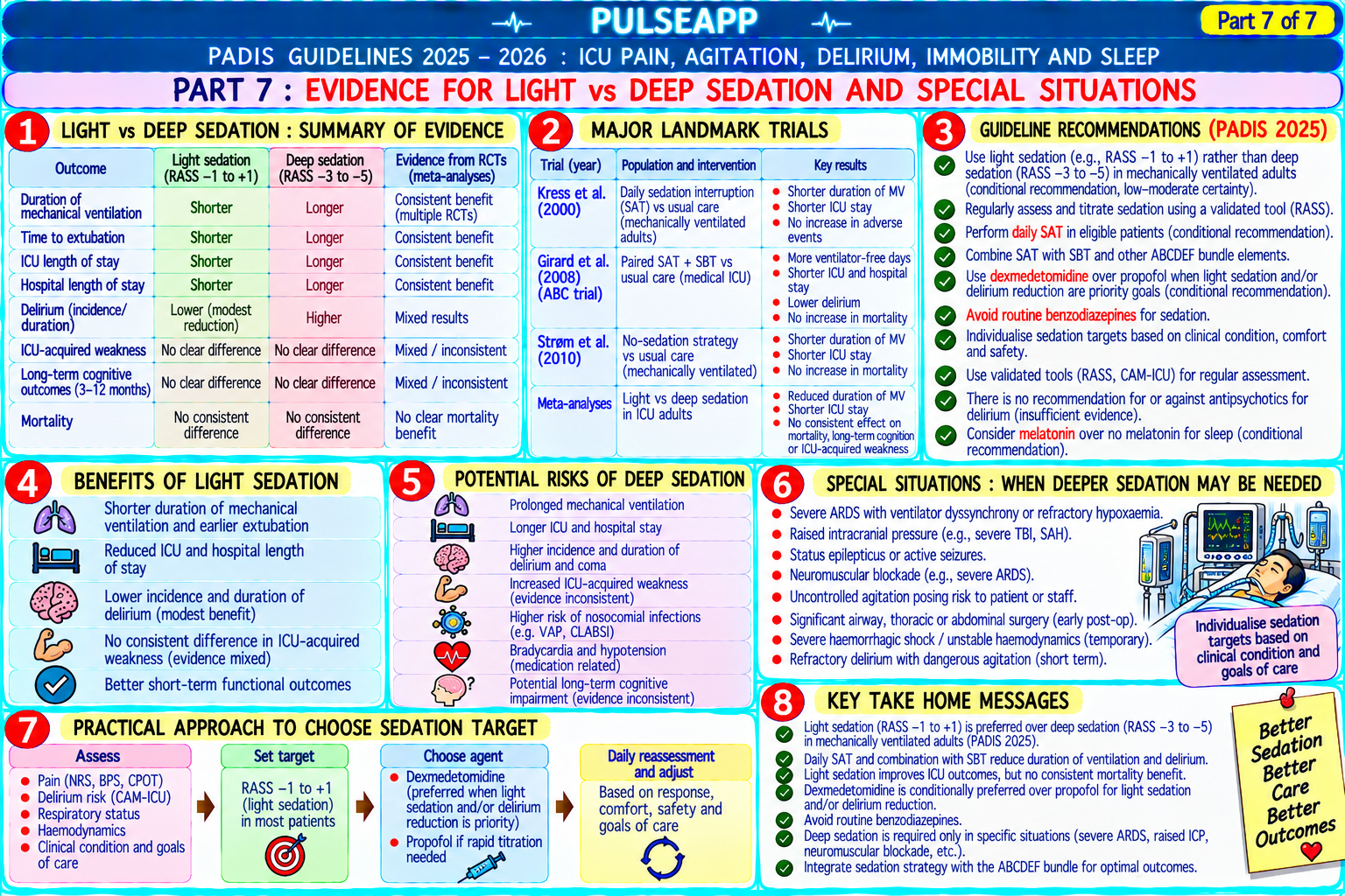
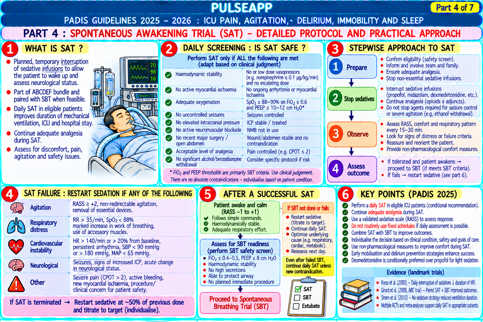
Describe the PADIS 2018 guidelines framework for ICU pain, agitation, delirium, immobility, and sleep. Outline the RASS scale, the Spontaneous Awakening Trial (SAT), sedation targets, and the CAM-ICU delirium assessment. Discuss the evidence for light sedation over deep sedation.
collections Question Diagrams & Reference Images (1)
Question Reference Diagram
zoom_in View Image
description Clinical Response
⚙ Core Concept
The PADIS 2018 guidelines represent the most important ICU bundle for improving patient outcomes — the shift from deep sedation (the traditional default) to light sedation (RASS target 0 to −2) combined with daily spontaneous awakening trials and early rehabilitation has reduced ICU mortality, duration of mechanical ventilation, delirium, and long-term cognitive impairment. (Devlin JW et al. — Crit Care Med 2018 PADIS; Ely EW — SAT + SBT NEJM 2008; Barr J — PAD Guidelines; Miller's Anaesthesia 9th Ed)
A. PADIS Framework — Five Domains2 marks

Domain Key Recommendation Assessment Tool

P — Pain Assess pain routinely; use analgesia-first approach (treat pain before sedation); non-opioid NRS (Numeric Rating Scale 0–10) for analgesics first (paracetamol, NSAIDs if appropriate, regional techniques); opioids titrated to communicative; BPS (Behavioural Pain Scale) or VAS/NRS ≤3/10 CPOT (Critical-Care Pain Observation Tool) for non-communicative

A — Agitation Light sedation target (RASS 0 to −2) preferred over deep sedation (RASS −3 to −5); reduces RASS (Richmond Agitation-Sedation Scale −5 to (Sedation) ventilator days, ICU stay, delirium; dexmedetomidine preferred over benzodiazepines for non- +4) deeply sedated patients

D — Delirium Routine delirium screening; multicomponent non-pharmacological bundle (reorientation, sleep CAM-ICU (Confusion Assessment Method for hygiene, early mobilisation, family involvement, natural lighting); avoid benzodiazepines (delirium ICU) — validated for intubated patients; takes <2 risk); antipsychotics (haloperidol) do not reliably prevent delirium but may treat hyperactive minutes delirium symptoms

I — Immobility Early mobilisation within 24–48 hours of ICU admission where safe; reduces ICU-acquired ABCDEF bundle: Awaken + Breathe + (Rehabilitation) weakness, delirium, and ventilator days; structured progressive rehabilitation (passive ROM → Coordinate + Delirium + Early mobility + Family active assisted → active → sitting → standing → walking)

S — Sleep Promote sleep-wake cycling; minimise nighttime interruptions; reduce light and noise; avoid Richards-Campbell Sleep Questionnaire (RCSQ) benzodiazepines and propofol for sleep (both suppress N3 and REM sleep); melatonin or for patient self-report dexmedetomidine may promote more physiological sleep architecture

B. Richmond Agitation-Sedation Scale (RASS)2 marks

Score Level Description +4 Combative Overtly combative, violent, immediate danger to staff +3 Very agitated Pulls or removes tubes/catheters; aggressive +2 Agitated Frequent non-purposeful movement, fights ventilator +1 Restless Anxious but movements not aggressive or vigorous 0 Alert and calm Spontaneously alert and calm — TARGET for most ICU patients −1 Drowsy Not fully alert; sustained awakening (eye opening >10 seconds to voice) −2 Light sedation Brief awakening to voice (eye opening <10 seconds); acceptable light sedation target −3 Moderate sedation Movement or eye opening to voice; no eye contact −4 Deep sedation No response to voice; movement or eye opening to physical stimulation −5 Unarousable No response to voice or physical stimulation

C. Spontaneous Awakening Trial (SAT) + SBT Protocol2 marks

Daily SAT (Kress JB — NEJM 2000; Ely EW NEJM 2008): all sedation stopped (or significantly reduced) every morning for up to 4 hours to assess the patient's neurological status and wakefulness; if the patient demonstrates adequate spontaneous breathing, awakens appropriately, and does not require immediate re-sedation for agitation/self-extubation → proceed to SBT; Ely NEJM 2008 (SAT + SBT): combining daily SAT with daily SBT reduced 1-year mortality by 14% and reduced ventilator days by 3 days vs SBT alone

SAT safety screen: active seizures; active alcohol withdrawal; open abdomen; FiO₂ >0.5 or PEEP >8 (too sick for SAT); actively receiving therapeutic neuromuscular blockade; resting agitation before SAT (cannot safely stop sedation in already-agitated patient)

Failed SAT: agitation, anxiety, pain, RR >35, SpO₂ <88%, new arrhythmia or haemodynamic instability during the SAT → restart sedation at half the previous dose

D. Delirium Assessment — CAM-ICU2 marks

Four CAM-ICU features (ALL four must be assessed):

Feature 1: ACUTE ONSET OR FLUCTUATING COURSE — new change or fluctuation in RASS score or GCS within the last 24 hours? (Yes → proceed)

Feature 2: INATTENTION — "Squeeze my hand every time you hear the letter A" test (reading a series of letters including multiple As); errors in squeezing on non-A letters or not squeezing on A letters = positive; OR picture recognition test in non-English speakers

Feature 3: ALTERED LEVEL OF CONSCIOUSNESS — current RASS other than 0; if yes → CAM-ICU positive (delirium without needing Feature 4)

Feature 4: DISORGANISED THINKING — can patient answer 4 simple yes/no questions correctly? Can patient follow 2 commands? CAM-ICU positive = delirium if Features 1 + 2 present AND either Feature 3 OR Feature 4 is positive

Types of delirium: hyperactive (agitated, pulling lines, fighting ventilator — obvious); hypoactive (quiet, withdrawn, blunted — frequently missed; worse prognosis than hyperactive); mixed (fluctuating between both)

Management: non-pharmacological first (reorientation, early mobilisation, sleep hygiene, family presence, remove unnecessary lines and catheters); pharmacological: haloperidol 0.5–2 mg IV BD (most widely used; evidence weak for prevention but useful for symptom management of hyperactive delirium); dexmedetomidine (MENDS trial — less delirium than midazolam; SEDCOM — less delirium than midazolam)

E. Evidence for Light vs Deep Sedation2 marks

Multiple RCTs (Kress NEJM 2000, Girard NEJM 2008, Strøm BJA 2010) consistently demonstrate: light sedation (RASS 0 to −2) vs deep sedation (RASS −3 to −5) produces: shorter mechanical ventilation duration (2–3 days fewer); shorter ICU stay; fewer delirium days; LOWER 1-year mortality in some analyses; no increase in patient recall of unpleasant experiences (patients sedated to RASS −2 do not remember being ventilated in the vast majority of cases) Analgesia-first approach (Strøm BJA 2010): protocol of morphine boluses for pain + paracetamol + no background sedation → significantly less sedation exposure, shorter MV time, and no increase in patient distress or self-extubation vs standard propofol-based sedation; analgesia-first is now endorsed by PADIS 2018 as the foundation of ICU pain and sedation management
🎤 Viva Corner
Q. An ICU nurse asks why you prefer dexmedetomidine over midazolam for ICU sedation. What is your evidence-based explanation?
The preference for dexmedetomidine over benzodiazepines (including midazolam) for non-deeply sedated ICU patients is supported by two major randomised trials and the PADIS 2018 guidelines. SEDCOM trial (Riker RR, JAMA 2009; n=375 intubated ICU patients): dexmedetomidine vs midazolam for sedation targeting RASS −2 to +1; dexmedetomidine patients spent significantly less time at too-deep sedation levels, had 22% fewer delirium days, and had shorter time to extubation (3.7 vs 5.6 days median); there was no difference in mortality, but the reduction in delirium duration is a clinically meaningful outcome. MENDS trial (Pandharipande PP, JAMA 2007): dexmedetomidine vs lorazepam; dexmedetomidine patients had significantly more days alive without delirium or coma. The mechanistic reasons for the superiority: dexmedetomidine produces its sedation primarily through the locus coeruleus (NREM-sleep-like sedation pattern on EEG) without significantly suppressing the cortex or limbic system — patients retain more normal sleep architecture including N3 slow-wave sleep; benzodiazepines suppress GABA-A receptors broadly, producing a pharmacological coma pattern that suppresses normal sleep stages (both N3 and REM sleep are suppressed by benzodiazepines), contributing to sleep deprivation and delirium; additionally, benzodiazepine accumulation (particularly lorazepam, with its active 6-glucuronide metabolite accumulating in renal failure) prolongs sedation unpredictably. PADIS 2018 recommends: for ventilated ICU patients not requiring very deep sedation — use dexmedetomidine over benzodiazepines (strong recommendation); for patients requiring deep sedation (RASS −3 to −5) — propofol or benzodiazepines are acceptable.
★ Examiner's Pearl
RASS scale (−5 to +4; target 0 to −2 for most ICU patients) must be reproduced with descriptors for each level. PADIS 2018 five domains (Pain/Agitation/Delirium/Immobility/Sleep) with the primary recommendation for each is the comprehensive framework. SAT + SBT (Ely NEJM 2008 — 14% mortality reduction + 3 fewer ventilator days) is the landmark evidence for the daily awakening strategy. CAM-ICU four features with the combination for positive diagnosis (features 1+2 PLUS either 3 or 4) must be reproduced correctly.
Devlin JW et al. PADIS Clinical Practice Guidelines 2018 (Crit Care Med 2018;46:e825-e873). Ely EW et al. NEJM 2008;358:1861-1869. Riker RR et al. SEDCOM trial (JAMA 2009;301:489-499). Kress JP et al. Daily interruption of sedation in ICU (NEJM 2000;342:1471-1477). Barr J et al. PAD guidelines (Crit Care Med 2013;41:263-306).
lock Protected Clinical Document • Pulse App • Screenshots Strictly Prohibited
QUESTION 113 person Asked by .
bookmark_add

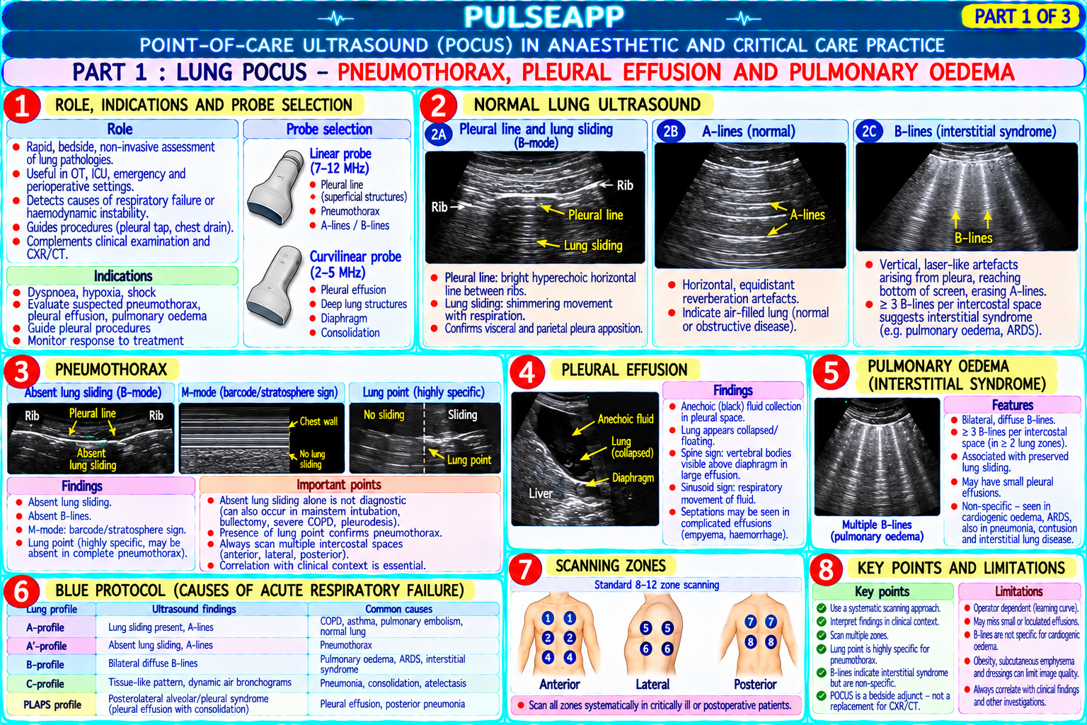
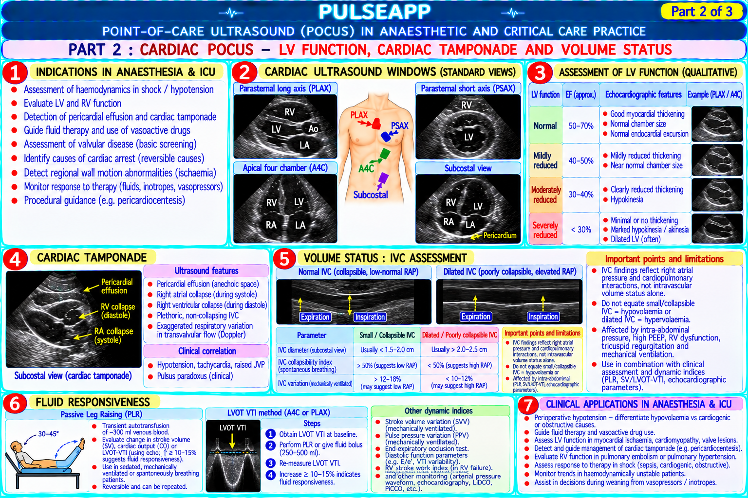
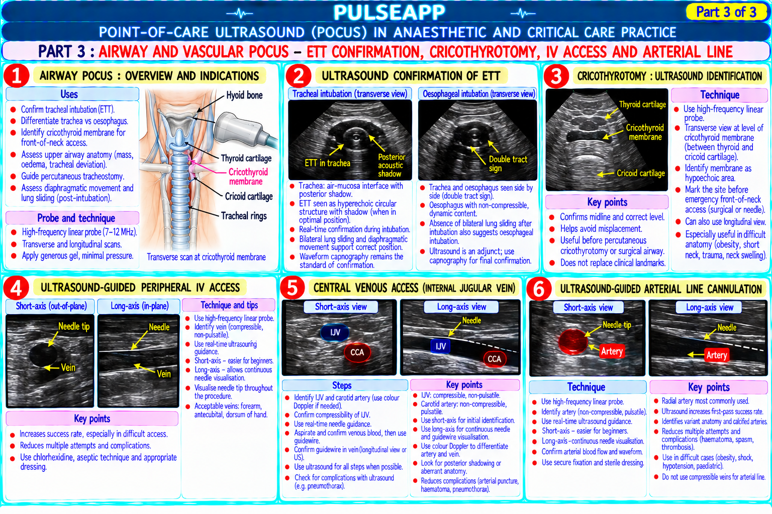
Describe the use of point-of-care ultrasound (POCUS) in anaesthetic and critical care practice. Outline: lung POCUS (pneumothorax, pleural effusion, pulmonary oedema); cardiac POCUS (LV function, cardiac tamponade, volume status); airway POCUS (ETT confirmation, cricothyrotomy); and vascular POCUS (IV access, arterial line).

description Clinical Response
⚙ Core Concept
POCUS is transforming anaesthesia and critical care — the bedside ultrasound provides real-time physiological information that was previously only available from invasive monitoring or delayed radiology. Lung POCUS can diagnose pneumothorax faster and more accurately than a chest X-ray; cardiac POCUS guides immediate haemodynamic decisions; and airway POCUS confirms ETT position within seconds. (Volpicelli G — International Liaison Committee 2012; Mayo PH; Lichtenstein DA; Perera P — 5Es cardiac POCUS; Miller's Anaesthesia 9th Ed)
A. Lung POCUS3 marks

Finding Ultrasound Sign Interpretation Normal lung Lung sliding (shimmering movement of pleural line with each breath Normal aerated lung; lung sliding confirms visceral pleura is moving → no — the visceral and parietal pleura moving against each other); A- pneumothorax at this site lines (horizontal reverberation artefacts from the pleural line — equidistant, repeating) Pneumothorax ABSENT lung sliding (the pleural line is static — visceral pleura is Absent lung sliding + absent B-lines + barcode sign on M-mode = separated from the parietal pleura by air); B-lines are absent; on M- pneumothorax; more sensitive than CXR for detecting pneumothorax; the mode: "Barcode sign" (static horizontal lines instead of the normal lung point (the anatomical border where the pneumothorax ends — sliding "seashore sign" in a healthy lung) reappears) confirms and localises the pneumothorax Pleural Anechoic (dark) fluid above the diaphragm in the dependent pleural Volume of effusion can be estimated by POCUS; guides thoracocentesis effusion space; the "curtain sign" — lung collapses away from the dependent needle placement under real-time visualisation pleural space; can be guided real-time drainage Pulmonary B-lines (formerly "lung rockets"): vertical, hyperechoic, laser-like Diffuse bilateral B-lines = cardiogenic pulmonary oedema OR ARDS; focal oedema (B- artefacts arising from the pleural line, extending to the far field, unilateral B-lines = pneumonia or contusion; the pattern (bilateral vs focal) and lines) moving with lung sliding, erasing A-lines; 3 or more B-lines in one clinical context distinguish cardiogenic from non-cardiogenic view = interstitial syndrome Consolidation Hypoechoic tissue-like appearance replacing the normal air-filled Pneumonia; pulmonary contusion; lobar collapse; the presence of dynamic air pattern; hepatisation of the lung; air bronchograms (hyperechoic bronchograms (moving with breathing) suggests patent bronchi and potentially spots moving with respiration within the consolidation) reversible atelectasis

B. Cardiac POCUS — "5 Es" of Emergency Cardiac Ultrasound3 marks

Question Finding Clinical Decision

Is there Pericardial effusion: anechoic fluid surrounding the heart; cardiac Effusion with RV diastolic collapse → tamponade → emergency Effusion? tamponade: diastolic collapse of the right ventricle and right atrium (the pericardiocentesis or pericardial window; PEA in the context of pericardial first sign of haemodynamically significant tamponade) effusion → tamponade until proven otherwise

Is cardiac LV function: visual estimation of LV ejection fraction (EF); hyperdynamic Severely reduced LV function in shock → cardiogenic shock → inotropes Ejection (EF >70%: walls touch in systole), normal (40–70%), or reduced (EF (dobutamine, milrinone); NOT give more fluid; hyperdynamic LV in adequate? <40%: poor wall motion) hypotensive patient → distributive shock → vasopressors Are the RV:LV size ratio; normally RV is smaller; RV dilation (RV:LV >0.6 in any Massive PE; acute cor pulmonale; RV MI; D-sign in a shocked hypoxic Equalities of view) + D-sign (interventricular septal flattening — septal bowing into patient → consider pulmonary embolism → consider thrombolysis RV and LV LV) suggests massive PE or RV failure appropriate? Are the Exits Aortic and pulmonary valve assessment; IVC size and collapsibility (a IVC <2 cm collapsing with inspiration → low CVP → likely hypovolaemia patent? surrogate for CVP and volume status) (volume responsive); IVC >2.5 cm non-collapsing → raised CVP → cardiogenic or obstructive cause of shock Is the motion Regional wall motion abnormalities (RWMA) — segments of the LV wall New RWMA in a patient with chest pain + ECG changes → STEMI → Expected? that are hypokinetic or akinetic = myocardial ischaemia or previous MI activate cath lab; RWMA in post-cardiac surgery patient → graft failure

C. Airway POCUS2 marks

ETT confirmation: probe placed transversely on the anterior neck at the level of the trachea; normal: trachea shows a single hyperechoic curve with posterior acoustic shadowing (air column in the trachea); ETT in trachea: the ETT appears as two hyperechoic curves (the anterior and posterior walls of the ETT) within the trachea — the "double lumen" sign; oesophageal intubation: the oesophagus (soft tissue structure posterior to the trachea) shows a "snowstorm" pattern with air entering it; real-time confirmation: as the ETT cuff is inflated, the hyperechoic cuff appears in the trachea; this is an immediate, real-time confirmation of correct ETT placement

Subglottic anatomy for cricothyrotomy: probe placed longitudinally on the anterior midline neck; identify: thyroid cartilage (superior hyperechoic curved structure); cricothyroid membrane (CTM — the relatively flat area between the thyroid and cricoid cartilages); cricoid cartilage (inferior hyperechoic curved structure); the CTM is identified as the hypoechoic (soft tissue) region between the two cartilage landmarks; ultrasound-guided identification of the CTM before difficult airway cases reduces the risk of misidentification (particularly in obese patients where the CTM is difficult to identify by external palpation)

D. Vascular POCUS2 marks

Central venous access: real-time ultrasound guidance for internal jugular (IJV) or subclavian vein cannulation; reduces: first-attempt failure (by 57%), arterial puncture (by 78%), haematoma, pneumothorax (for subclavian); the vein (anechoic, compressible, distends with Valsalva) is distinguished from the artery (pulsatile, non-compressible, round, thick-walled); the IJV typically lies anterolateral to the common carotid artery

Peripheral IV access: ultrasound-guided peripheral IV in patients with difficult access (obesity, prior IV drug use, multiple previous venepunctures); allows cannulation of deep veins (basilic, brachial) that are not visible or palpable

Arterial line: real-time ultrasound guidance for radial, femoral, or brachial artery cannulation; reduces first-attempt failure and haematoma; the artery is identified as a pulsatile, round, non-compressible structure

DVT diagnosis: compressibility test of the femoral and popliteal veins; normal vein compresses completely with probe pressure; DVT: vein fails to compress (blood clot within the lumen prevents collapse); quick bedside assessment for high-risk ICU/trauma patients

🎤 Viva Corner
Q. How does lung POCUS diagnose pneumothorax faster and more accurately than a chest X-ray?
Lung POCUS diagnoses pneumothorax by detecting the absence of the normal ultrasound signature of aerated lung at the pleural surface. The most sensitive and specific sign is absent lung sliding: in a normal lung, the visceral pleura (on the lung surface) moves against the parietal pleura (on the chest wall) with each breath, producing a shimmering, sparkling movement visible on ultrasound at the bright pleural line; this "lung sliding" confirms that the visceral pleura is in contact with the parietal pleura — which is only possible if no air has separated them. In pneumothorax, free air in the pleural space interposes between the two pleural surfaces, preventing them from touching; the visceral pleura no longer moves against the parietal pleura; the pleural line appears completely static — no sliding; this ABSENT LUNG SLIDING is the primary sign of pneumothorax on ultrasound. On M-mode, the normal seashore sign (granular pattern below the pleural line from moving lung parenchyma) is replaced by the barcode sign (parallel horizontal lines) because nothing is moving below the pleural line. The advantages over CXR: speed — the POCUS examination takes 30–60 seconds vs 10–20 minutes for a CXR (requisition, portable machine, positioning, exposure, development, interpretation); sensitivity — lung POCUS has sensitivity of 88–98% for pneumothorax vs only 40–50% for supine portable CXR (the standard in ICU, emergency, and trauma settings where upright CXR is not possible); the supine CXR misses anterior pneumothoraces because air collects anteriorly in the supine patient and does not produce the classic deep sulcus sign reliably; POCUS examines the most anterior portions of the chest (where a supine pneumothorax accumulates first) with the probe applied to the anterior chest wall — exactly where the air is. The lung point (the specific anatomical location where absent sliding transitions to present sliding) can precisely localise the size and extent of the pneumothorax.
★ Examiner's Pearl
Lung sliding = normal aerated lung (visceral + parietal pleura touching); ABSENT lung sliding = pneumothorax (air separating the two pleural surfaces). B-lines (vertical hyperechoic lines = interstitial fluid/oedema) vs A-lines (horizontal reverberation artefacts = normal air-filled lung) — this distinction between the two major artefact patterns is the most tested POCUS finding. The 5 Es of cardiac POCUS (Effusion, Ejection, Equalities, Exits, Expected motion) provides a memorable framework covering the most clinically important cardiac diagnoses.
Volpicelli G et al. International evidence-based recommendations for lung POCUS (Intensive Care Med 2012;38:577-591). Lichtenstein DA et al. A-lines and B-lines (Chest 2008;133:1659-1665). Perera P et al. The RUSH examination (Emerg Med Clin North Am 2010;28:29-56). Mayo PH. Critical care ultrasound (Intensive Care Med 2009). Miller RD et al. Miller's Anaesthesia, 9th Ed.
lock Protected Clinical Document • Pulse App • Screenshots Strictly Prohibited
QUESTION 114 person Asked by .
bookmark_add

Describe the DAS 2015 Difficult Airway Guidelines including the four-plan intubation algorithm (Plans A, B, C, D). Outline the Cannot Intubate, Cannot Oxygenate (CICO) emergency management. Describe the criteria and technique for extubation of the at-risk airway.

description Clinical Response
⚙ Core Concept
The DAS (Difficult Airway Society) 2015 guidelines provide the UK national framework for managing the difficult airway — from predicted difficult intubation (AFOI planning) to the unanticipated difficult airway in a paralysed, unconscious patient (the most dangerous scenario), and the CICO emergency (Cannot Intubate Cannot Oxygenate — a potentially fatal emergency requiring immediate front-of-neck access). (Frerk C et al. — DAS 2015 Anaesthesia; Henderson JJ; Difficult Airway Society; Miller's Anaesthesia 9th Ed)
A. The DAS 2015 Four-Plan Algorithm — Unanticipated Difficult Intubation4 marks

Plan Goal Technique Maximum Attempts

Plan A — Safe tracheal Position optimised (ramped, HELP, head-up); direct laryngoscopy OR video laryngoscopy as first Maximum 3 intubation Primary intubation on line (DAS 2015 does not mandate video laryngoscopy for all but recommends considering it from the attempts; if all 3 fail → Intubation the first attempt start in any anticipated difficulty); bougie as standard adjuvant (not as a rescue — should be the first- DECLARE FAILED choice adjuvant with any suboptimal view); maximum 3 attempts at intubation in Plan A, each with INTUBATION and move to optimisation (position, blade, assistant pressure) Plan B

Plan B — Maintain INSERT a supraglottic airway device (SAD — i-gel or LMA ProSeal as second-generation devices Maximum 2 SAD insertion Oxygenation oxygenation preferred); if SAD achieves adequate ventilation → continue surgery if immediately life-saving; if attempts; if both fail → move via SAD after failed elective → WAKE THE PATIENT UP (allow full recovery, plan definitive airway management before to Plan D (CICO) tracheal rescheduling); if SAD fails to ventilate → CICO → Plan D intubation; buy time

Plan C — Attempt face- Resume face-mask ventilation (two-person, two-handed technique); CALL FOR HELP (senior As many attempts as needed Final mask ventilation anaesthesiologist, ENT surgeon, ODP, nursing support); if face-mask ventilation maintains SpO₂ to buy time; the goal is to call Attempt at while preparing >90% → time to prepare for Plan D or wake the patient up; if SpO₂ still falling → CICO emergency for help and prepare for Oxygenation for CICO if SAD CICO also fails; ensure the team is assembled

Plan D — Emergency Scalpel cricothyrotomy: palpate the cricothyroid membrane (CTM); single horizontal stab incision No limit — do it as fast as CICO oxygenation via with a No.10 scalpel through the CTM; rotate the blade 90° (to widen the incision); insert a bougie possible; every second of Emergency a surgical (caudally into the trachea); railroad a size 6.0 cuffed ETT or specific cricothyrotomy tube over the CICO = hypoxic brain injury; — Front of airway when bougie; confirm ETCO₂ waveform; inflate cuff; ventilate; this is the DAS recommended technique — scalpel-bougie Neck ALL other the "scalpel-bougie" method (has replaced needle cricothyrotomy as the preferred CICO technique cricothyrotomy can be Access methods have for adults in UK practice based on superior reliability) completed in 30–60 seconds (FONA) failed by a trained practitioner

B. Cannot Intubate Cannot Oxygenate (CICO) — Definition and Immediate Response2 marks

CICO is defined as: failure to intubate the trachea despite optimal positioning and multiple attempts (Plans A and B have failed) AND failure to maintain adequate oxygenation by face mask or SAD; SpO₂ is falling despite all efforts; this is an IMMEDIATE LIFE-THREATENING emergency

The DAS 2015 CICO declaration: any patient in whom SpO₂ cannot be maintained >90% using ALL available non-surgical airway means is in CICO; the 90% threshold is chosen because brain injury risk becomes significant and quickly progressive below this level; do NOT delay FONA waiting for absolute zero — act at 90% and falling The ONLY correct response to true CICO is immediate FRONT OF NECK ACCESS (FONA) using the scalpel-bougie technique; no other intervention is appropriate once CICO is confirmed

C. Extubation of the At-Risk Airway2 marks

The difficult airway does not end at intubation — extubation of a patient who was difficult to intubate carries SIGNIFICANT RISK (loss of the established airway in a patient who may be more difficult to re-intubate after surgical trauma, oedema, bleeding)

DAS Extubation Guidelines (2012): every patient who was difficult to intubate should have an explicit extubation plan that includes: can the patient be safely re-intubated if needed post-extubation? (assess risk); what is the procedure for safe extubation? Extubation technique for at-risk airway:

Awake extubation: patient is fully awake, following commands, with intact reflexes, confirmed T4 on TOF, and normothermia BEFORE the ETT is removed; this is the safest option for the at-risk airway

Airway exchange catheter (AEC): a long hollow catheter (e.g., Cook AEC) is inserted through the ETT into the trachea before the ETT is removed; the ETT is removed over the AEC; the AEC remains in the trachea as a guide for immediate re-intubation if needed; can be used for jet ventilation as a bridge if reintubation is necessary; the AEC should remain in place for 30–60 minutes until the patient is confirmed stable and airway oedema risk has passed

Cuff leak test before extubation: deflate the ETT cuff and check if the patient can breathe around it (leak should be audible); no leak suggests airway oedema → consider IV dexamethasone 8 mg → delay extubation 24 hours → repeat cuff leak test before extubating

D. Awake Fiberoptic Intubation (AFOI) — The Technique for Predicted Difficult Airway2 marks

Indications: predicted difficult airway by assessment (Mallampati IV, limited mouth opening, limited neck extension, significant facial/cervical pathology); fixed anatomical obstruction; unstable cervical spine; patient preference after discussion Technique:

Sedation: dexmedetomidine (ideal — cooperative, rousable, no respiratory depression) OR midazolam + low-dose remifentanil TCI (careful — respiratory depression risk)

Topical airway anaesthesia: nebulised lidocaine 4% (4 mL × 4 min before procedure) + transtracheal injection of lidocaine 4% 2 mL through CTM (cough response then suppresses tracheal reflexes) + spray-as-you-go technique (lidocaine 2% sprayed via the working channel of the fibrescope as you advance)

Approach: nasal (better tolerated, steadier platform, passes naturally behind the soft palate → vocal cords) or oral (with Berman or Ovassapian airway guide)

Confirm ETT placement: ETCO₂ waveform; visualise carina through the fibrescope before removing it to confirm tracheal (not oesophageal) position

🎤 Viva Corner
Q. An RSI has been performed for emergency laparotomy. Three intubation attempts have all failed. An i-gel has been placed and is maintaining SpO₂ 96%. The surgeon says the surgery is immediately life-saving. Do you proceed with surgery through the i-gel?
This is a critical decision that hinges on the balance between the surgical urgency and the airway risk. The i-gel is maintaining SpO₂ at 96% — this means we are NOT in a CICO situation; the patient IS being adequately oxygenated; this is a "Cannot Intubate, CAN Oxygenate" situation. The DAS 2015 guidance for this specific situation: if the surgery is immediately life-saving (emergency laparotomy for bowel perforation, bleeding, ischaemia — procedures where surgical delay = death) AND oxygenation is maintained via the SAD, it may be acceptable to proceed with surgery through the second-generation SAD with very careful management. The igel as a second-generation device has an oropharyngeal leak pressure of 24–30 cmH₂O — sufficient for most controlled ventilation during laparotomy in a nonobese patient if peak airway pressures are kept below this. Before proceeding: test the seal pressure (gently squeeze the bag and note the pressure at which gas escapes around the cuff — if >25 cmH₂O, there is a reasonable safety margin); confirm we can ventilate adequately; if the patient is morbidly obese or expected to have high airway pressures (bowel distension, pneumoperitoneum) → the i-gel may not provide adequate ventilation and the seal may break → the airway would be lost mid-surgery in an uncontrolled manner. If the team has experience with awake fiberoptic intubation and the patient's SpO₂ is maintained: I would very strongly consider using this opportunity (patient oxygenated, some time available) to perform an awake FOI through the i-gel (Aintree intubating catheter technique) or to perform awake fibreoptic intubation with the patient partially recovered from NMB (give sugammadex if rocuronium was used) — this converts the failed intubation into a controlled, secured airway before the surgical stress of laparotomy. If the surgeon confirms the surgical urgency is absolute and FOI cannot be attempted in time: proceed with surgery through the i-gel with: surgeon ready for immediate surgical cricothyrotomy or emergency tracheostomy if i-gel seal fails; minimum required pneumoperitoneum pressure; avoid steep Trendelenburg; continuous airway pressure monitoring; awake extubation plan at the end of surgery (airway exchange catheter placement before removing the SAD).
★ Examiner's Pearl
The four-plan DAS algorithm (A: intubation 3 attempts max; B: SAD 2 attempts max; C: call for help + face mask; D: FONA/scalpel cricothyrotomy) must be reproduced in order with the decision points (declare failed intubation after 3 attempts on Plan A; declare CICO at SpO₂ <90% falling despite Plans A+B+C). Scalpel-bougie cricothyrotomy (scalpel stab → rotate → bougie → size 6.0 ETT) has replaced needle cricothyrotomy as the preferred CICO technique in adults in DAS 2015. AEC for extubation of the at-risk airway (placed through the ETT before removal; allows re-intubation over the AEC if needed; jet ventilation capability) is the specific extubation technique.
Frerk C et al. DAS difficult airway management guidelines 2015 (Anaesthesia 2015;70:1105-1117). Henderson JJ et al. DAS extubation guidelines 2012 (Anaesthesia 2012;67:318-340). DAS unanticipated difficult airway (updated algorithm 2015). Miller RD et al. Miller's Anaesthesia, 9th Ed.
lock Protected Clinical Document • Pulse App • Screenshots Strictly Prohibited
QUESTION 115 person Asked by .
bookmark_add

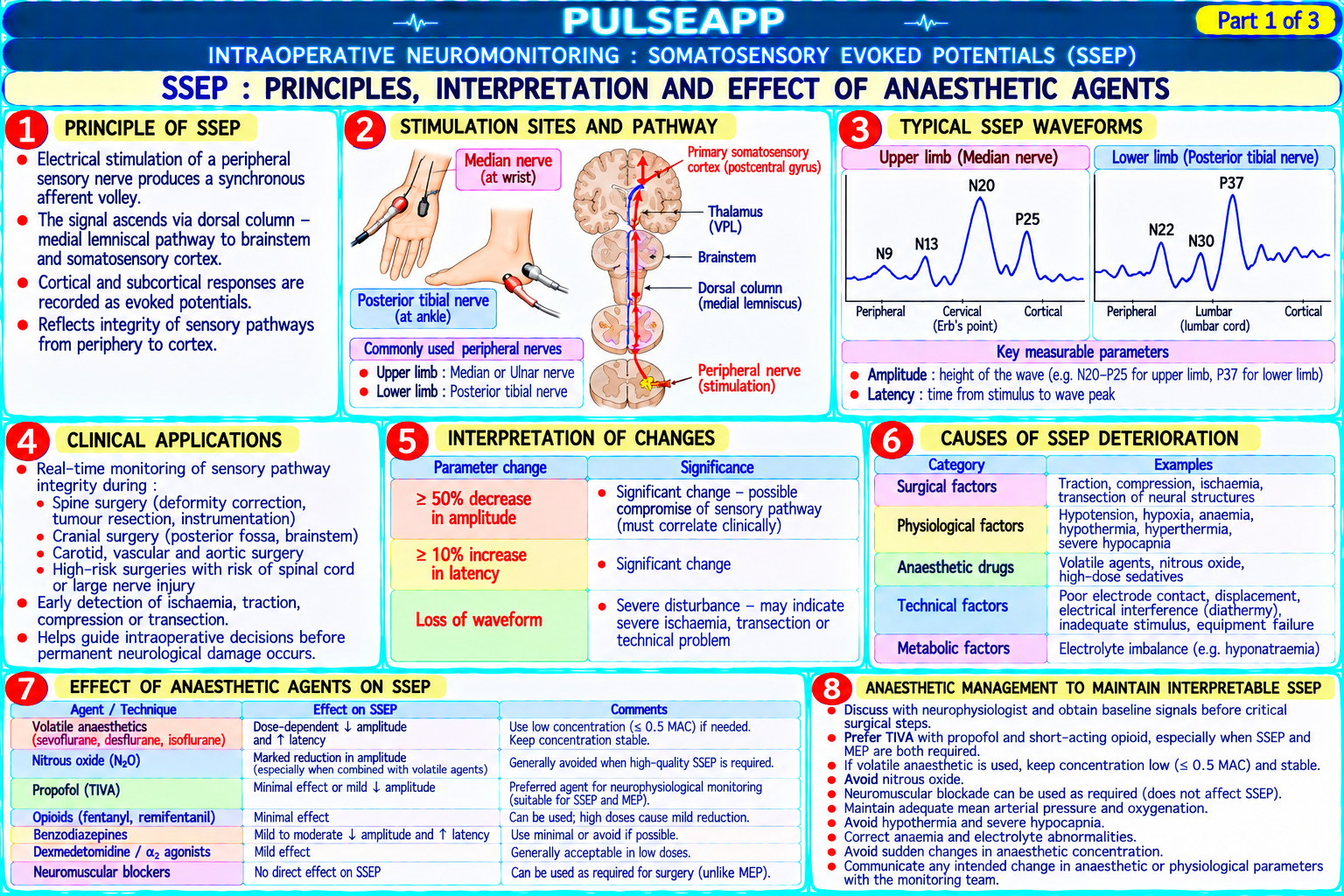
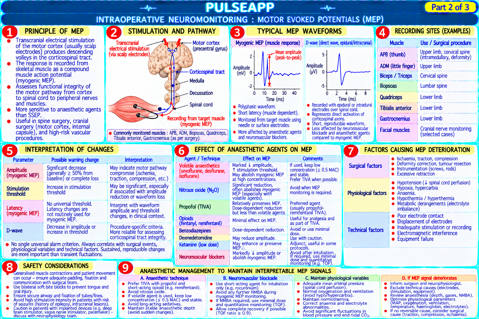
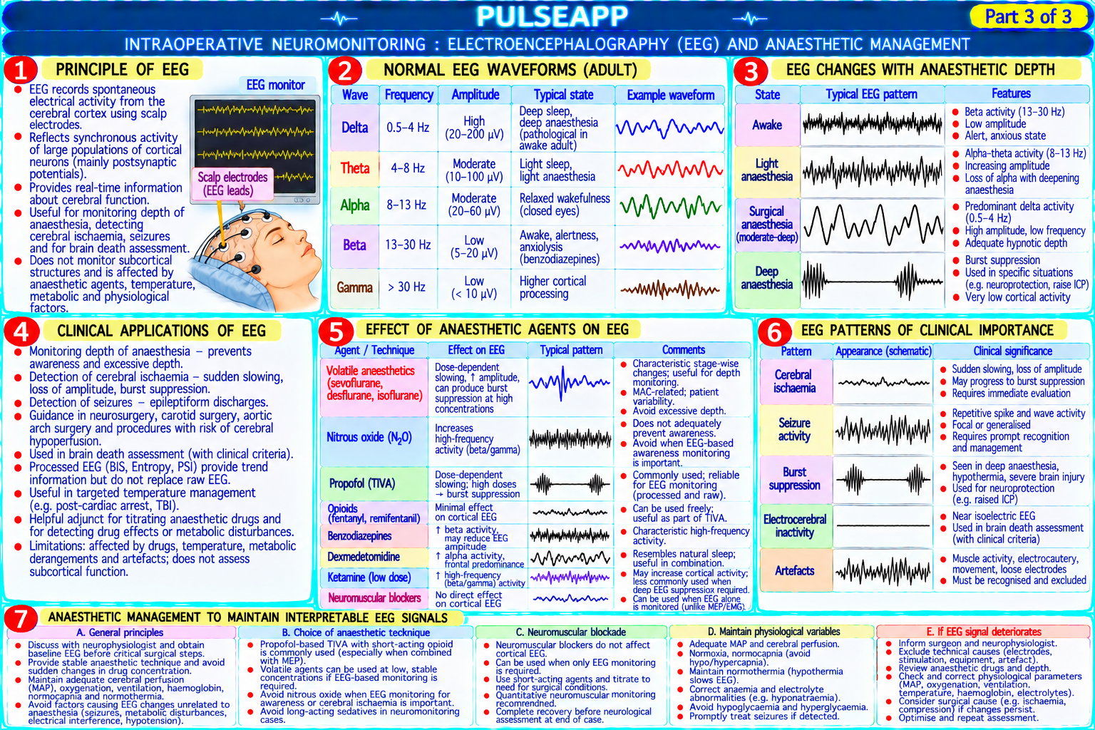
Describe the principles of somatosensory evoked potentials (SSEP), motor evoked potentials (MEP), and electroencephalography (EEG) used in intraoperative neuromonitoring. Outline how each modality is affected by anaesthetic agents and describe the anaesthetic management required to maintain interpretable signals.

collections Question Diagrams & Reference Images (1)
Question Reference Diagram
zoom_in View Image
description Clinical Response
⚙ Core Concept
Intraoperative neuromonitoring (IONM) provides continuous real-time assessment of spinal cord and cerebral function during high-risk surgical procedures — detecting neurological injury before it becomes permanent. Anaesthetic agents profoundly suppress evoked potential amplitudes and alter signal morphology, requiring specific TIVA-based techniques to maintain interpretable recordings. (Sloan TB; Jameson LC; Lotto ML; Banoub M — Anesthesiology 2004; Miller's Anaesthesia 9th Ed)
A. SSEP — Somatosensory Evoked Potentials3 marks

Principle: a peripheral nerve (median nerve at wrist for upper limb; posterior tibial nerve at ankle for lower limb) is stimulated with small electrical pulses; the signal travels via the dorsal column (posterior spinal cord) → brainstem → thalamus → somatosensory cortex; scalp electrodes record the cortical response; the response is averaged over hundreds of stimuli to extract from background EEG noise

What it monitors: the integrity of the POSTERIOR spinal cord (dorsal column — the sensory pathway); does NOT monitor the ANTERIOR spinal cord (corticospinal tracts — the motor pathway); therefore a pure anterior cord injury (from anterior spinal artery occlusion) may be missed by SSEP alone but detected by MEP

Signal parameters: latency (time from stimulus to cortical response; normally 15–25 ms for upper limb SSEP); amplitude (height of the waveform peak); alert criteria: ≥50% amplitude reduction OR ≥10% latency increase from baseline = significant change requiring surgical intervention

Surgical applications: scoliosis surgery (most common — monitoring for distraction-related cord injury); aortic surgery (monitoring during aortic cross-clamp); posterior fossa surgery; carotid endarterectomy (cerebral perfusion monitoring)

B. MEP — Motor Evoked Potentials3 marks

Principle: transcranial electrical stimulation (TES) or magnetic stimulation (TMS) is applied to the motor cortex (electrodes placed on the scalp at C3/C4 positions); the electrical impulse travels via the corticospinal tract (ANTERIOR spinal cord) → anterior horn cells → peripheral motor nerve → muscle; the response is recorded as a compound muscle action potential (CMAP) from muscles (tibialis anterior, abductor hallucis for lower limbs; thenar muscles for upper limbs)

What it monitors: the integrity of the ANTERIOR spinal cord (corticospinal tract — the motor pathway); complementary to SSEP — monitors the pathway that SSEP misses; together SSEP + MEP provide comprehensive spinal cord monitoring covering both sensory and motor pathways

Alert criteria: ≥80% CMAP amplitude reduction from baseline; loss of all MEP signals; new asymmetry between left and right limb responses = significant alert requiring surgical pause and intervention

Critical anaesthetic consideration: MEPs are ABOLISHED by neuromuscular blocking agents (NMBs); the CMAP is recorded from muscles and requires intact neuromuscular junction; even partial NMB (TOF ratio 0.5) significantly suppresses MEP amplitude; during MEP monitoring: do NOT use NDMR boluses; if NMB is essential for intubation, use a short-acting agent and allow complete recovery before MEP baseline is established; NMB infusions are completely incompatible with MEP monitoring

Safety consideration: TES can cause patient movement at the time of stimulation (the transcranial stimulus causes brief generalised muscle contraction); the surgeon must be warned before each stimulus train; particularly important during procedures near the spinal cord (sudden movement during posterior instrumentation could cause injury) — TES is temporarily paused during the most critical surgical steps

C. EEG Monitoring — Depth of Anaesthesia and Cerebral Ischaemia2 marks

Processed EEG (BIS, Entropy, SEDLine): the raw EEG is processed by mathematical algorithms (bispectral analysis, spectral entropy) to produce a dimensionless number (BIS 0–100; 40–60 = appropriate depth for GA; >60 = at risk of awareness; <40 = overly deep); BIS monitoring is the standard processed EEG tool for TIVA awareness prevention

Raw EEG for cerebral ischaemia: during carotid endarterectomy (CEA), 8-lead or 16-lead raw EEG is recorded continuously; the EEG changes predictably with ischaemia: first, high-frequency activity decreases; then delta waves emerge; then progressive slowing; then burst suppression; finally, isoelectric EEG; these changes occur within 1–3 minutes of ischaemia onset and precede irreversible injury → if detected, the surgeon can immediately insert a carotid shunt to restore perfusion; EEG sensitivity for detecting significant CEA-related ischaemia: approximately 90%

D. Anaesthetic Management for IONM2 marks

Agent Effect on SSEP/MEP/EEG IONM Compatibility Volatile agents Dose-dependent suppression of SSEP amplitude (20–50% at 1 MAC); MEP Can be used at low doses (<0.5 MAC) alongside TIVA; above (sevoflurane, amplitude suppressed 50–80% at 1 MAC; high-frequency EEG suppressed 0.5 MAC → signals may become uninterpretable; AVOID for isoflurane, MEP-intensive monitoring desflurane) Nitrous oxide Significant MEP amplitude suppression (additional 50% reduction when added to AVOID in IONM cases; use air-O₂ mixture instead (N₂O) propofol); SSEP amplitude also reduced Propofol (IV Dose-dependent EEG suppression (dose-dependent BIS reduction); SSEP Well tolerated for IONM; TIVA with propofol is the preferred infusion) amplitude modestly reduced but maintained at clinical doses; MEP amplitude technique reduced but maintained at doses used for maintenance TIVA Remifentanil (IV Minimal direct effect on SSEP or MEP at clinical doses; provides analgesia Excellent — ideal TIVA combination: propofol 2–4 mcg/mL + infusion) without significant IONM signal interference remifentanil 2–6 ng/mL Ce provides adequate anaesthesia with minimal IONM signal suppression NMBs (non- ABOLISH MEP (muscle-recorded CMAPs cannot be generated through a CONTRAINDICATED during MEP monitoring; only acceptable depolarising) blocked NMJ); no effect on SSEP (recorded from scalp, not from muscles) for SSEP-only monitoring Ketamine INCREASES cortical excitability → ENHANCES SSEP and MEP amplitude; Beneficial adjuvant for IONM; sub-anaesthetic ketamine (0.5 antagonises the amplitude-suppressing effects of other agents mg/kg bolus + 0.25 mg/kg/hr infusion) as part of a balanced TIVA technique improves IONM signal quality

🎤 Viva Corner
Q. During scoliosis surgery, the neurophysiologist reports that MEP amplitudes have decreased by 80% bilaterally. What are the immediate steps?
An 80% bilateral MEP amplitude reduction during scoliosis surgery is a CRITICAL alert — this meets the standard threshold (≥80% reduction or complete loss) for potential motor pathway compromise and requires immediate systematic response. Step 1: immediately inform the surgeon — the surgical team must stop all spinal instrumentation and distraction immediately; the first action is to pause the mechanical cause. Step 2: confirm the signal change is real — check: are the stimulating electrodes still correctly positioned? Have any leads been dislodged? Has the anaesthetic changed (has the volatile agent concentration drifted up, or was a bolus of propofol or NMB given in the last few minutes — these can suppress MEPs without any surgical cause)? Confirm with the neurophysiologist that baseline recordings are stable and this is a genuine new change. Step 3: correct reversible causes — (a) MEAN ARTERIAL PRESSURE: ensure MAP ≥80 mmHg (spinal cord perfusion pressure; if MAP has drifted down from blood loss → IV fluid bolus + vasopressor noradrenaline → raise MAP immediately — this is one of the most common reversible causes); (b) HAEMOGLOBIN: if significant blood loss has occurred → transfuse to maintain Hb ≥80 g/L to optimise O₂ delivery to the spinal cord; (c) TEMPERATURE: confirm normothermia (hypothermia suppresses evoked potentials); (d) ANAESTHETIC DEPTH: confirm no recent propofol bolus, volatile agent drift, or N₂O added; (e) ANAEMIA or hypoxia: check SpO₂ and recent ABG. Step 4: if signals do not recover after 5–10 minutes of optimisation → the surgeon performs the wake-up test (Stagnara test): reduce anaesthetic to allow partial waking; ask the patient to move their feet; if the patient can move feet bilaterally → spinal cord is functionally intact; if not → immediate partial implant removal or rod release; the wake-up test is the gold standard clinical confirmation of spinal cord function when IONM signals are ambiguous or lost. Step 5: once signals recover (whether spontaneously or after intervention) → resume surgery at a lower distraction level; continue intensive IONM monitoring to the end of the case.
★ Examiner's Pearl
SSEP monitors POSTERIOR (dorsal column — sensory) pathway; MEP monitors ANTERIOR (corticospinal — motor) pathway — they are complementary and together provide comprehensive spinal cord monitoring. NMBs ABOLISH MEP (muscle-recorded) but do NOT affect SSEP (scalp-recorded) — this distinction is the most commonly tested pharmacological fact in IONM. TIVA with propofol + remifentanil is the preferred anaesthetic for IONM; volatile agents and N₂O suppress signals dose-dependently and should be avoided or minimised. Alert criteria: SSEP ≥50% amplitude drop or ≥10% latency increase; MEP ≥80% amplitude reduction.
Banoub M et al. Pharmacologic and physiologic influences affecting sensory evoked potentials (Anesthesiology 2003;99:716-737). Sloan TB. Anesthetic effects on electrophysiologic recordings (J Clin Neurophysiol 1998;15:217-226). Lotto ML et al. Effects of anaesthetic agents on motor evoked potentials (Anaesthesia 2004;59:1216-1225). Miller RD et al. Miller's Anaesthesia, 9th Ed.
lock Protected Clinical Document • Pulse App • Screenshots Strictly Prohibited
QUESTION 116 person Asked by .
bookmark_add

Describe the perioperative management of phaeochromocytoma resection. Explain the rationale for alpha-blockade before beta-blockade. Outline intraoperative management of hypertensive crises, strategies during tumour manipulation, and post-resection hypotension management.

collections Question Diagrams & Reference Images (1)
Question Reference Diagram
zoom_in View Image
description Clinical Response
⚙ Core Concept
Phaeochromocytoma is a catecholamine-secreting tumour of the adrenal medulla (or extra-adrenal paraganglioma). Surgical resection carries exceptional anaesthetic risk from massive catecholamine surges during tumour manipulation — causing hypertensive crises, arrhythmias, and cardiovascular collapse. Systematic pre-operative alpha-blockade (then beta-blockade), meticulous intraoperative BP control, and preparation for post-ligation hypotension are the cornerstones of safe perioperative management. (Kinney MA; Prys-Roberts C; Lenders JW — J Hypertens 2014; Endocrine Society Guidelines; Miller's Anaesthesia 9th Ed)
A. Pathophysiology1 mark

Phaeochromocytoma secretes adrenaline and/or noradrenaline (and dopamine in some extra-adrenal tumours) — episodically or continuously; during tumour manipulation at surgery, massive catecholamine surges produce: extreme hypertension (SBP up to 300+ mmHg); tachycardia and arrhythmias; diaphoresis; headache; pallor; hyperglycaemia; after tumour vein ligation: sudden catecholamine withdrawal → profound vasodilation → hypotension

B. Pre-operative Preparation — Alpha-Blockade First Principle3 marks

Alpha-blockade MUST precede beta-blockade: if beta-blockers are given before alpha-blockers → the vasodilatory β₂ effects are blocked leaving only α₁ vasoconstriction unopposed → PARADOXICAL SEVERE HYPERTENSION; alpha-blockers first allow β₂-mediated vasodilation to counterbalance the α₁ vasoconstriction of the catecholamines, making blood pressure more manageable before beta-blockade is added Phenoxybenzamine (irreversible non-selective α-blocker): historically the gold standard; oral 10–20 mg BD, titrated up over 1–3 weeks; produces noncompetitive (irreversible) alpha-blockade — unresponsive to high catecholamine surges during surgery (the block persists regardless of catecholamine level); well-established safety record; side effects: orthostatic hypotension, nasal stuffiness (nasal α₁ vasoconstriction blocked), reflex tachycardia (α-blockade → vasodilation → reflex HR increase → this is why beta-blocker is then added); disadvantage: long duration of action causes prolonged hypotension postoperatively Doxazosin (selective α₁-blocker; competitive): increasingly preferred in many centres; shorter half-life → post-operative hypotension less prolonged; oral titration starting at 2 mg OD; competitive blockade — may be overcome by very high catecholamine surges (but manageable intraoperatively with vasoactive drugs)

Beta-blockade: added after adequate alpha-blockade (at least 1–2 weeks) ONLY if reflex tachycardia or arrhythmias develop; propranolol or atenolol; DO

NOT start beta-blockade until alpha-blockade is established

Adequacy of pre-operative blockade criteria: BP controlled <130/80 for 24 hours; orthostatic hypotension (confirms adequate vasodilation from alphablockade); nasal congestion; no ECG changes of ischaemia; HR 60–80 bpm (if on beta-blocker) High-salt diet + IV fluid loading: pre-operatively; chronic catecholamine excess causes contracted plasma volume; normalising volume with high-Na diet and adequate hydration reduces the magnitude of post-tumour-ligation hypotension

C. Intraoperative Management3 marks

Surgical Haemodynamic Response Management Phase Induction Hypertensive surge from laryngoscopic stimulus + Fentanyl 3–5 mcg/kg before laryngoscopy; lignocaine 1.5 mg/kg IV 90 sec before; avoid and surgical stress succinylcholine if possible (may stimulate catecholamine release via ganglionic stimulation); laryngoscopy ketamine AVOIDED (stimulates catecholamine release); propofol induction (least haemodynamically stimulating) Tumour MASSIVE catecholamine surge → SBP may reach Pre-prepared syringes: phentolamine (non-selective α-blocker) 1–5 mg IV bolus for acute manipulation 250–300 mmHg; tachycardia; arrhythmias hypertension; sodium nitroprusside (SNP) 0.25–8 mcg/kg/min infusion for rapid BP titration; labetalol for combined α/β effect; nicardipine 1–10 mg/hr; magnesium sulphate 2–4 g IV (inhibits catecholamine release from adrenal medulla — may reduce surges) Tumour vein SUDDEN catecholamine withdrawal → profound Alert surgeon 2–3 minutes before ligation; pre-load with IV crystalloid 500 mL; immediately ligation and vasodilation → precipitous hypotension (SBP may fall to start noradrenaline infusion (0.1–1.0 mcg/kg/min) — typically required for 12–24 hours postremoval 50 mmHg in seconds); this is the most dangerous operatively; dopamine or vasopressin as adjuncts; discontinue ALL antihypertensive infusions haemodynamic event the moment the tumour vein is ligated Post- Continued vasodilatory hypotension from ICU monitoring 24–48 hours; vasopressor infusion as required; blood glucose monitoring (risk operative catecholamine withdrawal; hypoglycaemia (insulin of hypoglycaemia post-resection); hydrocortisone 50 mg QDS if bilateral adrenalectomy period secretion resumes when catecholamine stimulation of performed (adrenal insufficiency) α₂ islet cell receptors is removed); continued alphablocker effect

D. Anaesthetic Considerations3 marks

Monitoring: arterial line (mandatory — beat-to-beat BP); central venous catheter (vasoactive drug delivery + CVP monitoring); TOE (where available — useful for assessing LV function and preload in real-time); temperature monitoring

AVOID in anaesthesia for phaeochromocytoma: succinylcholine (ganglionic stimulation may trigger catecholamine release); ketamine (direct catecholaminereleasing effect); morphine (histamine release may destabilise BP); atracurium high doses (histamine release); metoclopramide (BLOCKS central dopamine receptors → may worsen BP in dopamine-secreting tumours); halothane (sensitises myocardium to catecholamine-induced arrhythmias); pancuronium (catecholamine-releasing sympathomimetic properties)

PREFERRED agents: propofol (induction + maintenance or TIVA); fentanyl/remifentanil (opioid component); isoflurane or sevoflurane at low MAC if volatile desired; rocuronium (NMB without histamine release or catecholamine stimulation); vecuronium (similarly safe)

🎤 Viva Corner
Q. During laparoscopic phaeochromocytoma resection, BP surges to 280/140 mmHg when the surgeon manipulates the tumour. What do you do immediately?
This is an anticipated hypertensive crisis during phaeochromocytoma resection — expected with tumour manipulation — and the drugs should be pre-drawn and ready before surgery began. Immediate steps: alert the surgeon "Stop manipulating the tumour" — most BP surges during phaeochromocytoma surgery are reduced significantly when the surgeon stops manipulating the adrenal gland; this alone may bring the BP down within 30–60 seconds. Simultaneously: phentolamine 2–3 mg IV bolus (non-selective α-blocker; onset 1–2 minutes; duration 10–15 minutes; ideal for rapid acute management of catecholamine-induced hypertension; repeat in 5 minutes if BP remains elevated); OR sodium nitroprusside (SNP) infusion — if already running, increase the rate; if not running, start at 0.5–1 mcg/kg/min and titrate upward rapidly (SNP has a very short onset and is highly titratable for acute intraoperative BP control, but requires careful monitoring for cyanide toxicity with prolonged high-dose use); OR nicardipine 5 mg IV bolus → infusion (calcium channel blocker; smooth, sustained reduction in BP; increasingly preferred over SNP). For arrhythmias: if tachycardia is the primary concern with the hypertension → esmolol 0.5 mg/kg IV then 50–300 mcg/kg/min infusion (only after confirming adequate alpha-blockade is in place — never esmolol without alpha-blockade in phaeochromocytoma); lignocaine for ventricular arrhythmias; amiodarone for sustained VT. Ensure adequate anaesthetic depth (arousal from light anaesthesia can worsen the catecholamine response — consider increasing propofol or volatile agent if depth is uncertain). Check the infusion lines and vasodilator drugs are ready for the moment the tumour vein is ligated (hypotension will follow immediately after ligation).
★ Examiner's Pearl
Alpha-blockade before beta-blockade — the consequence of reversing this order (paradoxical hypertension from unopposed α₁ vasoconstriction when β₂ is blocked) is the most tested pharmacological safety fact. Three-phase haemodynamic pattern: manipulation → hypertensive surge → vein ligation → precipitous hypotension → ICU vasopressor support. Drugs to AVOID: ketamine (catecholamine release), succinylcholine (ganglionic stimulation), morphine (histamine), halothane (sensitises to arrhythmias), metoclopramide (dopamine-secreting tumours).
Lenders JW et al. Phaeochromocytoma and paraganglioma — endocrine society clinical practice guideline (J Clin Endocrinol Metab 2014;99:1915-1942). Kinney MA et al. Perioperative management of phaeochromocytoma (J Cardiothorac Vasc Anesth 2002;16:359-369). Prys-Roberts C. Phaeochromocytoma — recent progress in its management (BJA 2000;85:44-57). Miller RD et al. Miller's Anaesthesia, 9th Ed.
lock Protected Clinical Document • Pulse App • Screenshots Strictly Prohibited
QUESTION 117 person Asked by .
bookmark_add

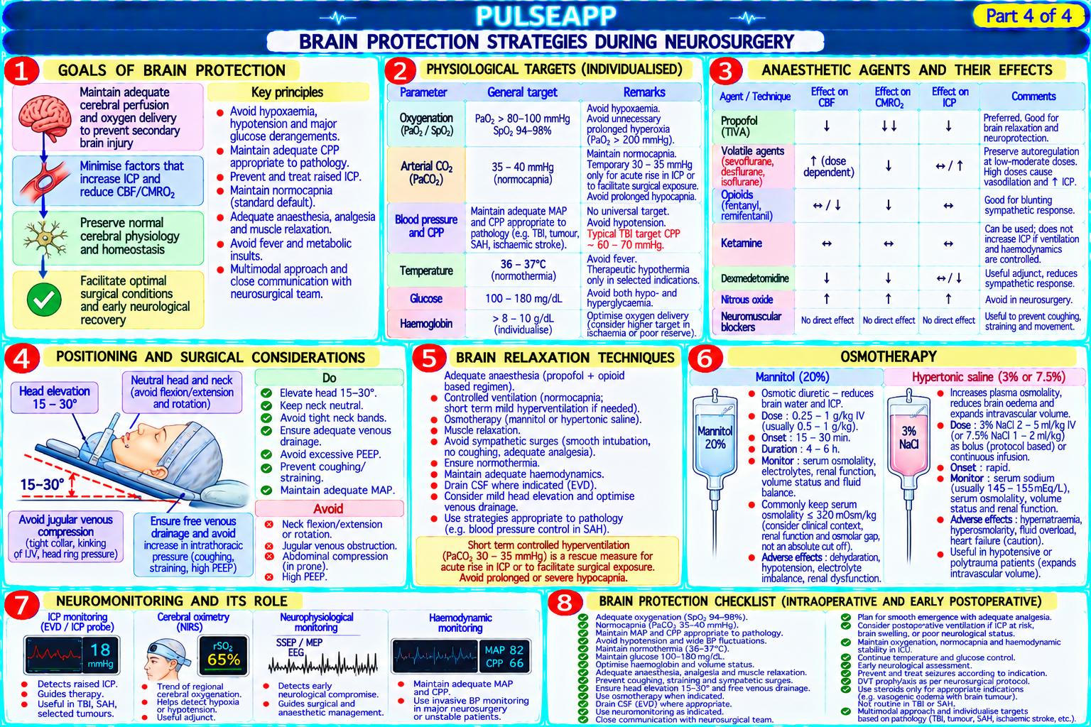
Describe the Monro-Kellie doctrine and the determinants of intracranial pressure (ICP). Outline the cerebral autoregulation curve and its clinical significance. Describe the perioperative management of raised ICP including osmotherapy, positioning, and CO₂ targets. Discuss brain protection strategies during neurosurgery.

collections Question Diagrams & Reference Images (1)
Question Reference Diagram
zoom_in View Image
description Clinical Response
⚙ Core Concept
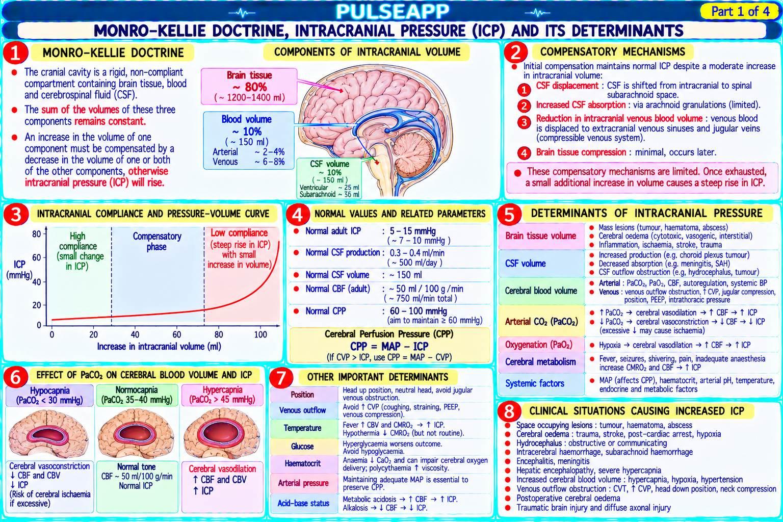
Intracranial pressure management is fundamental to neuroanaesthesia — the skull is a rigid box of fixed volume, and any increase in its three components (brain 80%, CSF 10%, blood 10%) must be compensated by reduction in another component or ICP rises exponentially. Understanding autoregulation, CO₂ reactivity, and cerebrovascular physiology allows rational design of anaesthetic techniques that optimise brain physiology during neurosurgery. (Miller's Anaesthesia 9th Ed; Drummond JC; Patel PM; Cottrell JE — Neuroanesthesia)
A. Monro-Kellie Doctrine and ICP Determinants2 marks

Monro-Kellie doctrine: the skull is a rigid, non-expandable box containing three components: brain parenchyma (~80% of volume); CSF (~10%); cerebral blood volume (CBV, ~10%); the total volume within the skull is constant; an increase in any one component must be accompanied by a compensatory decrease in another — otherwise ICP rises; normal ICP = 5–15 mmHg ICP determinants:

Cerebral blood volume (CBV): most rapidly modifiable component; PaCO₂ is the most potent regulator (see below); volatile agents increase CBV by causing cerebral vasodilation

CSF: produced by choroid plexus at 0.35 mL/min (450–500 mL/day); reabsorbed at the arachnoid granulations; drainage via EVD (external ventricular drain) is the most effective means of rapidly reducing ICP in acute raised ICP

Brain tissue: cerebral oedema (vasogenic — blood-brain barrier disruption; cytotoxic — cellular swelling from ischaemia); mass lesions (tumour, haematoma, abscess)

Intracranial compliance: the relationship between volume and pressure is exponential — initially, small volume additions are compensated without ICP rise (the compensated phase: CSF is displaced into the spinal subarachnoid space; veins compress); eventually the compensatory reserve is exhausted — any further increase in volume causes a steep, rapid ICP rise (the decompensated phase — a surgical sponge or a cough can now fatally raise the ICP)

B. Cerebral Autoregulation and CO₂ Reactivity2 marks

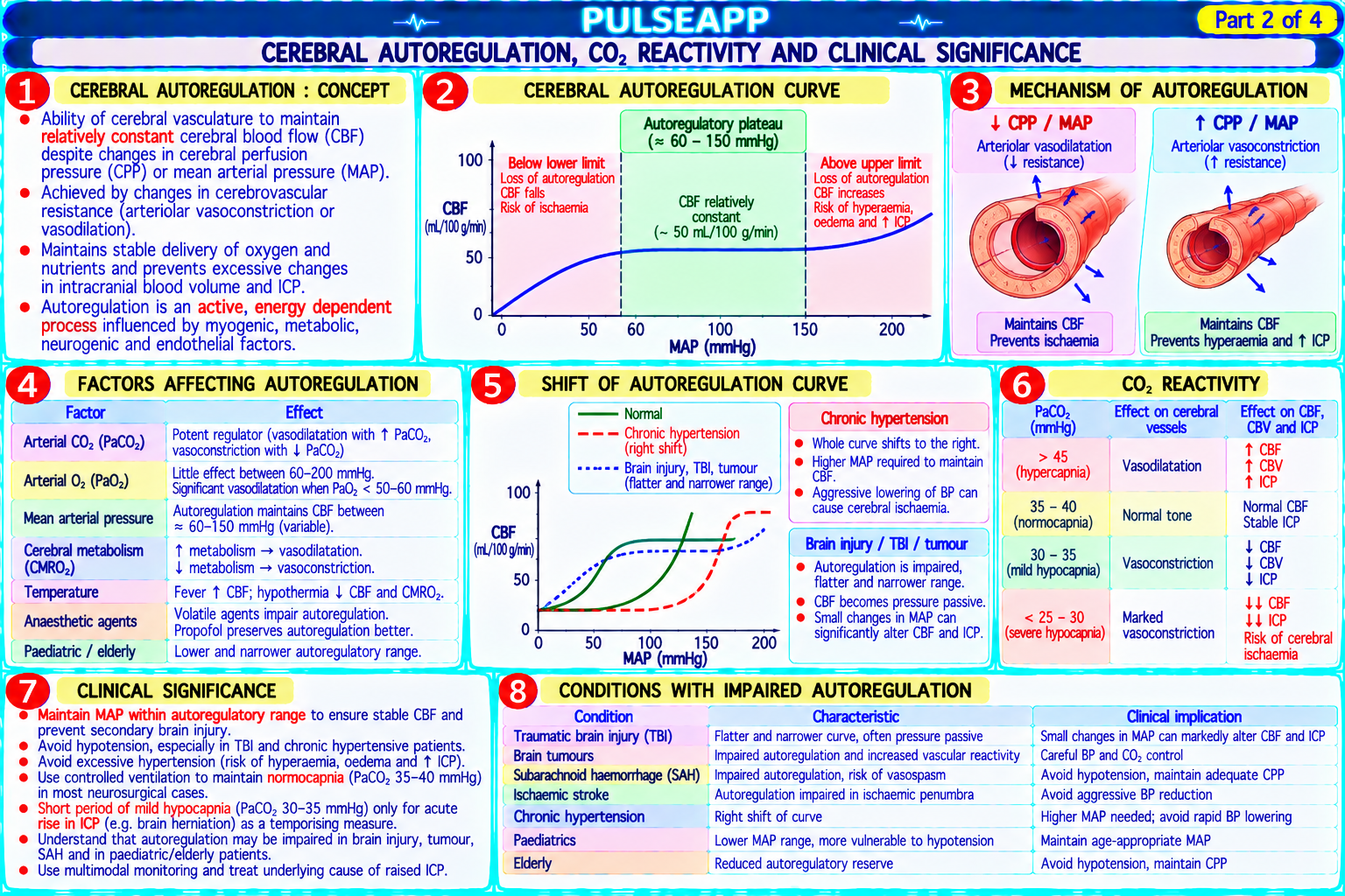
Cerebral autoregulation: the capacity of cerebral arterioles to maintain constant cerebral blood flow (CBF ≈ 50 mL/100g/min) over a wide range of MAP (approximately 60–150 mmHg) by intrinsic myogenic and metabolic vasoconstriction/dilation; below MAP 60 mmHg → autoregulation fails → CBF falls proportionally (ischaemia); above MAP 150 mmHg → autoregulation overwhelmed → CBF rises → cerebral hyperaemia → vasogenic oedema; volatile anaesthetic agents impair autoregulation dose-dependently at >0.5 MAC

CO₂ reactivity: the most powerful modulator of CBF in the physiological range; a rise in PaCO₂ of 1 mmHg → approximately 4% increase in CBF (cerebral vasodilation from CO₂-mediated perivascular acidosis → smooth muscle relaxation); a fall in PaCO₂ of 1 mmHg → approximately 3% decrease in CBF (cerebral vasoconstriction); PaCO₂ of 35–40 mmHg = optimal for most neuroanaesthesia (normocapnia); hyperventilation to PaCO₂ 30–35 mmHg → reduces CBF → reduces CBV → reduces ICP (useful for acute ICP crisis but not sustained — effect wanes after 4–6 hours as CSF bicarbonate equilibrates) CPP = MAP − ICP; target CPP ≥60–70 mmHg in TBI (Brain Trauma Foundation Guidelines); if MAP maintained but ICP rises, CPP falls → ischaemia; both MAP maintenance and ICP reduction are required simultaneously

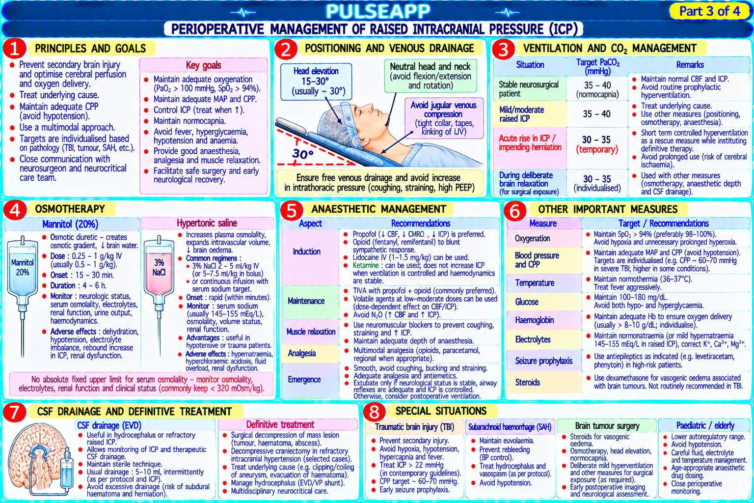
C. Perioperative Management of Raised ICP3 marks

Intervention Mechanism Target Head Head-up 30° → ↓ venous pooling in cerebral veins → ↑ cerebral venous drainage → ↓ CBV → ↓ ICP; head 30° head-up; neutral neck position; positioning must be in neutral position (neck rotation → compresses IJV → ↑ cerebral venous pressure) avoid venous compression from tight cervical collars or ETT tape Mannitol 20% mannitol 0.25–1 g/kg IV over 15–20 min; osmotic gradient draws water from brain parenchyma into the Acute ICP crisis (herniation); bridge to (osmotherapy) vasculature → ↓ brain water content → ↓ ICP; onset: 15–30 min; duration: 4–6 hours; monitor serum definitive treatment; use as a bolus, not osmolality (maintain <320 mOsm/kg; above this → risk of renal tubular damage) infusion Hypertonic Osmotic mechanism (similar to mannitol); additionally raises serum Na → ↑ osmolality → ↓ brain water; does 3% NaCl: 150–200 mL IV over 20 min; saline (3%, NOT cause osmotic diuresis (unlike mannitol — which can cause hypovolaemia with repeated doses); target serum Na 145–155 mEq/L; 7.5%, 23.4%) growing evidence that hypertonic saline is as effective or superior to mannitol for ICP reduction 23.4% NaCl: 30 mL IV for acute herniation Controlled PaCO₂ 35–40 mmHg for routine neuroanaesthesia (normocapnia); PaCO₂ 30–35 mmHg for acute ICP crisis Routine: 35–40 mmHg; acute ICP ventilation — (deliberate hyperventilation — reduces CBF/CBV/ICP temporarily); avoid PaCO₂ <30 mmHg (profound crisis: 30–35 mmHg (temporary bridge CO₂ target vasoconstriction → ischaemia) only) CSF drainage External ventricular drain placed in the lateral ventricle → direct CSF removal → immediate ICP reduction; Drain set at 10–15 cmH₂O above the (EVD) most effective intervention for acute hydrocephalus-related ICP rise foramen of Monro; allows controlled CSF removal Steroids Dexamethasone 8–16 mg loading → 4 mg QDS; reduces vasogenic oedema around brain tumours Brain tumours with vasogenic oedema; (disrupted BBB); NOT effective for cytotoxic oedema (TBI, ischaemia) does NOT improve outcome in TBI (CRASH trial)

D. Brain Protection Strategies3 marks

Temperature management: every 1°C fall in temperature → 6–7% reduction in cerebral metabolic rate for O₂ (CMRO₂); mild hypothermia (33–35°C) was studied for brain protection — but the landmark IHAST trial (Todd MM — NEJM 2005; n=1000 aneurysm surgery patients) found that intraoperative mild hypothermia did NOT improve neurological outcome vs normothermia in good-grade subarachnoid haemorrhage surgery; current recommendation: strict normothermia; prevent hyperthermia (every 1°C above normal → 6–7% increase in CMRO₂ → increased vulnerability to ischaemia)

Glucose management: hyperglycaemia worsens neurological outcomes after cerebral ischaemia (lactate accumulation in anaerobic metabolism; increased infarct volume); NICE-SUGAR trial confirmed tight glucose control (<6 mmol/L) increases mortality; current recommendation for neurological patients: maintain glucose 6–10 mmol/L (avoids both hyperglycaemia and hypoglycaemia — both are harmful)

Volatile anaesthetics vs TIVA: volatile agents (particularly isoflurane) provide PRECONDITIONING and POSTCONDITIONING of the brain against ischaemia (volatile-induced ischaemic tolerance — via mitochondrial K+ATP channel activation and other mechanisms); BUT volatile agents also cause cerebral vasodilation → ↑ CBV → ↑ ICP; TIVA (propofol) reduces CMRO₂ and ICP without the vasodilation effect; choice depends on the specific neurosurgical context: raised ICP → TIVA preferred; operations where cerebral ischaemia risk is high (aneurysm clipping, carotid surgery) → volatile preconditioning may be beneficial

Burst suppression: propofol or thiopentone infusion titrated to burst suppression on EEG (complete electrical silence interrupted by brief bursts of activity) represents maximal CMRO₂ reduction — used during temporary aneurysm clipping (to maximally reduce metabolic demand during the period of reduced blood flow); thiopentone 3–5 mg/kg bolus → infusion to achieve burst suppression; propofol 100–200 mcg/kg/min similarly

🎤 Viva Corner
Q. A patient undergoing elective craniotomy for a cerebral AVM develops sudden ICP elevation (known from pre-placed ICP monitor) from 12 to 35 mmHg during incision. What do you do?
An acute ICP rise from 12 to 35 mmHg during craniotomy incision in a known AVM patient requires immediate, systematic management while informing the surgical team. Normal ICP is <20 mmHg; 35 mmHg is a significant elevation requiring intervention. Immediate systematic management: first — check the MAP: if MAP is low (hypovolaemia, anaesthetic-related vasodilation), the CPP (= MAP − ICP) may already be critically compromised; ensure MAP ≥80 mmHg with vasopressors (phenylephrine or noradrenaline) or IV fluid to optimise CPP before reducing ICP; second — head position: confirm 30° head-up elevation with neutral neck (no rotation, no tight ETT tape or cervical collar compressing the IJV); third — ventilation: check ETCO₂; if PaCO₂ has drifted above 40 mmHg (hypoventilation, circuit disconnection), immediately increase minute ventilation to target PaCO₂ 33–35 mmHg; CO₂ reduction will produce rapid cerebral vasoconstriction and reduce CBV and ICP within minutes; this is the fastest available intervention; fourth — anaesthetic depth: confirm adequate depth with BIS monitoring; a light anaesthetic → sympathetic activation → increased CBF and ICP; ensure BIS 40–60 and increase propofol or volatile concentration if BIS is high; fifth — mannitol: if ICP remains elevated after optimising the above, give mannitol 0.5 g/kg (e.g., 40 mL/kg of 20% mannitol) over 20 minutes; onset: 15–30 minutes; will draw water from brain parenchyma and reduce cerebral oedema; sixth — inform the surgeon: the surgical team needs to know the ICP is elevated; the surgeon may elect to proceed more rapidly with the craniotomy (opening the dura will immediately relieve the ICP) OR may place a temporary EVD or perform a decompressive craniectomy if ICP is uncontrollable; seventh — if the ICP is associated with acute herniation signs (Cushing triad: hypertension + bradycardia + Cheyne-Stokes breathing) or acute pupillary dilation → give hypertonic saline 23.4% 30 mL IV as the most aggressive osmotic intervention and immediately inform the neurosurgeon of a critical emergency requiring urgent decompression.
★ Examiner's Pearl
Monro-Kellie doctrine (fixed skull volume; brain 80% + CSF 10% + blood 10%) with the compliance curve (exponential rise once compensatory reserve is exhausted) is the fundamental conceptual framework. CO₂ reactivity (1 mmHg PaCO₂ rise → 4% CBF increase; 1 mmHg fall → 3% decrease) with the normocapnia target (35–40 mmHg for routine; 30–35 mmHg for acute ICP) are the specific numerical facts. IHAST trial (mild hypothermia did NOT improve aneurysm surgery outcome) is the landmark negative trial that changed practice away from routine intraoperative hypothermia.
Drummond JC, Patel PM. Neurosurgical anesthesia (in Miller's Anaesthesia 9th Ed). Todd MM et al. IHAST trial (NEJM 2005;352:135-145). Brain Trauma Foundation. Guidelines for TBI Management. Cottrell JE, Patel P. Cottrell and Patel's Neuroanesthesia, 6th Ed.
lock Protected Clinical Document • Pulse App • Screenshots Strictly Prohibited
QUESTION 118 person Asked by .
bookmark_add

Describe the modes of neuromuscular monitoring — TOF, PTC, DBS, and tetanic stimulation. Explain clinical interpretation of each. State the sugammadex doses for reversal at each level of block and the definition of adequate recovery (TOF ratio ≥0.9).

description Clinical Response
⚙ Core Concept
Residual neuromuscular blockade (RNMB) at extubation is one of the most common preventable perioperative complications — causing pharyngeal dysfunction, airway obstruction, and silent pulmonary aspiration. The gold standard for confirming adequate recovery is quantitative acceleromyography (TOF ratio ≥0.9) — NOT clinical tests (head lift, hand grip) which are insensitive to clinically significant residual block. (Murphy GS; Brull SJ — Anesthesiology; Naguib M; Miller's Anaesthesia 9th Ed)
A. Neuromuscular Monitoring Modes4 marks

Stimulus Pattern What it Detects Clinical Use Mode TOF (Train- 4 supramaximal 0.2 TOF count (T1–T4 present or absent) during profound/moderate Standard monitoring throughout anaesthesia; TOF count of-Four) ms pulses at 2 Hz block; TOF ratio (T4/T1 amplitude) during recovery guides dosing; TOF ratio guides extubation decision (2 per second), delivered every 12– 15 seconds

PTC (Post- 50 Hz tetanic Post-tetanic potentiation: the tetanic stimulus increases ACh Monitoring PROFOUND block when TOF count = 0; PTC 1–2 Tetanic stimulus for 5 mobilisation at the nerve terminal → more ACh available → = very deep block; PTC 10–15 = moderately deep; as PTC Count) seconds → pause 3 subsequent stimuli produce detectable responses even when increases → TOF count will soon reappear seconds → then TOF count = 0; PTC counts (1–15) the number of post-tetanic TOF twitches DBS 2 short bursts of 50 Fade between the 2 responses is more easily detectable by Allows detection of moderate residual block (TOF ratio 0.6– (Double- Hz tetanic tactile assessment than TOF fade; DBS₃,₃ is the standard; DBS 0.9) that is MISSED by visual/tactile TOF assessment; useful Burst stimulation (3 ratio ≈ TOF ratio at the end of anaesthesia when qualitative assessment of fade Stimulation) pulses per burst), is needed but quantitative acceleromyography is not available separated by 750 ms Tetanic 50 or 100 Hz Fade during tetanic stimulation indicates residual non- Sensitive indicator of residual block; painful in the awake stimulation continuous depolarising block (normal: sustained contraction; residual patient → only for use under deep anaesthesia or general stimulation for 5 block: fading contraction during tetanus) anaesthesia seconds Single Single 0.2 ms T1 height as % of baseline; T1 suppression during block Calibrating baseline before NMB; monitoring during very deep twitch supramaximal pulse relaxant block when all 4 TOF twitches are absent at 0.1–1 Hz

B. Interpretation — Levels of Block2 marks

Level of Block TOF Count PTC Clinical Intense/Ultra- 0/4 (no twitches) PTC = 0 Maximum surgical relaxation (intubating conditions, rigid abdomen); used for specific procedures deep block (retroperitoneal surgery, ophthalmic surgery) Profound block 0/4 PTC 1–5 Good surgical relaxation; recovery will occur within 10–30 min depending on drug and dose Deep block 0–1/4 PTC >10 Acceptable for most abdominal surgery; spontaneous recovery approaching Moderate block 1–3/4 N/A (TOF count Inadequate for adequate closure of abdomen; reversal with neostigmine may be attempted but not at TOF present) 1/4 Shallow block 4/4 with fade N/A Patient can breathe but may have residual dysfunction; TOF ratio 0.4–0.9 = clinical RNMB; requires reversal Adequate 4/4 with TOF N/A Safe for extubation; pharyngeal and airway reflexes are clinically intact at TOF ratio ≥0.9 recovery ratio ≥0.9

C. Sugammadex Reversal Dosing2 marks
✅ Sugammadex Dosing — Based on TOF Count or PTC
Clinical State Sugammadex Dose Routine reversal — TOF 1–4 present 2 mg/kg IV Routine reversal — TOF 1–4 present 2 mg/kg IV (shallow/moderate block) Deeper block — PTC ≥1 but TOF count 0 (deep 4 mg/kg IV block) Immediate reversal — within 3 minutes of 16 mg/kg IV — rescues a "cannot intubate, cannot oxygenate" rocuronium RSI where the airway cannot be rocuronium 1.2 mg/kg (RSI dose) secured; reversal to normal NMJ function within 3 minutes Mechanism: sugammadex is a modified γ-cyclodextrin that forms a tight 1:1 encapsulation complex with rocuronium (highest affinity), vecuronium, and pancuronium; the complex is pharmacologically inactive; it is renally excreted; free drug in the NMJ is drawn into the plasma by the concentration gradient as the plasma concentration is eliminated by excretion → complete reversal; does NOT work for succinylcholine or benzylisoquinolinium NMBs (atracurium, cisatracurium, mivacurium) TOF ratio ≥0.9: the threshold for extubation safety; below 0.9, upper airway muscle dysfunction is clinically significant; at TOF ratio 0.7, pharyngeal muscle function is impaired enough to cause aspiration; the adductor pollicis (thumb adductor — the standard monitoring muscle) recovers SLOWER than laryngeal and diaphragm muscles but FASTER than pharyngeal muscles → TOF ratio ≥0.9 at the thumb provides a conservative safety margin for pharyngeal function
D. Clinical Tests and Their Limitations2 marks

Sustained head lift for 5 seconds: requires TOF ratio ≥0.6 only — INSENSITIVE; a patient failing the head lift test has very significant RNMB, but a patient who can lift their head for 5 seconds may still have TOF ratio as low as 0.6 → still unsafe to extubate Hand grip strength, ability to open eyes, tongue depressor test — all insensitive; do not reliably detect TOF ratio 0.6–0.9 (the clinically dangerous range of

RNMB)

Tactile and visual TOF fade assessment: cannot reliably detect fade when TOF ratio >0.4; the eye and finger cannot detect fade at TOF ratio 0.4–0.9 → qualitative monitoring misses the most dangerous range of residual block

Conclusion: quantitative TOF monitoring (acceleromyography — Mechanosensor®, TOFscan®, TetraGraph®) is the only reliable method to confirm TOF ratio ≥0.9; this is now the standard of care recommendation (PORC prevention guidelines; 2020 European recommendations)

🎤 Viva Corner
Q. Your patient at the end of laparoscopic cholecystectomy has TOF count of 3/4 by tactile assessment. You are about to give neostigmine. Is this safe and what dose do you give?
Neostigmine CAN be given at TOF count 3/4, but this requires careful consideration. The standard recommendation for neostigmine reversal is that it should only be given when TOF count is at least 1/4 (and ideally 3–4/4) — neostigmine administered at deeper levels of block (TOF count 0–1/4) does not produce reliable reversal and may cause cholinergic complications without reversing the block. At TOF count 3/4 by tactile assessment, neostigmine 50 mcg/kg IV with glycopyrrolate 10 mcg/kg IV (to prevent muscarinic bradycardia) is appropriate and would be considered standard practice. Dose: for TOF count 3/4, neostigmine 50 mcg/kg (maximum 5 mg) + glycopyrrolate 0.2 mg per 1 mg neostigmine. However, there are important caveats: tactile TOF assessment of 3/4 is not reliable — the human finger cannot detect fade at TOF ratio 0.4–0.9; a patient assessed as TOF 3/4 by palpation may in fact have a TOF ratio of only 0.4–0.6, and neostigmine at this level of block may not achieve complete reversal (TOF ratio ≥0.9) reliably within 10 minutes; the ceiling of neostigmine's reversal capacity is approximately TOF ratio 0.9 — but the time to reach 0.9 from TOF count 3/4 by palpation may be longer than the clinical impression suggests. The better approach: if quantitative acceleromyography is available → confirm the actual TOF ratio before the decision to extubate; if TOF ratio is 0.6–0.9 after neostigmine → cannot extubate safely; consider sugammadex 2 mg/kg (will reliably achieve TOF ratio ≥0.9 from any level where TOF count ≥1). The key message: neostigmine at TOF 3/4 is a reasonable clinical practice, but should be followed by quantitative TOF ratio measurement before extubation — not by clinical tests (head lift, hand grip) which are insensitive surrogates.
★ Examiner's Pearl
Sugammadex three doses (2 mg/kg for TOF 1–4; 4 mg/kg for PTC ≥1, TOF 0; 16 mg/kg for immediate post-RSI reversal) must be reproduced correctly and linked to the clinical state. PTC: the only monitoring mode that provides information during profound block (TOF count = 0) — low PTC (1–5) = very deep; high PTC (>10) = recovery approaching. TOF ratio ≥0.9 is the extubation threshold — NOT head lift (only requires ratio ≥0.6) or tactile assessment (cannot detect fade above ratio 0.4).
Murphy GS et al. Residual neuromuscular blockade and critical respiratory events in the PACU (Anesth Analg 2008;107:130-137). Naguib M et al. Advances in neurobiology of the NMJ (Anesthesiology 2002;96:202-231). Fuchs-Buder T et al. Good clinical research practice for pharmacodynamic studies (Anesthesiology 2007;106:A19). Miller RD et al. Miller's Anaesthesia, 9th Ed.
lock Protected Clinical Document • Pulse App • Screenshots Strictly Prohibited
QUESTION 119 person Asked by .
bookmark_add

Describe capnography waveform analysis — phases I–IV, the clinical significance of waveform abnormalities (bronchospasm, oesophageal intubation, cardiac arrest, rebreathing). Outline the limitations of pulse oximetry (COHb, MetHb, low-perfusion states). Describe pressure-volume loops and their use in optimising mechanical ventilation.

collections Question Diagrams & Reference Images (3)
Question Reference Diagram
zoom_in View Image
Question Reference Diagram
zoom_in View Image
Question Reference Diagram
zoom_in View Image
description Clinical Response
⚙ Core Concept
Capnography (end-tidal CO₂ monitoring) is mandatory monitoring for all intubated patients — it confirms correct ETT placement, detects disconnection immediately, and provides continuous quantitative information about ventilation, circulation, and metabolism. Its clinical applications extend far beyond simple tracheal intubation confirmation. (Bhavani-Shankar K — Anesth Analg 1995; Miller's Anaesthesia 9th Ed; AAGBI monitoring standards)
A. Capnography Waveform — Four Phases3 marks

Phase Description Physiological Basis Phase I Flat baseline at CO₂ = 0 Dead space gas from the airways and breathing circuit; no CO₂ if circuit is functioning correctly; elevated baseline = (Inspiratory mmHg REBREATHING (inadequate CO₂ absorber, incompetent expiratory valve, insufficient fresh gas flow in Mapleson system) baseline) Phase II Rapid rise in CO₂ as Mixing of dead space and alveolar gas; slope of Phase II reflects the homogeneity of alveolar emptying; steeper = more (Expiratory exhaled gas transitions uniform; prolonged gradual slope = V/Q inhomogeneity (COPD, bronchospasm) upstroke) from dead space to alveolar gas Phase III Relatively flat plateau at Predominantly alveolar gas exhaled; the slope of the plateau is slightly upward in normal lungs; steep upward slope (shark fin (Alveolar or near ETCO₂ value pattern) = BRONCHOSPASM (inhomogeneous alveolar emptying — obstructed alveoli empty slowly and their higher CO₂ plateau) concentration is reached later in exhalation); in normal lungs: plateau slope ≤2 mmHg; in COPD/asthma: >5 mmHg Phase 0 Rapid fall in CO₂ as Inspiratory fresh gas washout of CO₂; abrupt fall; the point of maximum CO₂ before this fall = ETCO₂ value (Phase IV fresh gas is inspired — Inspiration)

B. Waveform Abnormalities and Their Clinical Significance3 marks

Waveform Appearance Diagnosis Pattern Oesophageal ETCO₂ absent or rapidly falling to zero (a few Immediate diagnosis of oesophageal intubation; this is the most important airway safety application intubation small waves diminishing over 5 breaths then of capnography; if no sustained ETCO₂ waveform → ETT is NOT in the trachea until proven nothing — CO₂ in the stomach from mask otherwise ventilation exhausted) Bronchospasm Phase II prolonged (gradual rise); Phase III Bronchospasm (or severe COPD exacerbation); inhomogeneous airway resistance → alveoli with — "Shark Fin" steep upward slope; sharp peak replacing the high resistance empty slowly → CO₂ concentration continues to rise throughout expiration → steep or "Reverse normal flat plateau; the overall waveform Phase III slope Shark" resembles a shark's dorsal fin Rebreathing Phase I baseline elevated above zero (ETCO₂ Inspired CO₂; causes: exhausted soda lime; incompetent expiratory valve; insufficient FGF in baseline >0 mmHg) Mapleson system; rebreathing increases ETCO₂ and PaCO₂ → respiratory acidosis Cardiac arrest Sudden sharp fall in ETCO₂ to near-zero ETCO₂ is a surrogate for cardiac output (CO₂ delivery to the lungs requires pulmonary blood flow); or severe ↓ CO (despite continued ventilation); the CO₂ sudden fall in ETCO₂ during surgery = cardiac arrest, massive PE, severe haemorrhage; during reaching the alveoli falls dramatically when CO CPR: ETCO₂ >10 mmHg correlates with adequate chest compression quality; ETCO₂ rise to >35 is absent mmHg during CPR = early sign of ROSC Curare notch Notch or dip within Phase III (the plateau) Insufficient neuromuscular blockade — the patient is making respiratory efforts during the Phase III (cleft in plateau; each effort draws fresh gas briefly, producing a dip in CO₂; a sign of inadequate relaxation plateau) or recovering block — assess TOF count

C. Pulse Oximetry Limitations2 marks

Limitation Mechanism Clinical Implication Carbon monoxide Standard 2-wavelength pulse oximeter cannot distinguish COHb from Require co-oximetry ABG to measure COHb directly; SpO₂ is poisoning (COHb) OxyHb (both absorb identically at 660 nm); SpO₂ reads FALSELY HIGH USELESS for diagnosis of CO poisoning; clinical suspicion even when functional O₂ saturation is critically low; patient appears well- must guide diagnosis in fires and closed-space exposure oxygenated while CO is occupying 40–60% of haemoglobin Methaemoglobinaemia MetHb absorbs at both 660 nm and 940 nm equally (absorption ratio Suspect if SpO₂ plateau at 85% despite increasing FiO₂; (MetHb) approaches 1.0); the pulse oximeter reads SpO₂ as approximately 85% causes: dapsone, prilocaine (>600 mg → EMLA cream large regardless of the actual MetHb level; MetHb >20% → SpO₂ stuck at ~85% area), nitrites, benzocaine, rasburicase; treat with methylene (does not accurately reflect the true severity) blue 1–2 mg/kg IV (reduces MetHb to functional Hb) Low-perfusion states Peripheral vasoconstriction (hypothermia, shock, vasopressors) reduces Unreliable in hypothermia or shock; move probe to a betterperipheral pulsatile flow → the SpO₂ signal becomes weak, inaccurate, or perfused site (ear, forehead reflectance probe) or use coabsent; the probe may read the venous plethysmographic wave rather than oximetry ABG as the gold standard the arterial signal if pulsatile flow is very low Nail polish and Dark nail polish absorbs at the measurement wavelengths → falsely low Ask about nail polish pre-operatively; remove or use alternative artificial nails SpO₂; remove or use side-of-finger placement to avoid the nail probe placement

D. Pressure-Volume Loops2 marks

P-V loops plot airway pressure (x-axis) against tidal volume (y-axis) during a single breath; modern ventilators display these in real-time; a normal P-V loop is roughly elliptical: the inspiratory limb shows increasing volume with increasing pressure; the expiratory limb shows passive deflation; hysteresis (the loop area) represents the energy consumed in overcoming airway resistance and lung viscoelasticity Clinical applications:

Upper inflection point (UIP): kink in the inspiratory limb at high pressures where compliance suddenly decreases (lung overdistension) → if tidal volume exceeds this point, barotrauma and volutrauma risk; PEEP should be set BELOW the UIP

Lower inflection point (LIP): kink at low pressures where compliance improves suddenly (alveolar recruitment) → PEEP should be set ABOVE the LIP to maintain open alveoli and prevent atelectrauma; the "open lung" strategy: PEEP between LIP and UIP Increased area of the loop (wider loop) = increased resistance (bronchospasm) — the pressure required to move the tidal volume is disproportionately high, creating a characteristic "peanut-shaped" loop in severe bronchospasm Birds-beak appearance at the beginning of inspiration = patient-triggered breath with flow limitation (auto-PEEP/gas trapping) — the loop does not start at zero pressure but at a positive pressure baseline

🎤 Viva Corner
Q. During CPR in theatre, the ETCO₂ reads 8 mmHg. What does this tell you and what action does it guide?
An ETCO₂ of 8 mmHg during CPR provides critical information about the quality of resuscitation and guides immediate action. Physiology: CO₂ is delivered to the alveoli only by pulmonary blood flow (cardiac output); during CPR, chest compressions generate a fraction of normal cardiac output (typically 25–30% at best); ETCO₂ during CPR directly correlates with the cardiac output generated by chest compressions and therefore serves as a real-time quality monitor of compression effectiveness. An ETCO₂ of 8 mmHg indicates poor compression quality — the threshold for adequate CPR quality is ETCO₂ ≥10 mmHg (with some guidelines using ≥15–20 mmHg as the target for optimal compressions); 8 mmHg means cardiac output from compressions is too low to deliver adequate CO₂ to the lungs. Action: switch compressor immediately (compressor fatigue is the commonest reason for low ETCO₂ — compressions lose rate and depth within 2 minutes of fatigue onset); ensure correct hand position (centre of sternum, heel of hand, straight elbows); compress at 100–120 per minute with at least 5 cm depth; allow full chest recoil between each compression; STOP any interruptions to compressions (minimise pulse checks); confirm ventilation is not excessive (overventilation does not raise ETCO₂ — it merely dilutes the alveolar CO₂ and may worsen haemodynamics by increasing intrathoracic pressure). ETCO₂ as a prognostic marker: if ETCO₂ remains persistently <10 mmHg after 20 minutes of optimal CPR → strongly predicts failure of ROSC (poor prognosis) and may guide the decision to terminate resuscitation. Conversely: a sudden, sustained rise in ETCO₂ to >35 mmHg during CPR → early ROSC indicator → immediately reassess pulse; this sign may precede any other clinical sign of return of spontaneous circulation by several minutes.
★ Examiner's Pearl
The four capnography phases with physiological basis must be reproduced (I: dead space/baseline; II: upstroke from dead-space-to-alveolar transition; III: alveolar plateau; 0/IV: inspiratory fall). Shark-fin capnogram in bronchospasm (steep upward Phase III slope from inhomogeneous alveolar emptying) is the most commonly tested waveform abnormality. ETCO₂ during CPR: ≥10 mmHg = adequate compressions; sudden rise to ≥35 mmHg = ROSC. Pulse oximetry falsely normal in CO poisoning (COHb absorbs identically to OxyHb at 660 nm — the most tested SpO₂ limitation).
Bhavani-Shankar K et al. Capnometry and anaesthesia (Can J Anaesth 1992;39:617-632). Idris AH et al. ETCO₂ as a monitor of CPR quality (Circulation 2012;125:e517-e519). Tremper KK. Pulse oximetry (Chest 1989;95:713-715). Miller RD et al. Miller's Anaesthesia, 9th Ed.
lock Protected Clinical Document • Pulse App • Screenshots Strictly Prohibited
QUESTION 120 person Asked by .
bookmark_add

Describe the physiological changes in morbid obesity relevant to anaesthesia. Outline pre-operative assessment for bariatric surgery including OSA screening. Describe optimal positioning (HELP position), drug dosing (IBW vs TBW), lung-protective ventilation, and post-operative analgesia in the obese patient.

collections Question Diagrams & Reference Images (3)
Question Reference Diagram
zoom_in View Image
Question Reference Diagram
zoom_in View Image
Question Reference Diagram
zoom_in View Image
description Clinical Response
⚙ Core Concept
Morbid obesity (BMI ≥40 kg/m²) creates a physiologically hostile environment for anaesthesia — reduced FRC (approaching closing capacity in the supine position), difficult airway, OSA-related sleep-disordered breathing, restrictive pulmonary physiology, and altered drug pharmacokinetics. Meticulous preoperative optimisation, the HELP position, lung-protective ventilation, and TIVA (lower PONV, earlier return to CPAP) are the key elements of safe bariatric anaesthesia. (Nightingale CE et al. — OAA/AAGBI 2015; Marrel J; Pelosi P; Miller's Anaesthesia 9th Ed)
A. Physiological Changes in Morbid Obesity2 marks

System Change Clinical Relevance Respiratory ↓ FRC (supine position → abdominal fat compresses diaphragm → FRC may fall below closing Rapid desaturation during apnoea (lowest FRC capacity → atelectasis); ↑ O₂ consumption; ↑ CO₂ production; restrictive pattern (↓ TLC, ↓ FVC); + highest O₂ demand); difficult preoxygenation; obstructive sleep apnoea in 70–80% of morbidly obese challenging ventilation; atelectasis-prone under GA Cardiovascular ↑ Blood volume (↑ CO, ↑ stroke volume); LVH (pressure and volume overload); ↑ HR; systemic and Cardiac assessment essential; diastolic failure pulmonary hypertension; diastolic dysfunction; obesity cardiomyopathy; ↑ atherosclerosis risk → sensitive to fluid overload; higher perioperative cardiac risk Airway Difficult mask ventilation (Mallampati IV, large tongue, retrognathia, short thick neck, excess Anticipated difficult airway; RSI or awake FOI; pharyngeal tissue); difficult laryngoscopy (reduced cervical flexion, excessive chest fat limiting video laryngoscopy as first-line laryngoscope handle movement); high risk of aspiration (↑ gastric volume, ↑ intragastric pressure, ↑ GERD) Gastric ↑ Gastric volume; ↑ intragastric pressure from abdominal fat; ↑ GERD; ↑ aspiration risk RSI for GA induction; anti-aspiration precautions; ranitidine or PPI pre-operatively

B. Pre-operative Assessment2 marks

OSA screening: STOP-BANG questionnaire (Snoring, Tired, Observed apnoea, Pressure, BMI >35, Age >50, Neck ≥40 cm, Male Gender); score ≥3 = high risk of OSA; ≥5 = high risk of severe OSA; all morbidly obese patients should be considered for pre-operative sleep study (polysomnography or home sleep test) and CPAP therapy optimisation before elective bariatric surgery

Respiratory: spirometry (to identify coexisting COPD or severe restriction); ABG (if suspect O₂ desaturation or CO₂ retention — obesity hypoventilation syndrome); CXR or CT chest for preoperative planning; resting SpO₂ on air (if <95% → investigate for OHS)

Cardiovascular: ECG; echocardiography if clinical suggestion of cardiomyopathy, pulmonary hypertension, or significant dyspnoea; establish exercise tolerance (METS)

Medications: CPAP therapy — patient should bring their CPAP machine to hospital for immediate post-operative use; ensure CPAP settings documented

C. HELP Position and Airway Management2 marks

HELP (Head-Elevated Laryngoscopy Position): 25–30° reverse Trendelenburg (head-up tilt of the whole table) + careful ramping of the upper body with a purpose-built ramp or folded blankets (the external auditory meatus should be at the same horizontal level as the sternal notch); this position: extends the intubation duration by reducing mediastinal and abdominal fat compression on the diaphragm → extends the safe apnoea time; improves laryngoscopic view by aligning the oral-pharyngeal-laryngeal axes more effectively; reduces FRC decrease during the apnoeic intubation phase

Pre-oxygenation: 100% O₂ for minimum 5 minutes in HELP position before induction; use 25° head-up for pre-oxygenation (even before ramping for intubation) — head-up position during pre-oxygenation significantly extends safe apnoea time in obese patients (Boyce JR ANESTH 2003); HFNO (high-flow nasal O₂ 60–70 L/min during apnoea — transnasal humidified rapid insufflation ventilatory exchange — THRIVE) extends safe apnoea time further

Airway strategy: video laryngoscopy as first-line intubation tool (DAS recommendation for obese patients — greater Cormack-Lehane grade I-II rate vs direct laryngoscopy); RSI for all obese patients due to aspiration risk; difficult airway trolley at the bedside; backup plan (SAD, AFOI) documented in pre-op assessment

D. Drug Dosing — IBW vs TBW2 marks

Use Drug Use TBW Use AdjBW IBW Succinylcholine No TBW (plasma pseudocholinesterase activity correlates with — TBW; underdosing on IBW → inadequate intubating conditions) Rocuronium No IBW (avoid overdose and prolonged block; the NMJ drug — (intubating binding is related to lean body mass not fat) dose) Propofol No No Lean Body Weight (LBW) or IBW for induction dose; TBW overestimates and induction causes overdose; IBW may underdose large individuals; LBW or dose-titrate to effect Propofol No No LBW with Schnider model (Schnider automatically calculates LBW from maintenance height/weight/age/gender) (TCI) Remifentanil No No IBW (or LBW) for remifentanil TCI — using TBW significantly overdoses Fentanyl No No LBW or dose cautiously — accumulates in fat; titrate to effect Morphine IBW No — Gentamicin / No No AdjBW = IBW + 0.4 × (TBW − IBW) antibiotics

E. Lung-Protective Ventilation and Post-op Care2 marks

LPV in obesity: tidal volume 6–8 mL/kg IBW (NOT TBW — using TBW produces dangerously high tidal volumes in obese patients); PEEP 8–12 cmH₂O (higher PEEP needed to prevent atelectasis in obese patients — the increased abdominal pressure elevates closing volume); FiO₂ 0.5 during surgery (highest FiO₂ worsens atelectasis via absorption atelectasis); intraoperative recruitment manoeuvres (sustained inflation 40 cmH₂O for 40 seconds) improves oxygenation and prevents atelectasis

Post-operative: CPAP immediately post-extubation for known OSA (patient's home CPAP settings); nursing in head-up or lateral position (not supine) in recovery; supplemental O₂; careful opioid avoidance (opioids worsen OSA — multimodal analgesia with TAP block, IV paracetamol, NSAIDs, dexamethasone 8 mg); TIVA with propofol preferred (lower PONV than volatile agents; earlier return to CPAP use)

🎤 Viva Corner
Q. Why is the tidal volume in an obese patient calculated using IBW rather than TBW, and what is the clinical consequence of using TBW?
The tidal volume for mechanical ventilation should be calculated from Ideal Body Weight (IBW) rather than Total Body Weight (TBW) in obese patients because the lung size — specifically the functional volume of the lung parenchyma available for gas exchange — correlates with height (and the lung size at ideal body weight), not with the patient's actual weight. Fat tissue does not participate in gas exchange; the lungs of a 180 kg morbidly obese person are not twice as large as a 90 kg person of the same height — they may actually be slightly smaller in functional terms due to compression atelectasis from the abdominal fat. The lung-protective ventilation threshold of 6 mL/kg is designed to prevent overdistension (volutrauma) of the alveoli — alveolar overdistension occurs when the tidal volume exceeds the functional capacity of the available aerated lung parenchyma; this threshold is determined by the size of the functional lung, which correlates with IBW not TBW. If TBW is used to calculate tidal volume for an obese patient: a 150 kg patient whose IBW is 70 kg → TBW-based TV at 6 mL/kg = 900 mL; IBW-based TV at 6 mL/kg = 420 mL; the 900 mL tidal volume would be delivered to a lung with the functional capacity of approximately 70 kg (IBW) → massive alveolar overdistension → volutrauma → inflammatory cytokine release → biotrauma → ALI/ARDS; plateau pressures would exceed 30 cmH₂O; this is clinically dangerous. The correct calculation: IBW (male) = 50 + 2.3 × (height in inches − 60); IBW (female) = 45.5 + 2.3 × (height in inches − 60); then TV = 6–8 mL/kg IBW.
★ Examiner's Pearl
HELP position (25–30° reverse Trendelenburg + ramping so external auditory meatus = sternal notch level) is the specific positioning for obese patients — components and goal (extend safe apnoea time, improve laryngoscopic view) must be described. Drug dosing table (succinylcholine = TBW; rocuronium intubating dose = IBW; propofol maintenance = LBW with Schnider; remifentanil = IBW; morphine = IBW) are the high-yield specific dosing facts. TV based on IBW not TBW (6 mL/kg IBW) is the lung protective ventilation principle with the rationale (lung functional volume = IBW-related, not TBW-related).
Nightingale CE et al. OAA/AAGBI guidelines for the management of morbidly obese patients during and after pregnancy 2015. Pelosi P et al. Effects of obesity on respiratory mechanics (Anesthesiology 1996;84:562-569). Boyce JR et al. A preliminary study of the optimal anesthesia positioning for obese patients (Obesity Surgery 2003;13:4-9). Miller RD et al. Miller's Anaesthesia, 9th Ed.

Showing 111120 of 337 questions

account_tree

Subcategory Tree

Explore Anesthesia subcategories

folder_special Anesthesia
Main
lock

Category Subscription

Subscribe to Anesthesia to unlock this module and all nested subcategories.

  • check_circle Access Anesthesia & all subcategories
  • check_circle Detailed, Peer-Reviewed Answers
  • check_circle High-yield visual aids & imaging
Get Category Subscription arrow_forward
Secure 256-bit SSL Connection

Anatomical Models

Explore high-fidelity 3D visualizations included in premium modules.

Case Reviews

Real-world clinical scenarios narrated by senior consultants.

Pulse App