Describe the adrenergic receptor pharmacology of vasopressors and inotropes. Compare noradrenaline, adrenaline, dopamine, vasopressin, phenylephrine, dobutamine, and milrinone — their receptor profiles, haemodynamic effects, and specific clinical indications.
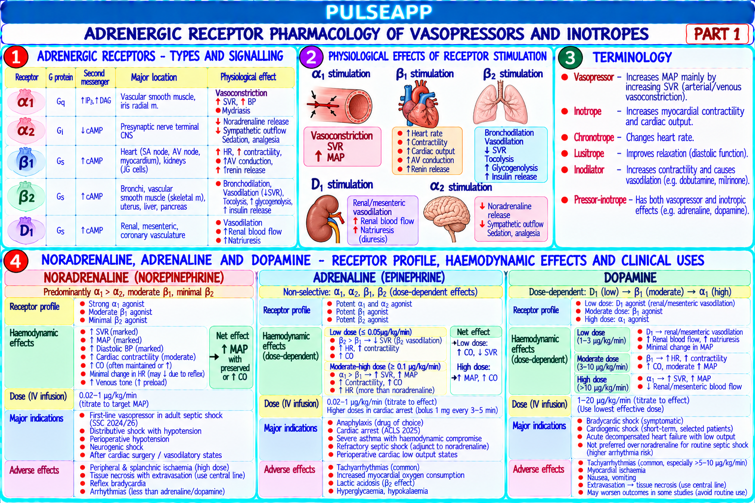
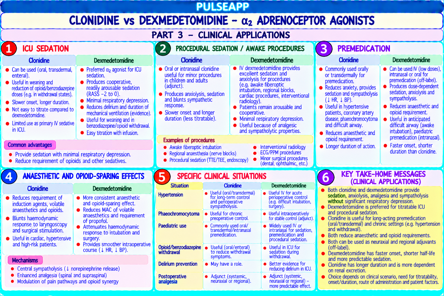
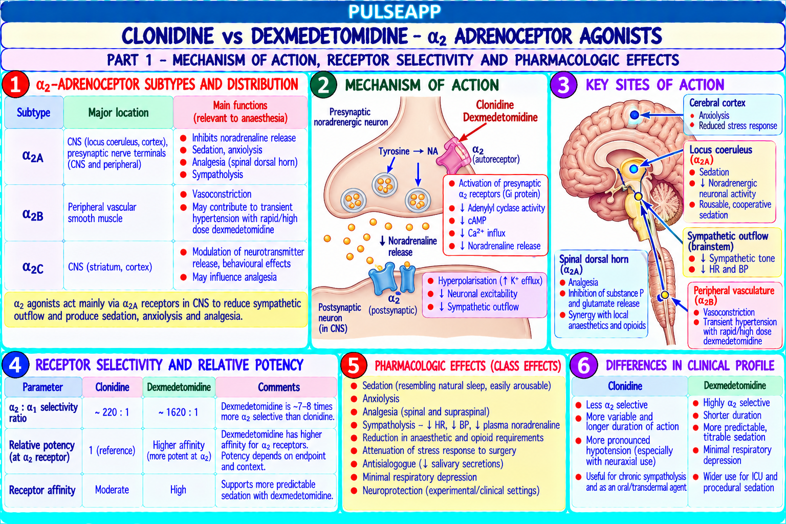
Receptor Location Effect When Stimulated α₁ adrenergic Peripheral vascular smooth muscle; skin, splanchnic, Vasoconstriction → ↑ SVR → ↑ MAP; pupil dilation; urethral sphincter contraction renal vessels α₂ adrenergic Presynaptic nerve terminals (central + peripheral); Central: sedation, analgesia, ↓ sympathetic outflow; peripheral: vasoconstriction (lower vascular smooth muscle potency than α₁) β₁ adrenergic Cardiac sinoatrial node, AV node, myocardium ↑ Heart rate (chronotropy), ↑ contractility (inotropy), ↑ AV conduction, ↑ myocardial O₂ demand β₂ adrenergic Vascular smooth muscle; bronchial smooth muscle; Vasodilation → ↓ SVR; bronchodilation; uterine relaxation; mast cell stabilisation; ↑ insulin uterus; mast cells release DA₁ Renal and splanchnic vasculature Vasodilation → ↑ renal/splanchnic blood flow; historically thought to provide "renal (dopamine) protection" — now disproven V₁ Vascular smooth muscle Vasoconstriction independent of catecholamine pathway; particularly useful in (vasopressin) catecholamine-resistant vasodilatory shock
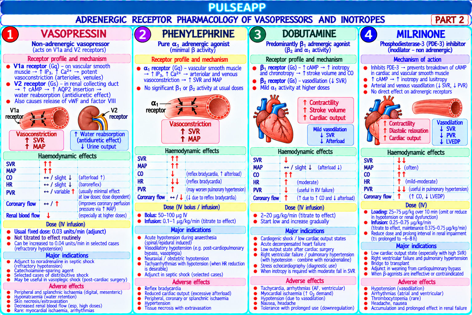
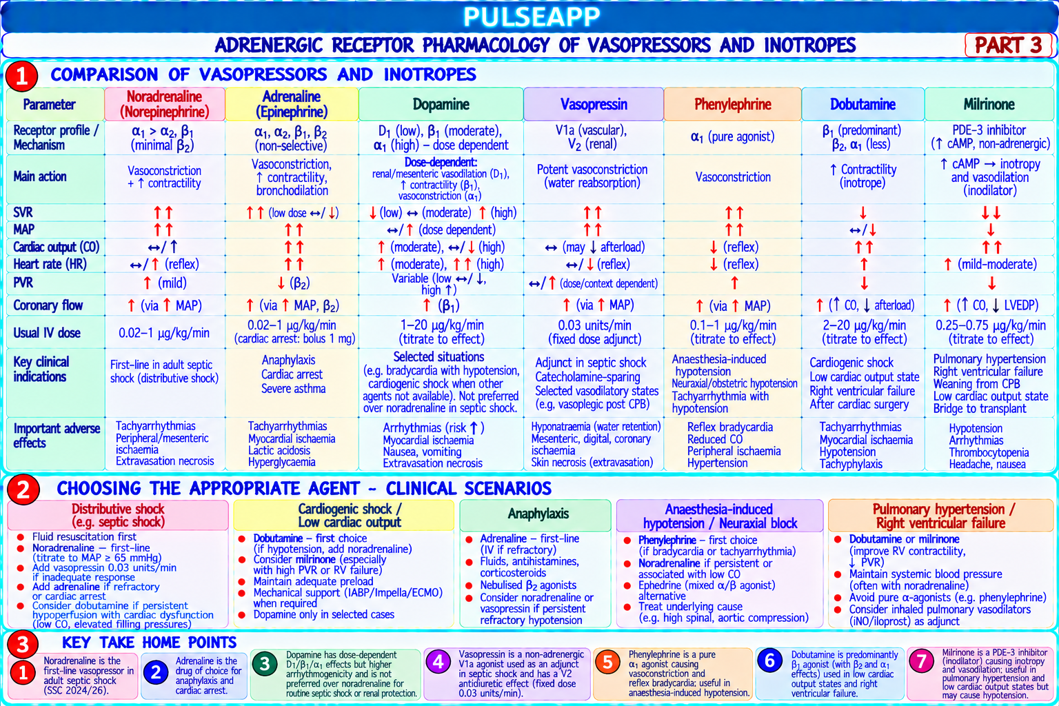
Drug Receptor Profile Haemodynamic Effect Primary Indication Key Safety Point Noradrenaline α₁ >> β₁; minimal β₂ ↑ SVR (↑ MAP); modest ↑ HR; no FIRST LINE for septic shock (SSC 2021); Peripheral ischaemia with (norepinephrine) significant vasodilation; maintains first line for most vasodilatory shock; extravasation (use central line); CO via afterload-mediated LV maintains MAP ≥65 mmHg; preferred over reflex bradycardia at high doses; optimization dopamine (De Backer NEJM 2010) splanchnic vasoconstriction at very high doses Adrenaline β₁ + β₂ at low doses; Low dose: ↑ CO, ↑ HR, ↓ SVR (β₂ Cardiac arrest (1 mg IV every 3–5 min); Tachyarrhythmias (high dose); (epinephrine) α₁ added at high dominant); High dose: ↑↑ SVR, anaphylaxis (0.5 mg IM or 50–100 mcg IV); metabolic acidosis (lactic acid from doses ↑↑ HR, ↑↑ MAP; universal potent severe refractory shock; low cardiac output β₂ glycogenolysis and impaired effect with hypotension tissue perfusion at high doses); myocardial ischaemia; hyperglycaemia Vasopressin V₁ receptor: vascular Vasoconstriction via V₁ Vasodilatory shock refractory to Coronary and mesenteric (ADH) smooth muscle (independent of adrenergic noradrenaline alone (adds V₁ vasoconstriction at high doses — vasoconstriction; V₂ receptors); ↑ SVR; ↑ MAP; no vasoconstriction via a different receptor ischaemia risk; hyponatraemia (V₂ receptor: renal water direct chronotropy or inotropy; pathway); vasodilatory shock post-CPB effect — SIADH-like); skin necrosis reabsorption reduces noradrenaline (vasopressin deficiency after bypass); with high doses and extravasation; requirements (catecholamine- relative vasopressin deficiency of septic typical ICU dose: 0.03–0.04 sparing) shock (vasopressin levels paradoxically low units/min (do not increase above in septic shock) this) Phenylephrine Pure α₁ agonist; no β ↑ SVR; ↑ MAP; reflex bradycardia Perioperative hypotension from vasodilation Reflex bradycardia; if used in activity (baroreceptor response to ↑ BP); (regional anaesthesia, volatile agents) — cardiogenic shock → worsens NO increase in HR or contractility first-line vasopressor for this; obstetric spinal cardiac output (↑ afterload on failing hypotension (preferred — no tachycardia, heart without ↑ inotropy); avoid in maintains uteroplacental blood flow); heart failure or low CO states preserves HR (useful if baseline tachycardia) Dopamine DA₁ (low dose <5 Low dose: ↑ renal/splanchnic Not recommended as first-line vasopressor Significantly more arrhythmias than mcg/kg/min); β₁ blood flow (historically "renal (De Backer NEJM 2010: dopamine → more noradrenaline; "low-dose dopamine" (medium dose 5–10); protection" — NOT proven); arrhythmias than noradrenaline + more for renal protection is NOT evidenceα₁ (high dose >10 Medium: ↑ CO; High: deaths in cardiogenic shock subgroup); only based and should be abandoned mcg/kg/min) vasoconstriction alternative if noradrenaline unavailable Dobutamine β₁ >> β₂; minimal α ↑ Contractility (↑ CO, ↑ SV); ↑ Cardiogenic shock (↑ CO in failing heart); Tachycardia and arrhythmias; Dobutamine β₁ >> β₂; minimal α ↑ Contractility (↑ CO, ↑ SV); ↑ Cardiogenic shock (↑ CO in failing heart); Tachycardia and arrhythmias; HR; ↓ SVR (β₂ vasodilation); net: acute decompensated heart failure (reduced worsens myocardial ischaemia (↑ O₂ ↑ CO, may ↓ BP if SVR falls more EF <35% + hypoperfusion); post-cardiac demand); tachyphylaxis with than CO rises surgery low output syndrome; often prolonged use; NOT for vasodilatory combined with noradrenaline (dobutamine ↑ shock (will ↓ SVR further) CO, noradrenaline maintains SVR) Milrinone PDE-III inhibitor ↑ Contractility + ↓ SVR + ↓ PVR; Acute decompensated heart failure Hypotension (significant vasodilation (phosphodiesterase the "inodilator" — lowers both (particularly in patients with β-blocker- — often requires concomitant type 3 inhibition → ↑ SVR and PVR while increasing induced receptor downregulation where vasopressor support); arrhythmias; cAMP in cardiac CO; does NOT act via adrenergic dobutamine is less effective); post-cardiac long t½ (2–3 hours) makes titration muscle → inotropy; ↑ receptors → useful when surgery LCOS (low cardiac output slow; renal excretion → accumulates cAMP in vascular downregulation of β-receptors syndrome); pulmonary hypertension (↓ PVR); in renal failure smooth muscle → has occurred (chronic HF) right heart failure vasodilation)
Type of Shock Primary Agent Second Agent Septic/Distributive shock (↑ CO, Noradrenaline 0.1–1 mcg/kg/min Vasopressin 0.03 units/min if noradrenaline >0.25 mcg/kg/min; ↓ SVR) hydrocortisone 200 mg/day if vasopressor-refractory Cardiogenic shock (↓ CO, ↑ SVR) Dobutamine (↑ CO, ↓ SVR) + Noradrenaline if hypotensive Milrinone if β-receptor downregulated; levosimendan (Ca²⁺ (↑ SVR to counteract dobutamine vasodilation) sensitiser — Europe) for acute-on-chronic decompensation Anaphylactic shock (↓ CO, ↓ Adrenaline (α₁ for vasoconstriction + β₂ for bronchodilation + Vasopressin for refractory anaphylaxis (V₁ vasoconstriction SVR, bronchospasm) β₁ for ↑ CO) — the ONLY appropriate first-line agent independent of epinephrine); noradrenaline if adrenaline inadequate Perioperative vasodilatory Phenylephrine (pure α₁ — restores SVR without Noradrenaline infusion for sustained hypotension hypotension (regional/volatile tachycardia) for hypotension alone; ephedrine if bradycardic agent) Right heart failure + pulmonary Milrinone (↓ PVR + ↑ RV contractility) + Vasopressin Inhaled NO (selective pulmonary vasodilation); prostacyclin hypertension (maintains systemic BP without worsening PVR) analogues