Anesthesia | Eklavya Medical
vaccines
medical_services Main Specialty Domain lock Subscription Required

Anesthesia

Clinical anesthesiology, local and general anesthesia delivery systems, monitoring, and perioperative care.

verified Verified Medical Faculty menu_book 337 Q&A Modules
lock Protected Clinical Document • Pulse App • Screenshots Strictly Prohibited
QUESTION 121 person Asked by .
bookmark_add

Classify acute transfusion reactions. Describe the pathophysiology, clinical features, diagnosis, and management of: ABO-incompatible haemolytic transfusion reaction (AHTR), transfusion-related acute lung injury (TRALI), transfusion- associated circulatory overload (TACO), and febrile non-haemolytic transfusion reactions (FNHTR).

collections Question Diagrams & Reference Images (1)
Question Reference Diagram
zoom_in View Image
description Clinical Response
⚙ Core Concept
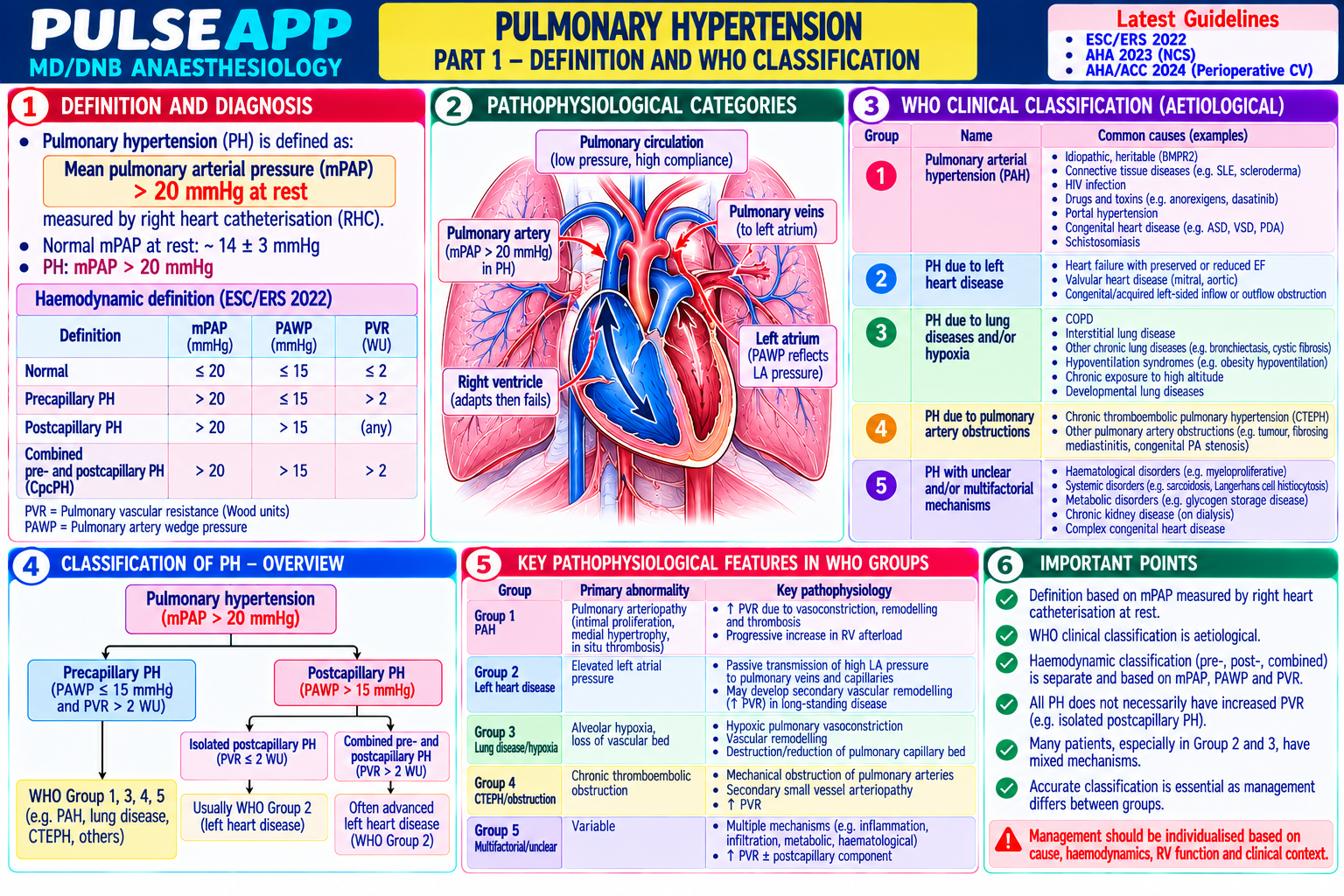
Transfusion reactions range from mild (FNHTR — urticaria) to potentially fatal (AHTR, TRALI) and must be rapidly distinguished from each other — because the management of each is different, and misidentification can lead to administering the wrong treatment. TRALI (pulmonary — non-cardiogenic) vs TACO (pulmonary — cardiogenic overload) is the most important clinical distinction to make, as their management is diametrically opposite (diuretics for TACO, contraindicated in TRALI). (Toy P — TRALI definition; Silliman CC; Taylor C — SHOT report 2023; BCSH Transfusion guidelines; Miller's Anaesthesia 9th Ed)
A. Classification of Acute Transfusion Reactions1 mark

Immunological: AHTR (ABO incompatibility); FNHTR (antibodies to white cell antigens); allergic/anaphylactic (IgE-mediated to plasma proteins); TRALI (antibody-mediated lung injury); platelet refractoriness

Non-immunological: TACO (circulatory overload); septic transfusion reaction (bacterial contamination — most common from platelet concentrates); air embolism; hypothermia; hyperkalaemia; hypocalcaemia (citrate toxicity)

B. Acute Haemolytic Transfusion Reaction (AHTR) — ABO Incompatibility3 marks

Pathophysiology: ABO-incompatible blood (e.g., type A blood given to a type O patient) → pre-formed IgM antibodies in the recipient (anti-A, anti-B) bind donor RBCs → complement activation → intravascular haemolysis → massive haemoglobin release → renal tubular haemoglobin precipitation → acute tubular necrosis → acute kidney injury; simultaneous: DIC (from tissue factor release from haemolysed RBCs + complement activation); systemic inflammatory cytokine storm; hypotension; shock

Clinical features: in the awake patient: burning pain at the IV site; lumbar/flank pain (renal haemoglobin deposition); fever and rigors; hypotension; haemoglobinuria (dark/red urine); in the ANAESTHETISED patient (most dangerous — all symptoms masked): unexplained hypotension; unexplained haemoglobinuria (dark urine in the catheter); oozing from surgical wounds (DIC); haemoglobin in the urine Management: STOP the transfusion immediately; send the blood bag and fresh patient blood sample to the transfusion laboratory (with the transfusion number documented to identify the specific unit) IV fluid resuscitation (maintain urine output ≥1 mL/kg/hr to prevent renal tubular precipitation of haemoglobin); monitor urine colour

Treat DIC: FFP, cryoprecipitate, platelets as guided by TEG/ROTEM; transfuse with compatible blood only Vasopressors for hypotension; monitor for AKI (creatinine, urine output, K⁺); renal replacement therapy if established AKI Notify blood bank, transfusion physician, and haematology; SHOT (Serious Hazards of Transfusion) reporting

C. TRALI vs TACO — Critical Distinction4 marks

Feature TRALI (Transfusion-Related Acute Lung Injury) TACO (Transfusion-Associated Circulatory Overload) Mechanism Non-cardiogenic pulmonary oedema; donor antibodies (anti-HLA or anti-neutrophil) in Cardiogenic pulmonary oedema; volume overload from the transfused blood activate recipient neutrophils → neutrophil sequestration in transfusion in a patient with limited cardiac reserve → ↑ pulmonary capillaries → neutrophil degranulation → capillary leak → interstitial and hydrostatic pressure in pulmonary capillaries → fluid alveolar oedema transudation into alveoli Risk factors Multiparous female donors (higher anti-HLA antibody prevalence); male donors Pre-existing heart failure (EF <40%); elderly; renal failure; increasingly used for plasma/FFP to reduce TRALI risk; surgery, mechanical rapid large-volume transfusion; anaemia (lower threshold for ventilation, active infection (priming the neutrophil "second hit") transfusion, larger volume gap) Clinical onset During or within 6 hours of transfusion; often within 1–2 hours During or within 6 hours of transfusion; often during the transfusion itself Respiratory Acute hypoxaemia (PaO₂/FiO₂ <300 mmHg); bilateral pulmonary infiltrates on CXR Acute hypoxaemia; bilateral pulmonary infiltrates; frothy pink features (non-cardiogenic pattern); frothy white sputum if severe sputum Haemodynamic Normal or LOW blood pressure (inflammatory vasodilation); JVP normal or low; NO ↑ Blood pressure (hypertension); ↑ JVP; ↑ CVP; evidence of evidence of cardiac overload; no response to diuretics (not cardiogenic) cardiac overload; responds to diuretics BNP/NT- Normal or mildly elevated Markedly elevated (BNP >250 pg/mL; NT-proBNP >1500 proBNP pg/mL) ECHO/TOE Normal or hyperdynamic LV; no pericardial effusion Reduced EF; dilated LV; ↑ LVEDP; pericardial effusion (if chronic HF) Management STOP transfusion; supportive: O₂ → NIV → IPPV with ARDS-protective ventilation if STOP or slow transfusion; furosemide 40–80 mg IV; O₂ → needed; NO diuretics (not fluid overload); NO steroids (no proven benefit); notify blood NIV; treat underlying cardiac failure; vasodilators if bank; save blood bag for donor antibody testing hypertensive Mortality 5–10%; leading cause of transfusion-related death (SHOT data); severe TRALI has 5%; lower than TRALI with appropriate management; higher 25–40% mortality with ARDS in severe cardiac failure 25–40% mortality with ARDS in severe cardiac failure

D. FNHTR2 marks

Febrile non-haemolytic transfusion reaction: temperature rise ≥1°C during or within 4 hours of transfusion; caused by cytokines in stored blood products (IL-6, IL-8, TNF — accumulate during storage) OR recipient antibodies reacting against donor HLA antigens on white cells; now less common since universal leucodepletion (removal of WBCs from blood products) in the UK

Management: STOP the transfusion; assess the patient (rule out AHTR — haemolysis; rule out septic reaction); if fever is mild and isolated (no haemoglobinuria, no haemodynamic compromise, no rigors): can cautiously restart at a slower rate after paracetamol 1 g oral/IV and 15–30 minutes observation; if fever >2°C, rigors, or any other concerning features → do NOT restart; full AHTR workup (DAT, blood cultures, transfusion laboratory)

🎤 Viva Corner
Q. An anaesthetised patient receiving their third unit of packed red cells develops sudden onset SpO₂ 82% despite FiO₂ 1.0, BP 90/50, and bilateral wheeze on auscultation. How do you differentiate TRALI from TACO and what is your immediate management?
This is an acute life-threatening transfusion reaction requiring simultaneous rapid assessment and management. Stop the transfusion immediately — the first action regardless of the specific diagnosis. The clinical picture: onset during the third unit of transfusion; severe hypoxaemia (SpO₂ 82% on 100% O₂ = PaO₂/FiO₂ likely <200 mmHg = severe); bilateral wheeze; hypotension (BP 90/50) — this haemodynamic pattern is KEY to the TRALI vs TACO distinction: TACO characteristically presents with HYPERTENSION (cardiac overload from volume overload raises BP), while TRALI presents with NORMAL BP or HYPOTENSION (inflammatory vasodilation); the BP 90/50 in this patient strongly favours TRALI over TACO. Additional differentiating information I would immediately obtain: JVP or CVP (TRALI = normal/low; TACO = raised); if TOE is available — LV function (TRALI = normal or hyperdynamic; TACO = reduced EF, dilated LV); stat BNP or NT-proBNP (TRALI = normal/mildly elevated; TACO = markedly elevated); CXR (both show bilateral infiltrates — not discriminating alone). Management — assuming TRALI (supported by hypotension): continue 100% O₂; if NIV or invasive ventilation is needed → ARDS-protective ventilation (6 mL/kg IBW TV, PEEP 8–12, FiO₂ to maintain SpO₂ ≥94%); for the hypotension: cautious fluid challenge 250 mL 0.9% NaCl (DO NOT give diuretics — furosemide would worsen TRALI hypotension catastrophically); vasopressors (noradrenaline) if fluid does not restore BP; DO NOT give steroids (no proven benefit and potential harm); notify the blood bank: stop all current transfusions; save the blood bag and IV set for TRALI investigation (donor anti-HLA/anti-neutrophil antibody testing); take fresh blood samples (FBC, DAT, renal function, LFT, BNP); send to transfusion laboratory. If I was wrong and this is actually TACO: the NIV/IPPV support is still appropriate; the absence of diuretics initially is less harmful than giving diuretics in true TRALI would be; once BNP results return and ECHO confirms reduced EF, furosemide 40–80 mg IV can be added — so the initial TRALI-first management is the safer default when BP is low and the diagnosis is uncertain.
★ Examiner's Pearl
TRALI vs TACO — the single most important clinical distinction: TRALI = hypotension (vasodilation), normal BNP, normal or hyperdynamic LV, NO response to diuretics; TACO = hypertension (overload), markedly elevated BNP, reduced EF, RESPONDS to diuretics. Giving furosemide to a TRALI patient worsens shock and mortality. AHTR in the anaesthetised patient: all symptoms are masked — unexplained hypotension + dark urine + surgical oozing (DIC) are the only signs. STOP transfusion is the FIRST action in ALL transfusion reactions regardless of type.
Toy P et al. Transfusion-related acute lung injury — definition and review (Crit Care Med 2005;33:721-726). Silliman CC et al. TRALI (Transfusion 2003;43:1-8). Taylor C et al. SHOT Annual Report 2023. BCSH Transfusion Guidelines. Miller RD et al. Miller's Anaesthesia, 9th Ed.
lock Protected Clinical Document • Pulse App • Screenshots Strictly Prohibited
QUESTION 122 person Asked by .
bookmark_add

Describe the cardiovascular and respiratory physiological effects of CO₂ pneumoperitoneum. Outline the additional challenges of steep Trendelenburg position and robotic-assisted radical prostatectomy (RARP) — airway, ICP, and access considerations.

description Clinical Response
⚙ Core Concept
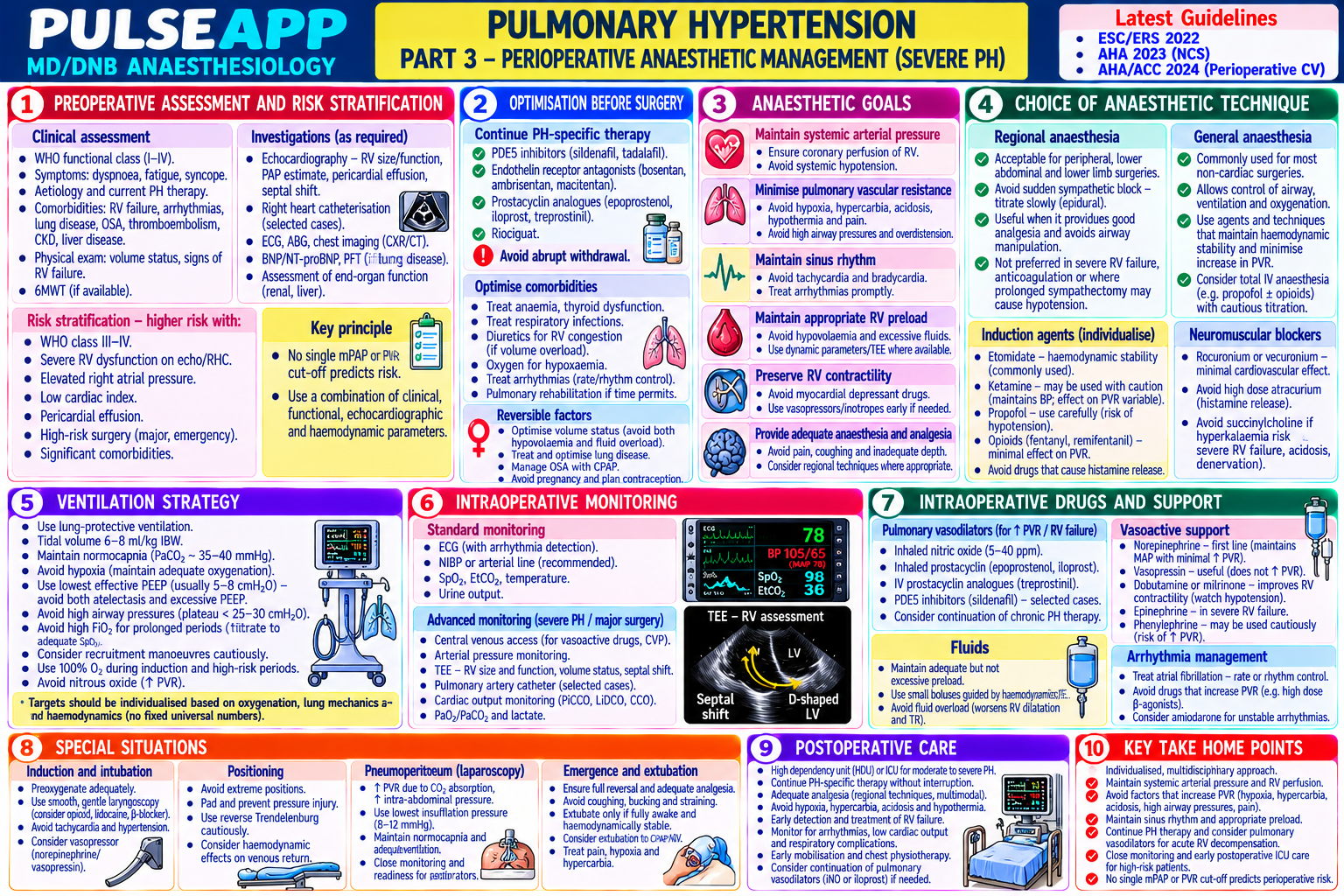
Laparoscopic and robotic surgery create compounding physiological challenges: CO₂ pneumoperitoneum raises intra-abdominal pressure and delivers CO₂ systemically, and steep Trendelenburg (30–45° head-down) redistributes blood volume cephalad, raises ICP, and progressively worsens pulmonary mechanics. RARP combines both with extreme positioning for up to 4 hours, limited intraoperative patient access, and specific post-operative complications. (Joris JL — laparoscopy physiology; Casati A; Miller's Anaesthesia 9th Ed)
A. CO₂ Pneumoperitoneum — Physiological Effects3 marks

System Intra-abdominal Pressure (IAP) Effect CO₂ Absorption Effect Cardiovascular IAP 15 mmHg → IVC compression → ↓ venous return → ↓ CO; reflex peripheral CO₂ absorption → hypercapnia → sympathetic activation → ↑ vasoconstriction (↑ SVR) maintains MAP initially; at IAP >20 mmHg → HR, ↑ SVR (partially counteracts IVC compression effect); pH ↓ decompensated ↓ CO; ↑ CVP paradoxically (from abdominal compression) from respiratory acidosis Respiratory Diaphragm pushed cephalad → ↓ FRC → ↓ compliance → ↑ peak airway pressure CO₂ absorbed from the peritoneum → ↑ PaCO₂; increase minute → atelectasis; cephalad displacement of the carina (risk of right main bronchus ventilation by 15–25% to maintain normocapnia; ETCO₂ intubation when ETT was positioned correctly pre-inflation) underestimates PaCO₂ by 3–5 mmHg in healthy patients (V/Q mismatch widens this gap in sick patients) Renal IAP → ↓ renal cortical perfusion (renal artery compression + ↑ renal venous — pressure) → ↓ GFR → ↓ urine output intraoperatively (not indicative of hypovolaemia if urine output returns to normal after desufflation) Surgical Gas embolism (CO₂ injected directly into vessel during Veress needle insertion or — complications trocar placement); capnothorax (CO₂ tracking through diaphragm → pneumothorax); subcutaneous emphysema (CO₂ tracking into subcutaneous tissue → dramatically elevated ETCO₂, palpable crepitus)

B. Steep Trendelenburg — Additional Effects2 marks

Further ↓ FRC (abdominal viscera push diaphragm cephalad + IAP from pneumoperitoneum combined); peak airway pressures may reach 35–45 cmH₂O in steep Trendelenburg with pneumoperitoneum — especially in obese patients; manage with pressure-controlled ventilation (avoids high pressures), ↑ PEEP (8– 12 cmH₂O), and accepting slightly higher ETCO₂ if permissive hypercapnia is tolerated Cephalad redistribution of blood volume → ↑ cerebral blood flow → ↑ ICP; risk of cerebral oedema with prolonged steep positioning; raised intraocular pressure (IOP) — particularly concerning in pre-existing glaucoma; facial and conjunctival oedema (from ↑ venous pressure); post-operative airway oedema (from prolonged head-down → laryngeal oedema) — assess for stridor before extubation; consider leaving the ETT in situ longer if significant facial oedema is noted Brachial plexus injury (steep Trendelenburg with shoulder braces pushing into the supraclavicular region) — avoid shoulder braces where possible; use nonslip mattress and table friction instead; position arms alongside the body, well padded

C. RARP-Specific Challenges2 marks

Extreme (30–45°) steep Trendelenburg maintained for 3–4 hours; robot is docked over the patient → complete loss of patient access (cannot reach the head, cannot change patient position, cannot perform emergency airway manoeuvres) during surgery; pre-dock airway security is paramount: confirm ETT position with ETCO₂ waveform, chest auscultation, and bilateral breath sounds BEFORE docking; check ETT tie/tape security; tape eyes (cannot monitor or treat complications of eye during the docked phase)

Specific risks: endobronchial intubation from cephalad ETT migration after Trendelenburg (auscultate after final positioning); CO₂ embolism from large venous plexuses in the pelvis; significant blood loss requiring transfusion (but cannot access patient for lines if robot is docked — ensure adequate IV access before docking); haemodynamic instability during extended pneumoperitoneum in elderly prostatectomy patients with ischaemic heart disease

Post-operative: post-extubation stridor from laryngeal oedema → treat with nebulised adrenaline 1 mg in 4 mL 0.9% NaCl; consider re-intubation if severe; PONV high from peritoneal insufflation and opioids → TIVA preferred; bladder irrigation post-RARP may absorb significant volumes (dilutional hyponatraemia monitoring)
🎤 Viva Corner
Q. During RARP, ETCO₂ suddenly rises from 38 to 68 mmHg and you notice crepitus on the patient's chest wall. What has happened and what do you do?
The sudden rise in ETCO₂ to 68 mmHg combined with chest wall crepitus (subcutaneous emphysema) indicates a CO₂-related complication of pneumoperitoneum: most likely extensive subcutaneous emphysema with CO₂ tracking from the peritoneal cavity through a trocar or peritoneal defect into the subcutaneous tissues of the abdominal wall, chest, and potentially the mediastinum. This is one of the commonest causes of sudden ETCO₂ rise during laparoscopy — CO₂ is absorbed rapidly from subcutaneous tissue (large surface area) far more rapidly than from the peritoneal cavity alone. Less likely but more dangerous: capnothorax (CO₂ tracking into the pleural space through a diaphragmatic defect); CO₂ embolism (would cause sudden fall in ETCO₂, not a rise). Immediate actions: inform the surgeon immediately; increase minute ventilation (increase respiratory rate and/or tidal volume within safe limits — plateau ≤30 cmH₂O) to bring ETCO₂ down; this buys time while the source is addressed. Ask the surgeon to: reduce the pneumoperitoneum pressure (lower insufflation IAP from 15 mmHg to 10 mmHg or lower) — this reduces CO₂ absorption; inspect the trocar sites for gas leak (trocar malposition); continue surgery if feasible at lower IAP. Obtain ABG to confirm respiratory acidosis severity (pH and PaCO₂); monitor haemodynamics (hypercapnia causes sympathetic stimulation → ↑ HR and BP); check for tension capnothorax (absent breath sounds on one side, ↓ SpO₂, haemodynamic compromise) — if tension capnothorax is suspected → needle decompression, convert to open surgery; extensive subcutaneous emphysema will resolve spontaneously within 1–2 hours of desufflation — no specific treatment required except ventilatory management of hypercapnia; post-operatively: monitor ETCO₂ in recovery until it normalises; warn the patient they may experience subcutaneous crepitus for 24–48 hours (benign, self-limiting).
★ Examiner's Pearl
Intraoperative CO₂ pneumoperitoneum at IAP 15 mmHg: paradoxical ↑ CVP (from abdominal compression transmitting to the central veins) + ↓ venous return (IVC compression) + ↑ SVR (reflex vasoconstriction) — the combination of ↑ CVP + ↓ CO is counter-intuitive and frequently tested. ETCO₂ underestimates PaCO₂ during laparoscopy (V/Q mismatch — PaCO₂ may be 3–10 mmHg higher than ETCO₂ depending on pre-existing lung disease). Sudden ETCO₂ rise during laparoscopy = subcutaneous emphysema (most common) vs CO₂ embolism (sudden fall) vs capnothorax — the ETCO₂ direction distinguishes these.
Joris JL et al. Hemodynamic changes during laparoscopic cholecystectomy (Anesth Analg 1993;76:1067-1071). Casati A et al. Laparoscopic surgery (Curr Opin Anaesthesiol 2003;16:581-586). Miller RD et al. Miller's Anaesthesia, 9th Ed.
lock Protected Clinical Document • Pulse App • Screenshots Strictly Prohibited
QUESTION 123 person Asked by .
bookmark_add

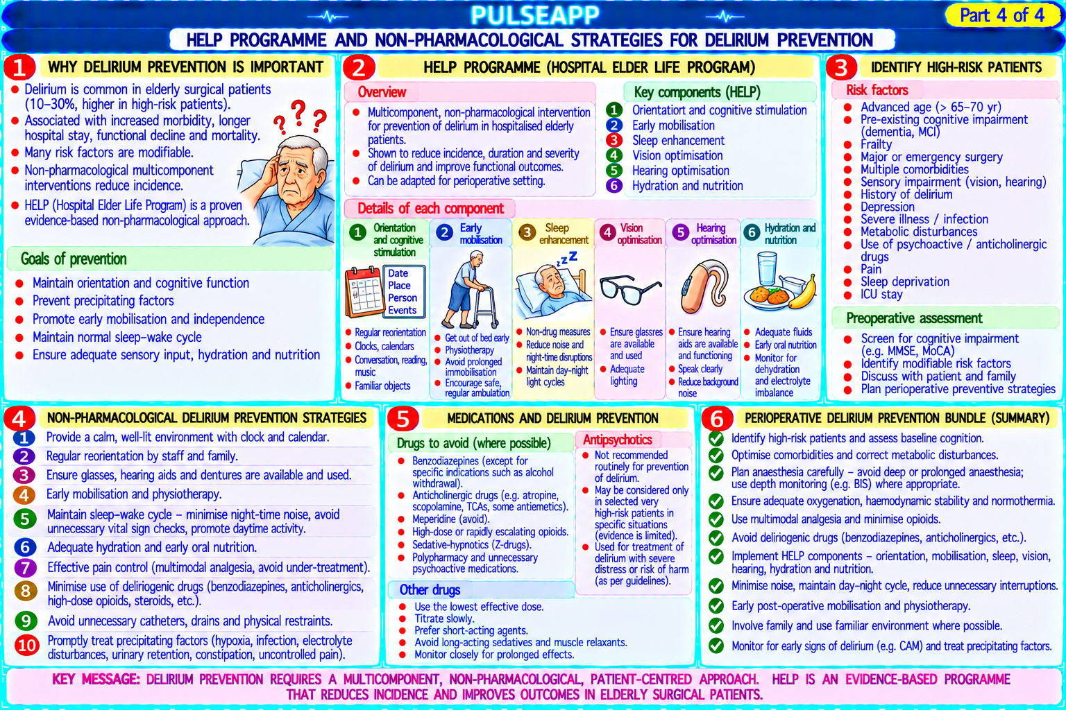
Describe the physiological changes of ageing relevant to anaesthesia. Outline the pharmacological implications (MAC reduction with age, altered PK). Discuss post-operative cognitive dysfunction (POCD) and the ISPOCD study. Describe the HELP programme and non-pharmacological strategies for delirium prevention.

collections Question Diagrams & Reference Images (1)
Question Reference Diagram
zoom_in View Image
description Clinical Response
⚙ Core Concept
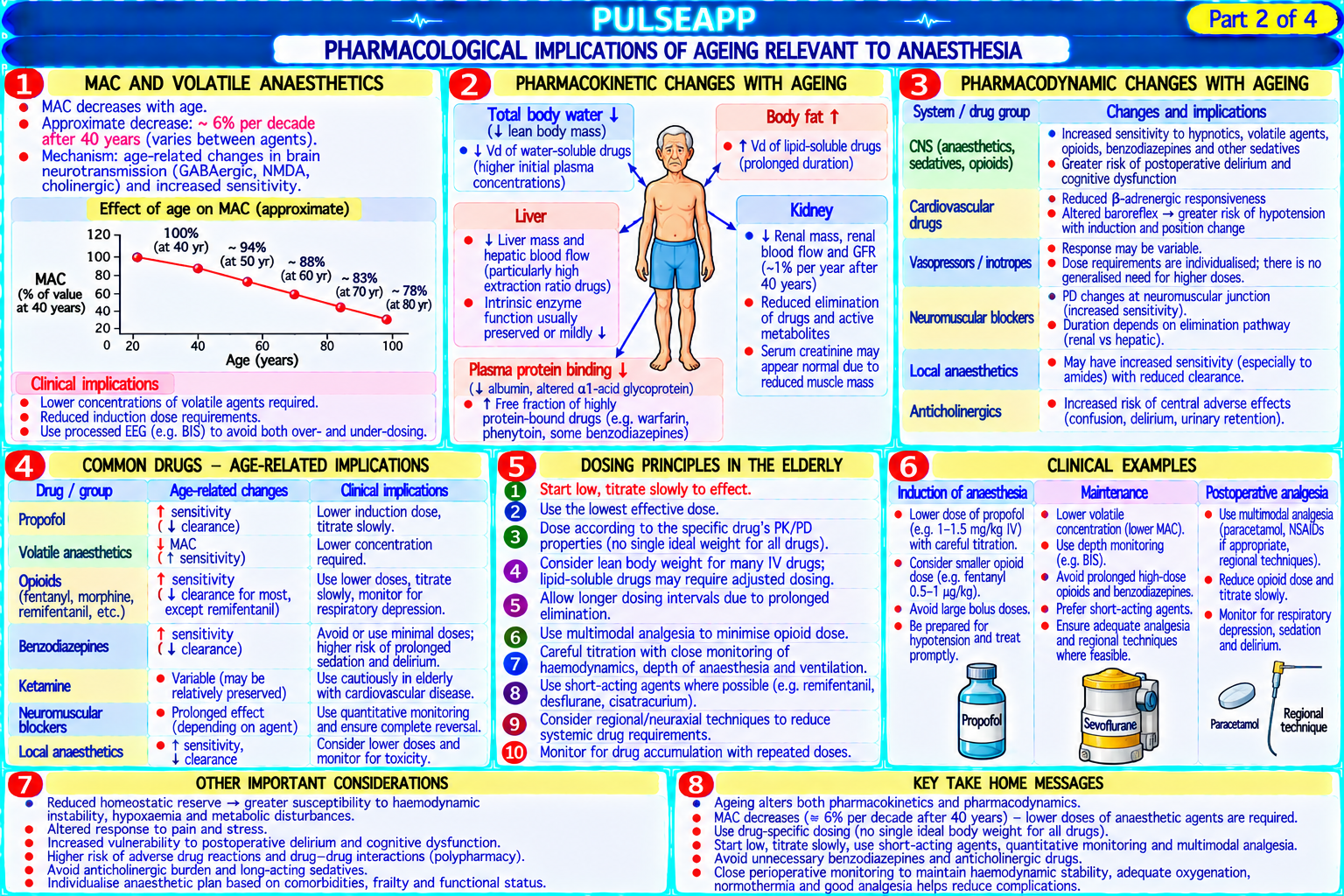
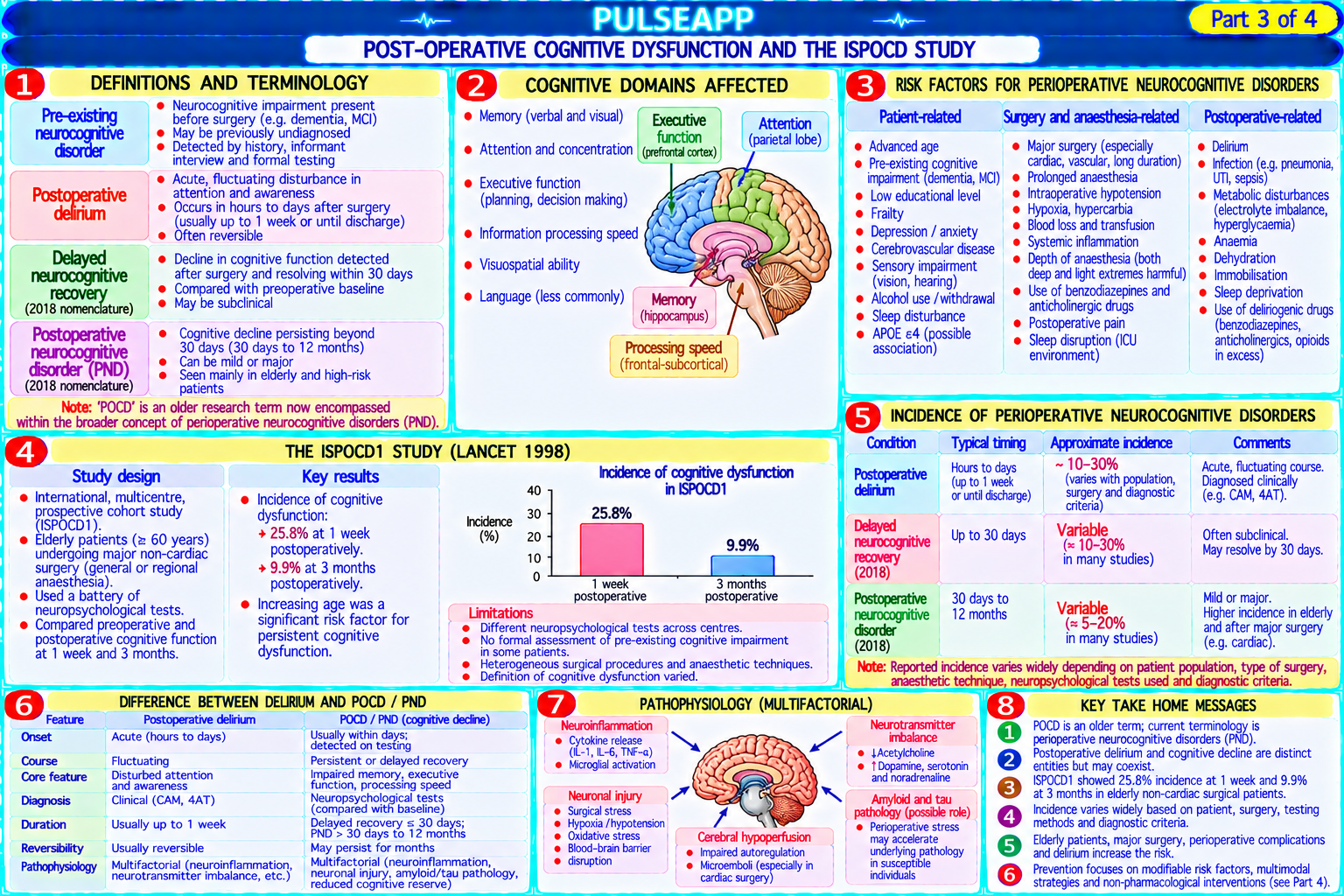
The elderly surgical patient presents compound challenges: multiple comorbidities, polypharmacy, reduced physiological reserve, altered drug pharmacokinetics (↓ albumin, ↓ renal clearance, ↓ hepatic flow), and particular vulnerability to post-operative delirium and cognitive dysfunction. POCD — a subtle decline in cognitive function persisting weeks to months post-surgery — affects 25% of patients over 60 after major surgery and is distinct from acute delirium. (Moller JT — ISPOCD1 Lancet 1998; Inouye SK — HELP program; Eckenhoff RG; Miller's Anaesthesia 9th Ed)
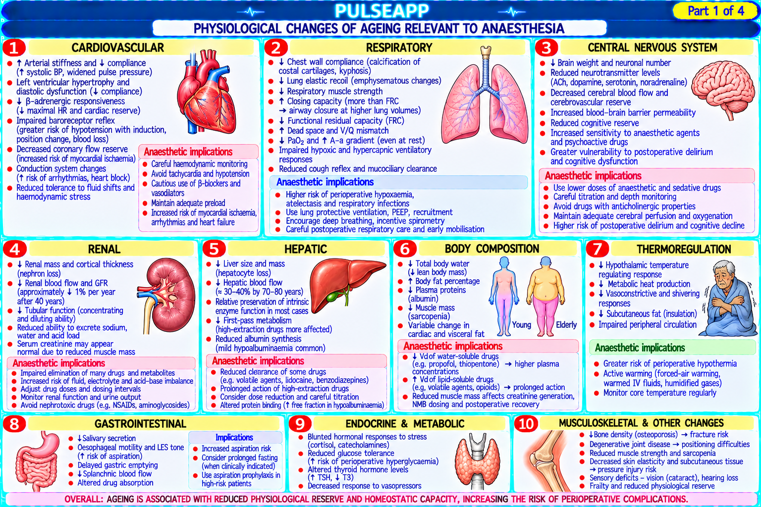
A. Physiological Changes of Ageing2 marks

System Key Changes Anaesthetic Implication Respiratory ↓ FVC, ↓ FEV1 (~1% per year after 30); ↓ FRC; ↑ closing capacity (exceeds FRC at Rapid desaturation during apnoea; ↑ atelectasis tendency; 65 yrs upright, 44 yrs supine); ↓ hypoxic and hypercapnic ventilatory response; ↓ impaired aspiration clearance; higher FiO₂ may be mucociliary clearance; ↓ cough reflex required during surgery Cardiovascular ↓ Maximum heart rate (220 − age); ↓ cardiac reserve; diastolic dysfunction; ↑ SVR; ↑ Haemodynamic fragility; hypotension more likely with systolic hypertension; ↑ atrial fibrillation risk; ↓ baroreceptor sensitivity; ↓ induction; poor tolerance of rapid volume shifts; AF risk responsiveness to catecholamines (β-receptor downregulation) with surgery; blunted compensatory tachycardia Renal/Hepatic ↓ GFR (by 50% at 80 vs 30 years); ↓ renal tubular function; ↓ hepatic blood flow (by ↑ Drug accumulation for renally cleared drugs (morphine, 40%); ↓ hepatic enzyme activity gabapentin, some NMBs); prolonged action of hepatically metabolised drugs; drug dose reduction essential Neurological ↓ Brain volume (cortical atrophy); ↓ neurotransmitter synthesis; ↓ pain perception Reduced anaesthetic requirements (↓ MAC); cognitive threshold variation; ↑ anxiety; ↑ delirium susceptibility vulnerability; pre-operative cognitive assessment (baseline) Pharmacokinetics ↓ Albumin → ↑ free fraction of protein-bound drugs (warfarin, diazepam, propofol); ↑ Dose reduction for most sedatives and analgesics; start body fat:muscle ratio → ↑ Vd for lipophilic drugs (benzodiazepines, volatile agents); ↓ low and titrate total body water → ↓ Vd for water-soluble drugs (NMBs, morphine)

B. MAC Reduction with Age2 marks

MAC decreases approximately 6% per decade of life after age 40; an 80-year-old requires approximately 25–30% less volatile agent than a 40-year-old for the same depth of anaesthesia; this is due to: ↓ neuronal density and ↓ synaptic connections in the aged brain; ↓ CNS neurotransmitter turnover; ↑ sensitivity of inhibitory pathways (GABA-A) and ↓ excitatory (NMDA) activity

Practical implications: overly deep anaesthesia is common in the elderly with standard adult volatile agent concentrations → hypotension, prolonged emergence, delirium; processed EEG (BIS target 40–60) is particularly valuable in elderly patients to avoid both over-sedation and underdosing

Propofol: Schnider TCI model automatically adjusts for age-related pharmacokinetic changes (reduces central compartment volume and clearance estimates with increasing age) → lower initial bolus and infusion rates are automatically calculated for elderly patients; still titrate carefully

C. POCD and the ISPOCD Study3 marks

POCD definition: Post-Operative Cognitive Dysfunction — a measurable decline in cognitive test scores (memory, concentration, executive function) in the weeks to months following surgery, compared to the individual's pre-operative baseline; it is DISTINCT from delirium (which is acute, fluctuating, and begins within days); POCD may persist for months or years in a subset of patients; associated with reduced quality of life, impaired return to work, and increased mortality ISPOCD1 (Moller JT, Lancet 1998; n=1218 patients ≥60 years having major non-cardiac surgery): POCD was present in 25.8% of patients at 1 week post-op and 9.9% at 3 months; risk factors identified: age ≥60; pre-existing cognitive impairment; lower educational attainment; second procedure and postoperative infections; anaesthetic technique (GA vs regional) was NOT significantly associated with POCD risk — this finding challenged the theory that GA causes POCD; ISPOCD2 confirmed these findings

Pathophysiology of POCD: neuroinflammation (surgical inflammation triggers neuroinflammatory cascades via the blood-brain barrier); sleep deprivation; hypotension episodes causing microischaemia; anaesthetic agents may contribute (some evidence that volatile agents and benzodiazepines increase neuroinflammation); exact mechanisms remain under investigation

D. HELP Programme — Delirium Prevention3 marks

The Hospital Elder Life Program (HELP; Inouye SK, NEJM 1999): a structured multi-component non-pharmacological intervention to prevent delirium in hospitalised elderly patients; implemented by trained volunteers and nursing staff; has been shown to reduce delirium incidence by 33% and falls by 32% in medical patients; widely adapted to surgical and ICU settings HELP Components (targeting modifiable delirium risk factors):

Cognitive orientation: daily reorientation (clock, calendar, date); cognitive exercises; encouraging family participation in orientation

Mobility: early mobilisation out of bed within 24 hours of surgery; daily walking programme if able; avoid physical restraints (increase delirium)

Vision: provision of glasses or magnifying glass; adequate room lighting; large-faced clocks and calendars visible

Hearing: provision of hearing aids; speaking clearly and loudly; amplifying devices

Sleep: non-pharmacological sleep promotion (warm milk, relaxation); avoid nighttime interruptions; noise and light reduction; scheduled medications to avoid nighttime disturbance; avoid benzodiazepines for sleep (worsen delirium)

Hydration/Nutrition: ensure adequate oral intake; dehydration is a major delirium trigger; dentures provided for eating

Pharmacological considerations: haloperidol 0.5–1 mg oral/IV for hyperactive delirium symptom management (not prevention); dexmedetomidine-based ICU sedation reduces delirium vs benzodiazepines; avoid benzodiazepines (particularly in elderly — increase delirium 3-fold); avoid anticholinergic drugs (hyoscine, diphenhydramine, TCAs — all worsen delirium); avoid polypharmacy

🎤 Viva Corner
Q. Does general anaesthesia cause POCD — what does the evidence say?
The question of whether general anaesthesia causes POCD has been extensively studied and remains nuanced. The short answer from the best available evidence is: general anaesthesia per se is probably NOT the primary cause of POCD, and patient factors (age, pre-existing cognitive impairment, comorbidities) and surgical factors (neuroinflammation from surgery, hypotension, infection) appear to be more important determinants. The landmark ISPOCD1 study (Moller JT, Lancet 1998; n=1218 patients ≥60 years) found that POCD was present in 25.8% at 1 week and 9.9% at 3 months after major non-cardiac surgery — but the anaesthetic technique (type of agent, depth of anaesthesia) was NOT significantly associated with the risk of POCD in multivariate analysis; the strongest risk factors were age, baseline cognitive function, education level, and post-operative complications. Multiple subsequent RCTs comparing GA vs regional anaesthesia (spinal/epidural) for various surgeries have generally failed to demonstrate a significant reduction in POCD with regional techniques. The REGAIN trial (hip fracture surgery, GA vs spinal, n=950 elderly patients) found no difference in survival or recovery of ambulatory function between GA and spinal anaesthesia at 60 days. However, there is emerging evidence that specific components of anaesthetic management matter: deep anaesthesia (BIS <40) appears to be associated with higher POCD risk than light anaesthesia (BIS 40–60) in some RCTs, suggesting that minimising anaesthetic depth (particularly avoiding burst suppression) in elderly patients is advisable; intraoperative hypotension (MAP <65 mmHg for prolonged periods) is an established risk factor for 30-day cognitive decline, and meticulous haemodynamic management may reduce POCD. The practical advice: maintain adequate MAP (avoiding prolonged hypotension), target light anaesthesia with BIS monitoring, avoid benzodiazepines, minimise opioids, use regional techniques where they provide proven benefit (reduced opioid requirement, better post-operative pain), and focus on the post-operative environment (HELP programme) as the most modifiable contributor to delirium and subsequent POCD.
★ Examiner's Pearl
MAC reduction with age: 6% per decade after age 40 (an 80-year-old needs ~25% less volatile agent than a 40-year-old). ISPOCD1 (Moller Lancet 1998): POCD in 25.8% at 1 week, 9.9% at 3 months; anaesthetic technique NOT significantly associated with POCD risk; age, baseline cognitive impairment, and post-operative complications are the main risk factors — this is the key finding that must be cited. HELP programme (Inouye NEJM 1999): 33% reduction in delirium with multicomponent non-pharmacological intervention — mobility, orientation, vision, hearing, sleep, hydration.
Moller JT et al. ISPOCD1 study — postoperative cognitive dysfunction (Lancet 1998;351:857-861). Inouye SK et al. HELP programme — multicomponent intervention for delirium (NEJM 1999;340:669-676). Eckenhoff RG et al. Inhaled anaesthetic enhancement of amyloid-beta oligomerization (Anesthesiology 2004;101:703-709). Miller RD et al. Miller's Anaesthesia, 9th Ed.
lock Protected Clinical Document • Pulse App • Screenshots Strictly Prohibited
QUESTION 124 person Asked by .
bookmark_add

Describe perioperative management of: beta-blockers (POISE trial), ACE-inhibitors/ARBs, statins, antiplatelet agents (aspirin and clopidogrel), and corticosteroids. Outline which drugs to continue, omit, or modify on the day of surgery and the evidence basis.

collections Question Diagrams & Reference Images (1)
Question Reference Diagram
zoom_in View Image
description Clinical Response
⚙ Core Concept
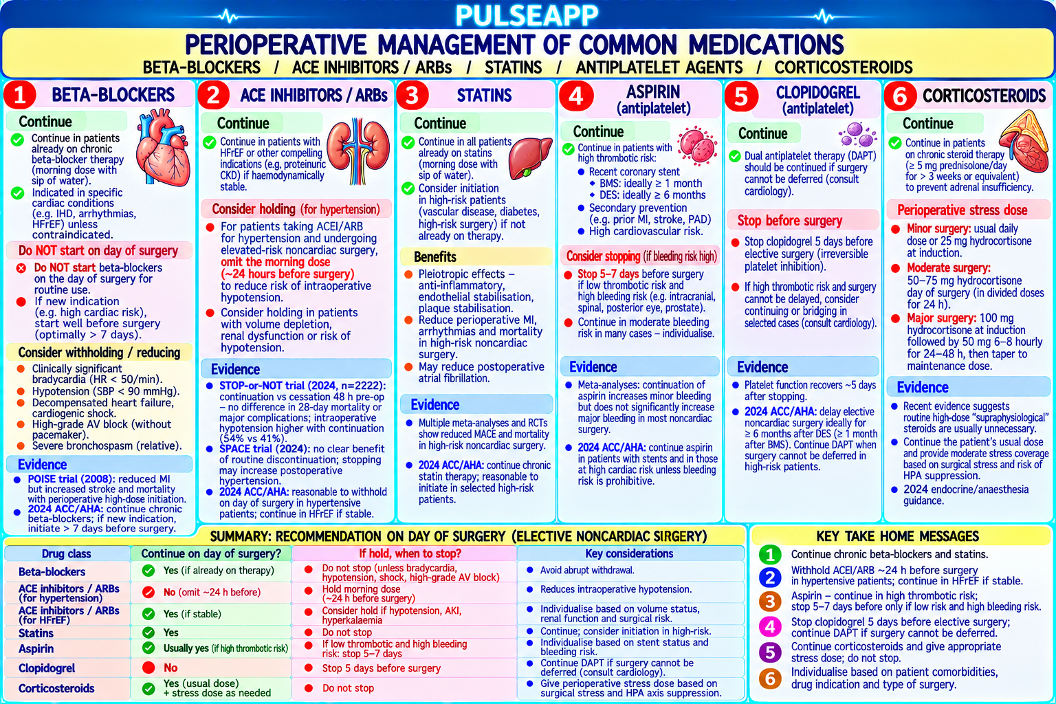
The perioperative management of chronic medications requires balancing the risks of continuing (bleeding, haemodynamic instability, drug interactions) against stopping (rebound effects, loss of cardioprotection, adrenal insufficiency). The POISE trial fundamentally changed perioperative beta-blocker prescribing — demonstrating that high-dose metoprolol reduced MI but increased stroke and overall mortality. (POISE trial NEJM 2008; Fleisher LA — AHA/ACC; ESC/ESA — non-cardiac surgery guidelines 2022; Miller's Anaesthesia 9th Ed)
A. Drug Management Table6 marks

Drug Class Continue or Omit? Evidence/Rationale Beta-blockers CONTINUE if the patient is already POISE trial (Devereaux PJ, NEJM 2008; n=8351): high-dose metoprolol succinate started 2–4h (chronic) chronically established on a beta-blocker; do before surgery in patients NOT previously on beta-blockers → ↓ MI (4.2% vs 5.7%), but ↑ stroke NOT stop suddenly (rebound tachycardia, (1.0% vs 0.5%), ↑ bradycardia, ↑ hypotension, ↑ all-cause mortality (3.1% vs 2.3%); lesson: do NOT ischaemia, hypertension — withdrawal start high-dose beta-blockers immediately before surgery in beta-blocker-naive patients; only start syndrome); convert to oral dose day-of- in selected high-risk patients with documented CAD if there is time for dose titration over weeks surgery (small sip of water) before surgery ACE-inhibitors / OMIT on the morning of surgery (for most ACE-I/ARBs block the renin-angiotensin system → impair the vasopressor response to anaestheticARBs patients having major surgery with general induced vasodilation → profound refractory intraoperative hypotension (particularly with neuraxial or anaesthesia); the ESC/ESA 2022 guideline volatile agents); hypotension in this setting is severe and poorly responsive to standard recommends withholding ACE-I/ARB on the vasopressors (phenylephrine, ephedrine) — may require vasopressin; the VISION study confirmed day of major non-cardiac surgery ↑ intraoperative hypotension with pre-operative ACE-I/ARB; omitting on the morning of surgery reduces but does not eliminate this risk; restart post-operatively when patient is eating and drinking (haemodynamically stable) Statins CONTINUE throughout the perioperative Abrupt statin withdrawal causes a rebound inflammatory state (↑ CRP, ↑ plaque instability, ↑ period; do NOT omit thrombotic risk); statins have pleiotropic benefits (anti-inflammatory, plaque-stabilising) that are lost acutely with withdrawal; observational studies suggest statin continuation is associated with lower perioperative cardiac events; if patient is NBM: some centres give their regular statin with a sip of water (safe for most statins which have t½ >12 hours); hold if IV formulation not available and patient is genuinely nil by mouth for >24 hours then restart as soon as oral route is available Aspirin (75 mg — CONTINUE for most cardiac-risk patients; Aspirin provides primary and secondary cardiovascular event prevention; POISE-2 trial (Devereaux antiplatelet) CONSIDER stopping 7 days before for very- PJ, NEJM 2014; n=10,010): perioperative aspirin did NOT reduce MACE (major adverse cardiac high bleeding risk surgery (neurosurgery, events) in non-cardiac surgery patients and increased major bleeding; however: patients already on posterior eye surgery, closed-cavity surgery) aspirin for established secondary prevention (post-MI, post-stent) should CONTINUE aspirin perioperatively — the POISE-2 findings apply to patients where aspirin was being considered as a

NEW perioperative intervention

Clopidogrel / STOP 5 days before surgery (ticagrelor: 5 P2Y12 inhibitors cause irreversible (clopidogrel, prasugrel) or reversible (ticagrelor) platelet P2Y12 inhibitors days; clopidogrel: 5 days; prasugrel: 7 days); inhibition; withholding for 5–7 days allows sufficient new platelet generation to restore haemostasis;

EXCEPTION: patients with recent (<1 year) for DES: cardiology/interventional cardiology must agree on minimum safe DAPT duration before drug-eluting stent (DES) or <6 weeks bare- elective surgery (typically ≥12 months for DES with 2nd generation stents — can consider at 6 metal stent — discuss with cardiology; months with cardiologist approval for truly essential elective surgery) premature DAPT interruption → stent thrombosis → MI with high mortality Corticosteroids CONTINUE regular dose; add stress-dose Patients on chronic steroids have HPA axis suppression → inadequate cortisol response to surgical (chronic >5 mg hydrocortisone for major surgery stress → adrenal insufficiency; stress-dose coverage: hydrocortisone 25–50 mg IV at induction + 25 prednisolone/day mg 8-hourly for 24 hours (minor surgery); 50–100 mg IV at induction + 50 mg 8-hourly for 48–72 >3 months) hours (major surgery); taper back to the patient's usual dose over 2–3 days; DO NOT abruptly stop steroids (Addisonian crisis)

B. Key Clinical Distinction — POISE Trial Summary1 mark

POISE trial key message: the HARM from starting beta-blockers (metoprolol 100 mg) in the immediate perioperative period in beta-blocker-naive patients OUTWEIGHS the cardiac benefit; do NOT routinely start beta-blockers within days of surgery; they may be started ONLY in high-risk patients if initiated weeks before surgery with careful dose titration; ALWAYS continue existing beta-blockers in patients already established on them

🎤 Viva Corner
Q. A patient on ramipril 5 mg OD for hypertension takes their morning tablet before coming to theatre. How does this affect your anaesthetic management?
This is a common scenario — the patient has taken their ramipril despite standard pre-operative instructions to omit ACE-inhibitors on the morning of surgery. The key clinical issue: ramipril blocks the renin-angiotensin system, which is the primary compensatory mechanism for maintaining blood pressure in the face of the vasodilatory effects of anaesthetic agents (both volatile and intravenous agents); when the RAS is blocked, the normal sympathetic and hormonal BP compensation is impaired → more profound and sustained intraoperative hypotension is expected, particularly at induction. Specific expectations: induction hypotension will likely be more severe than in a patient who omitted their ACE-I — prepare for a fall in MAP of 20–35 mmHg from baseline; this may occur within minutes of induction; standard vasopressors: phenylephrine works via α₁ (still functional with RAS blockade); ephedrine (β₁ and α₁ + indirect sympathomimetic) works partly; BUT the specific mechanism of severe ACE-I hypotension (bradykinin accumulation + impaired angiotensin II vasoconstriction) may make the hypotension poorly responsive to these agents and instead require: vasopressin (V₁ vasoconstriction — completely independent of the RAS) as an effective rescue; terlipressin. Pre-induction preparation: have phenylephrine and vasopressin drawn up; IV fluid pre-load 500 mL crystalloid before induction (higher preload helps buffer the hypotension); consider a lower induction dose of propofol; have the ephedrine/phenylephrine/vasopressin ready to administer; if neuraxial technique is planned → the hypotension will be even more profound with an ACE-I on board + sympathetic block from spinal/epidural → prepare vasopressor infusion before the block. Post-operatively: restart ramipril once the patient is eating, drinking, and haemodynamically stable — typically day 1 or 2 post-operatively.
★ Examiner's Pearl
POISE trial (Devereaux NEJM 2008): high-dose perioperative metoprolol → ↓ MI but ↑ stroke + ↑ all-cause mortality — do NOT start beta-blockers in the immediate perioperative period for beta-blocker-naive patients. ACE-I/ARB: OMIT on morning of surgery (refractory intraoperative hypotension via RAS blockade); vasopressin is the rescue vasopressor when standard agents fail. Statins: CONTINUE (withdrawal → rebound inflammation); corticosteroids: CONTINUE + stress dose hydrocortisone (HPA suppression). DAPT and DES: premature P2Y12 inhibitor withdrawal within 1 year of DES → stent thrombosis → MI — the most dangerous drug omission in perioperative medicine.
Devereaux PJ et al. POISE trial (NEJM 2008;358:1781-1794). ESC/EACTS Guidelines on Management of Patients Undergoing Non-cardiac Surgery 2022. Fleisher LA et al. ACC/AHA guideline on perioperative cardiovascular evaluation for noncardiac surgery 2014. Miller RD et al. Miller's Anaesthesia, 9th Ed.
lock Protected Clinical Document • Pulse App • Screenshots Strictly Prohibited
QUESTION 125 person Asked by .
bookmark_add

Describe the pericapsular nerve group (PENG) block — the anatomy of hip capsule innervation, the technique with ultrasound landmarks, and how it compares to the femoral nerve block (FNB) in hip arthroplasty analgesia. Cite the key RCT evidence (Lin et al. 2021 Anaesthesia).

description Clinical Response
⚙ Core Concept
The PENG (Pericapsular Nerve Group) block is an ultrasound-guided regional technique that targets the articular branches of the femoral nerve, accessory obturator nerve, and obturator nerve as they supply the anterior hip capsule — providing analgesia for hip arthroplasty and hip fracture WITHOUT the quadriceps weakness of a femoral nerve block. This motor-sparing property is particularly valuable for enabling early post-operative mobilisation. (Girón- Arango L — PENG block original description; Lin DY — Anaesthesia 2021; Mosaffa F; Miller's Anaesthesia 9th Ed)
A. Hip Capsule Innervation2 marks

The hip joint capsule has a rich nerve supply — primarily from the articular branches (sensory only) of three main nerves; these branches do NOT innervate the quadriceps (which are supplied by the motor branches of the femoral nerve — these are separate from the articular branches targeted by PENG)

Anterior capsule: articular branches of the FEMORAL NERVE (from the nerve to the rectus femoris and nerve to the vastus intermedius — these are pure articular sensory branches, distinct from the motor branches to the quadriceps); articular branches of the ACCESSORY OBTURATOR NERVE (when present — in 20–30% of individuals); articular branches of the OBTURATOR NERVE

Posterior capsule: branches of the sciatic nerve (superior gluteal nerve, nerve to the quadratus femoris)

PENG block targets: the anterior capsule innervation at the level of the anterior inferior iliac spine (AIIS) and ipsilateral eminence (IPE) — where these articular branches converge before entering the joint capsule

B. Ultrasound Technique2 marks

Patient position: supine; hip in neutral or mild external rotation

Probe: curved array (low frequency 2–5 MHz) or high-frequency linear probe; placed in a transverse plane at the level of the anterior hip; angled to align with the ilio-pubic eminence (IPE)

Landmarks: identify the anterior inferior iliac spine (AIIS) — the bony prominence of the ilium at the superior aspect; identify the ilio-pubic eminence (IPE) — the junction of the ilium and superior pubic ramus; the femoral artery and femoral nerve are identified medially; the psoas tendon is identified as the round, bright (hyperechoic) structure overlying the anterior hip capsule between the AIIS and IPE

Injection: in-plane needle approach; the injection target is the plane BETWEEN the psoas tendon anteriorly and the anterior hip capsule (the iliofemoral ligament) posteriorly; 20–25 mL bupivacaine 0.25% or ropivacaine 0.375% is deposited in this interfascial plane; the injectate spreads along the plane superficial to the hip capsule, bathing the articular branches of the femoral, accessory obturator, and obturator nerves as they course along the capsule

Motor-sparing mechanism: the injection does NOT target the femoral nerve trunk → the motor branches to the quadriceps are NOT blocked; only the articular sensory branches from the capsular surface are anaesthetised; patients retain full quadriceps power and can walk immediately post-operatively

C. PENG vs Femoral Nerve Block — Comparison2 marks

Feature PENG Block Femoral Nerve Block (FNB) Target Articular branches of femoral N, accessory obturator N, obturator N at AIIS/IPE Femoral nerve trunk at the femoral triangle (inguinal crease) level Motor NONE (motor-sparing — quadriceps function preserved) Quadriceps weakness (motor block of nerve to vastus lateralis, medialis, block intermedius) — falls risk; delays early mobilisation Analgesia Equivalent to FNB for anterior hip pain (surgical and fracture); may be less Good anterior hip analgesia; similar quality in many studies quality complete for posterior capsule pain (sciatic supply not included) Falls risk Minimal (quadriceps strength preserved) Significant — patients with FNB cannot be safely mobilised without supervised assistance; increased fall risk in hip fracture patients Adductor Partially preserved (obturator nerve articular branches blocked but main Not affected by FNB (FNB does not block the obturator nerve) strength obturator trunk usually not affected)

D. Key RCT Evidence — Lin et al. 2021 Anaesthesia1 mark

Lin DY et al. Pericapsular nerve group (PENG) block for hip arthroplasty (Anaesthesia 2021;76:1167–1173): RCT comparing PENG block vs placebo in total hip arthroplasty; PENG block significantly reduced: intraoperative opioid consumption; post-operative NRS pain scores at 24 hours; time to first analgesic request; PENG block group had preserved quadriceps function and achieved earlier mobilisation milestones vs placebo; no significant complications attributable to PENG block; conclusion: PENG block provides effective analgesia for THA with motor-sparing advantages consistent with an accelerated rehabilitation pathway

🎤 Viva Corner
Q. Why is the PENG block motor-sparing when the femoral nerve supplies both the quadriceps motor branches AND the articular branches of the hip — and we are injecting near the femoral nerve territory?
The key is the anatomical separation between the motor branches of the femoral nerve (which innervate the quadriceps) and the articular sensory branches (which supply the anterior hip capsule), and where the PENG block deposits the local anaesthetic relative to these two sets of branches. The femoral nerve, after passing under the inguinal ligament in the femoral triangle, immediately divides into its terminal branches: the anterior cutaneous branches (medial and intermediate cutaneous nerves of the thigh); the motor branches to the quadriceps muscles (nerve to rectus femoris, nerve to vastus lateralis, nerve to vastus medialis, nerve to vastus intermedius — these branches enter the quadriceps muscles in the thigh, well below the inguinal ligament); and crucially, the articular branches to the hip capsule — these are fine branches that arise from the nerve to the rectus femoris and the nerve to the vastus intermedius relatively close to the inguinal region, and they travel along the anterior aspect of the hip joint to supply the anterior capsule; these articular branches arrive at the ANTERIOR INFERIOR ILIAC SPINE and ILIO-PUBIC EMINENCE area, which is exactly where the PENG block deposits the local anaesthetic — in the interfascial plane between the psoas tendon and the hip capsule. This plane is above and anterior to where the motor branches have already diverged to travel toward the muscle bellies; by the time the local anaesthetic is deposited at the AIIS/IPE level, the motor branches are already traveling laterally and inferiorly toward the vastus muscles and are NOT exposed to the local anaesthetic spread in the PENG plane; only the fine articular branches that are still travelling along the capsular surface at this level are exposed to the drug. The motor branches have already 'left' this anatomical plane before the injection point — which is why the quadriceps strength is preserved.
★ Examiner's Pearl
PENG block injection target: the interfascial plane between the psoas tendon and the anterior hip capsule, at the level of the anterior inferior iliac spine (AIIS) and iliopubic eminence (IPE) — the two ultrasound bony landmarks must be named. Motor-sparing mechanism: the injection targets articular sensory branches (not the main femoral nerve trunk) → quadriceps motor branches are not blocked → no falls risk → early mobilisation possible. Lin et al. Anaesthesia 2021 is the key RCT to cite for evidence. Articular branches of the hip come from femoral N + accessory obturator N + obturator N — all three must be named.
Girón-Arango L et al. Pericapsular nerve group (PENG) block for hip fracture (Reg Anesth Pain Med 2018;43:859-863). Lin DY et al. PENG block for hip arthroplasty — RCT (Anaesthesia 2021;76:1167-1173). Mosaffa F et al. Comparison of PENG block with FNB in hip arthroplasty (Reg Anesth Pain Med 2022). Miller RD et al. Miller's Anaesthesia, 9th Ed.
lock Protected Clinical Document • Pulse App • Screenshots Strictly Prohibited
QUESTION 126 person Asked by .
bookmark_add

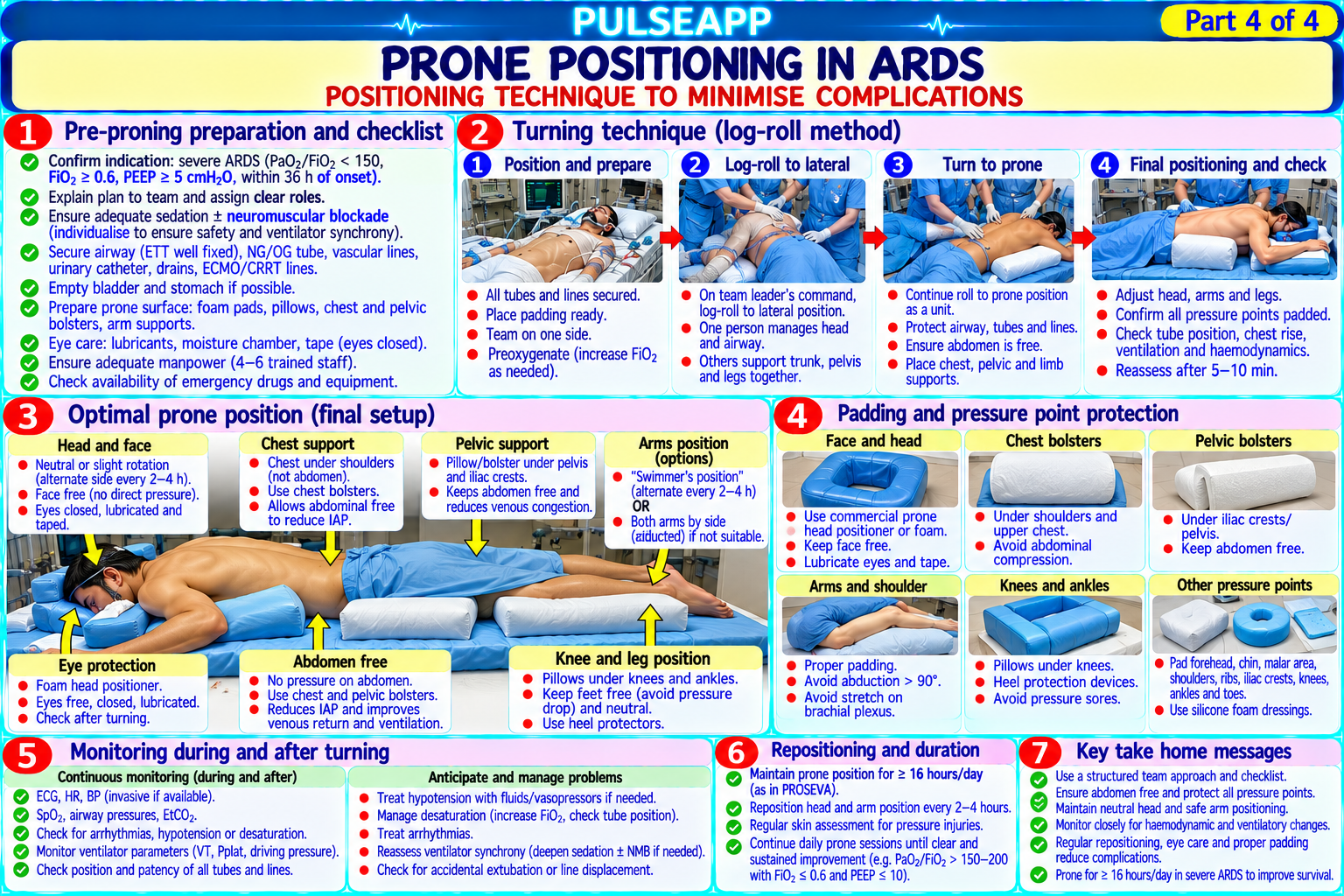
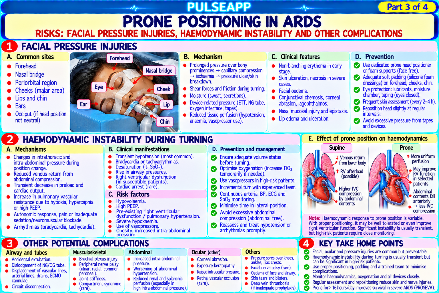
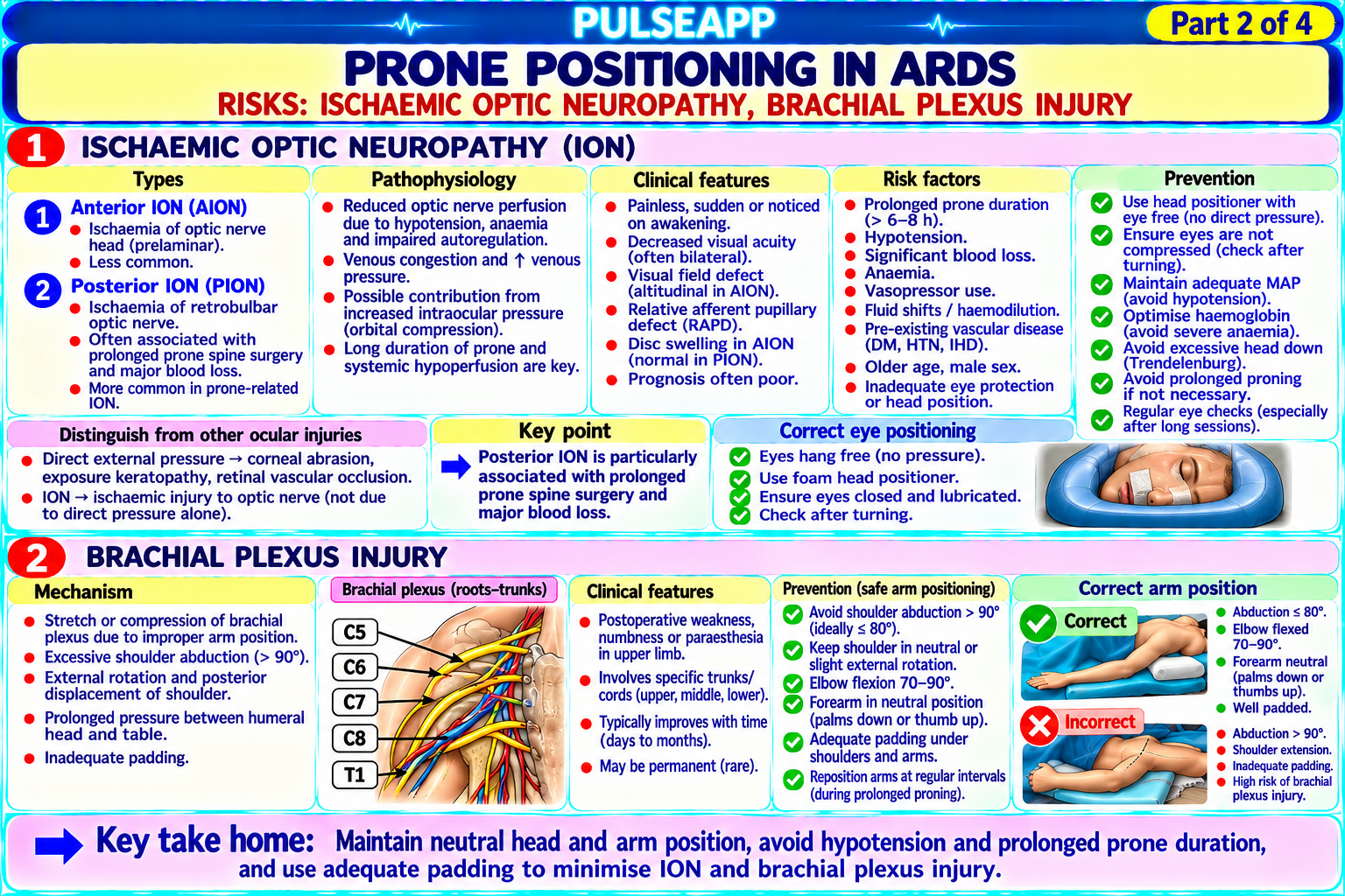
Describe the physiological benefits of the prone position in ARDS (PROSEVA trial). Outline the risks of prone positioning including: ischaemic optic neuropathy (ION), brachial plexus injury, facial pressure injuries, and haemodynamic instability during turning. Describe the positioning technique to minimise these complications.

collections Question Diagrams & Reference Images (1)
Question Reference Diagram
zoom_in View Image
description Clinical Response
⚙ Core Concept
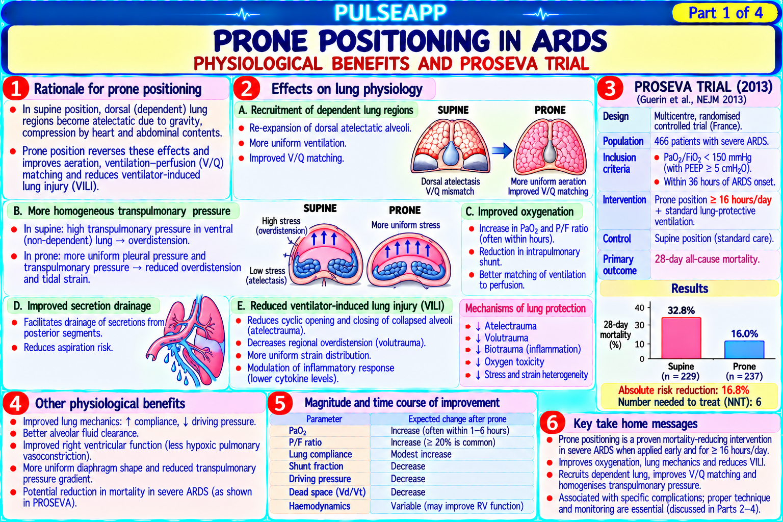
Prone positioning in severe ARDS has been shown to reduce 28-day mortality by 16% (PROSEVA trial, Guérin C, NEJM 2013) — a survival benefit equivalent to lung-protective ventilation and larger than any pharmacological intervention in ARDS. The physiological benefit is V/Q improvement from dorsal lung recruitment. However, prone positioning carries a significant risk of life-threatening complications (accidental extubation, haemodynamic collapse during turning) and position-related injuries (ION, brachial plexus, pressure ulcers) that require meticulous technique. (Guérin C et al. — PROSEVA NEJM 2013; Lee LA — ION in prone; Miller's Anaesthesia 9th Ed)
A. Physiological Benefits of Prone Positioning in ARDS3 marks

V/Q improvement mechanism: in ARDS, the dorsal (posterior) lung zones are the most severely consolidated and atelectatic (due to gravitational atelectasis — in the supine position, the heavy waterlogged lungs compress the dependent dorsal zones, which also receive the most blood flow due to gravity); in the prone position, the dorsal lung zones are now non-dependent → gravitational atelectasis is redistributed to the ventral (anterior) zones, which have less alveolar capacity; crucially, pulmonary blood flow redistributes less dramatically than ventilation in the prone position (the pulmonary vasculature is less responsive to gravity than the alveolar mechanics) → previously atelectatic dorsal lung regions open and are now both ventilated AND perfused → dramatic improvement in V/Q matching → ↑ PaO₂/FiO₂ ratio

More uniform stress distribution: the prone chest wall is stiffer (less compliant) than in the supine position — counterintuitively, this more uniform distribution of lung stress prevents overdistension of the non-dependent (now anterior/ventral) lung zones, which are already less consolidated; the dorsal zones, now nondependent, can be recruited at lower distending pressures; the result: more uniform alveolar inflation throughout the lung, less volutrauma to the small number of open alveoli

Improved secretion drainage: gravity assists secretion drainage from the posterior lung zones when prone (toward the main bronchus) → improved mucus clearance

B. PROSEVA Trial2 marks

Guérin C et al. PROSEVA (Proning Severe ARDS Patients) — NEJM 2013; n=466 patients with severe ARDS (PaO₂/FiO₂ <150 mmHg on FiO₂ ≥0.6, PEEP ≥5 cmH₂O): randomised to prone for at least 16 hours/day + lung-protective ventilation vs supine + LPV

Results: prone group: 28-day mortality 16.0% vs supine group 32.8% (absolute risk reduction 16.8%; relative risk 0.49; p<0.001); 90-day mortality 23.6% vs 41.0%; significantly greater improvement in PaO₂/FiO₂ in the prone group throughout the study; complications: higher rate of pressure ulcers, ETT displacement, and non-scheduled extubation in prone group (no difference in cardiac arrest during turning)

Indication for prone positioning: severe ARDS: PaO₂/FiO₂ <150 mmHg + FiO₂ ≥0.6 + PEEP ≥5 cmH₂O + adequate mechanical ventilation; initiated within 36 hours of ARDS diagnosis; minimum 16 continuous hours prone per session

C. Prone Position Complications and Prevention3 marks

Complication Mechanism Prevention Ischaemic Direct pressure on the orbit (from face-down position on an Head-specific prone support (Jackson frame, Mayfield head frame, padded optic unsupported face) → raised intraocular pressure → impaired optic horseshoe — ensuring NO pressure on the orbit or eyeball); confirm eye neuropathy nerve blood supply → optic nerve ischaemia → permanent position before draping (raise the head briefly and palpate to confirm the eye is (ION) blindness; prolonged prone anaesthesia (spine surgery >6 hours), not under any frame contact); check eyes every 30–60 minutes; maintain MAP hypotension, and anaemia increase risk; occurs in 0.1–0.2% of ≥80 mmHg; keep Hb ≥100 g/L; staged surgery if >6h anticipated prone spine cases but devastating when it does Brachial plexus Excessive arm abduction (>90°) in the prone position causes Arms alongside the body (preferred) or arms flexed at the elbow below injury traction on the brachial plexus (C5–T1 roots) between the clavicle shoulder level (<90° abduction); avoid the swimmer's position; pad all pressure and first rib; particularly the "swimmer's position" (one arm above the points (elbows, shoulders); check arm position after final prone positioning and head) → most common cause of brachial plexus injury in prone document spine surgery Facial pressure Prolonged pressure on face from headrest → ischaemic pressure Padded horseshoe or purpose-built prone head rest (ProneView, Dupaco); injuries ulcers on nose, cheeks, forehead, chin minimise face-to-surface contact area; check skin inspection whenever feasible during very long procedures Haemodynamic Turning from supine to prone (and back) → momentary venous Turning team of minimum 5 (ICU) or 6 (theatre) people; confirm ETT is secure compromise obstruction, arrhythmias from repositioning and line kinking, and has adequate length before turning; cross-clamp all unnecessary lines; during turning accidental ETT or IV line dislodgement senior anaesthesiologist controls the head and airway throughout the turn; have vasoactive drugs immediately available; continuous SpO₂, ETCO₂, and ECG monitoring during turning; post-turn: re-confirm ETCO₂ waveform, bilateral breath sounds, ETT position Accidental Most critical prone complication — re-intubating a prone patient is Secure the ETT with TWO separate securing methods (tape + tie, or a extubation extremely difficult; requires immediate log-roll back to supine before purpose-built tube-holder) before turning; ensure adequate ETT length (at any airway management least 3 cm beyond the teeth); a dislodged ETT in prone position = immediate log-roll back to supine then reintubation

D. Awake Prone Positioning (COVID-19 Era Innovation)2 marks

Self-proning in awake non-intubated patients with hypoxaemia (first described at scale during COVID-19 pandemic): patients with SARS-CoV-2 pneumonia who were receiving HFNO or NIV were asked to lie prone voluntarily for 4–8 hours per day; multiple observational studies showed significant improvement in SpO₂ and oxygenation; may delay or avoid intubation in selected patients

Limitations: not tolerated by all patients; requires patient cooperation; does not provide the sustained 16-hour prone sessions shown in PROSEVA; not proven to reduce intubation rates in RCTs; but a safe, low-resource intervention that may provide short-term oxygenation improvement in pre-intubation COVID-19 patients

🎤 Viva Corner
Q. After 3 hours of prone spine surgery, the patient's left SpO₂ probe is unreliable. When you check the eyes, you notice the left eye appears to be in contact with the headrest frame. What do you do?
This is a potential ischaemic optic neuropathy emergency — if the left eye has been in contact with or under pressure from the headrest frame for 3 hours, the patient may be developing ischaemic optic neuropathy from raised intraocular pressure impairing optic nerve blood flow. Immediate actions: alert the surgeon that the eye position needs to be urgently addressed — no further surgery should proceed until this is corrected; if feasible without turning the patient: carefully reposition the head support (have the assistant hold the head while you adjust the horseshoe or frame) to remove all pressure from the left orbit; physically confirm by touching around the frame that NO surface is in contact with the left orbit or eyeball; once head position is corrected, document the time of correction. If head repositioning is not possible without turning the patient back to supine: the surgeon must pause the operation and the patient must be temporarily repositioned to supine → allow the eye to be inspected and pressure relieved → re-prone after. Additional measures while the eye is at risk: raise MAP to ≥80 mmHg with vasopressors immediately if not already maintained there (low MAP worsens ION risk); ensure Hb is adequate (>80–100 g/L — transfuse if low); after surgery: immediate ophthalmology consultation; visual field testing and optic disc assessment; ION can present as painless monocular visual loss in the post-operative period — the patient must be warned pre-operatively of this risk, and post-operative visual assessment must be documented; if ION is confirmed: no proven effective treatment; consider raising MAP to increase optic nerve perfusion pressure (controversial); intraocular pressure measurement; ophthalmology ongoing management. Prevention is far superior to treatment — this case highlights the importance of checking eye position every 30 minutes during prolonged prone surgery and the critical role of an appropriate head frame that keeps all pressure off the orbits.
★ Examiner's Pearl
PROSEVA trial (Guérin NEJM 2013): prone ≥16h/day in severe ARDS (PaO₂/FiO₂ <150) → 28-day mortality 16% vs 32.8% in supine — the specific mortality figures must be cited. Prone positioning indication: PaO₂/FiO₂ <150 mmHg + FiO₂ ≥0.6 + PEEP ≥5 — the SPECIFIC numerical threshold. ION prevention: confirm NO orbital contact with the frame immediately after prone positioning and every 30–60 minutes — this is the single most important prone safety check. Accidental extubation in prone = immediately log-roll back to supine (cannot re-intubate prone without extreme skill and equipment).
Guérin C et al. PROSEVA — prone positioning in severe ARDS (NEJM 2013;368:2159-2168). Lee LA et al. ION in prone position spine surgery (Anesthesiology 2006;105:652- 659). Mezidi M, Guérin C. Prone positioning in ARDS (Best Pract Res Clin Anaesthesiol 2020;34:123-137). Miller RD et al. Miller's Anaesthesia, 9th Ed.
lock Protected Clinical Document • Pulse App • Screenshots Strictly Prohibited
QUESTION 127 person Asked by .
bookmark_add

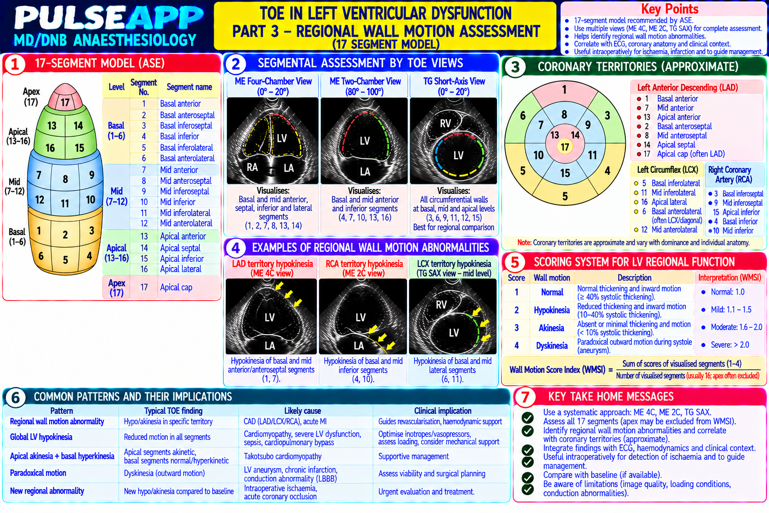
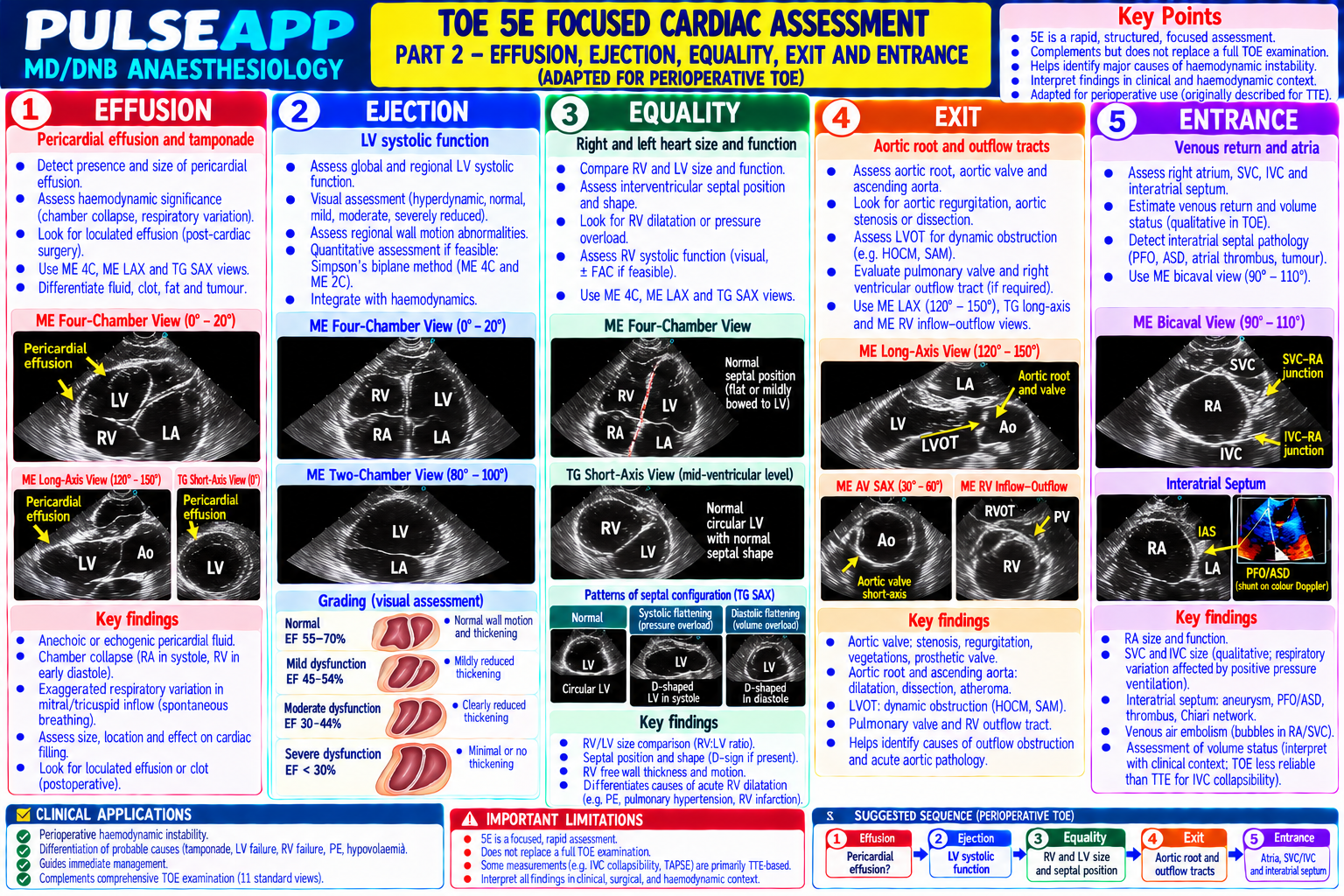
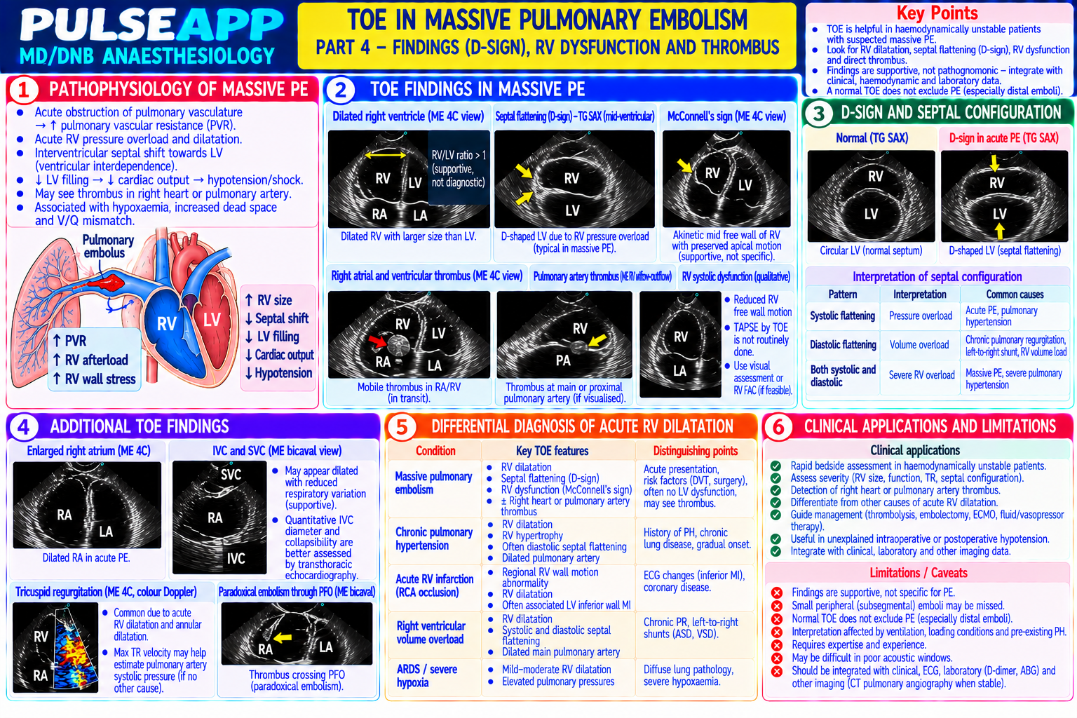
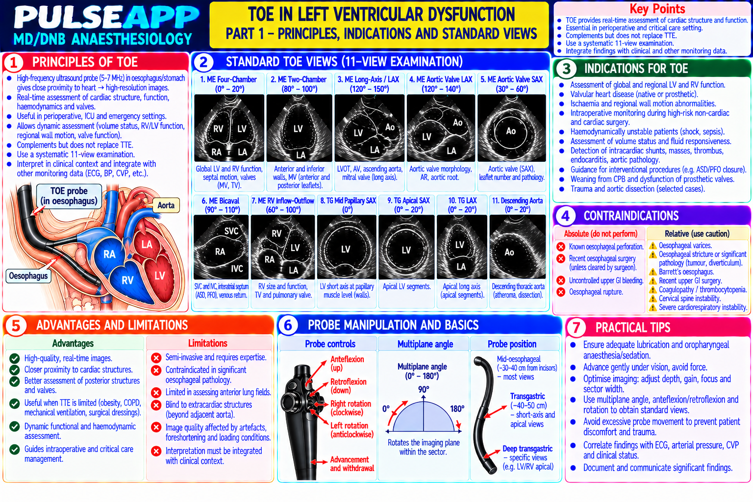
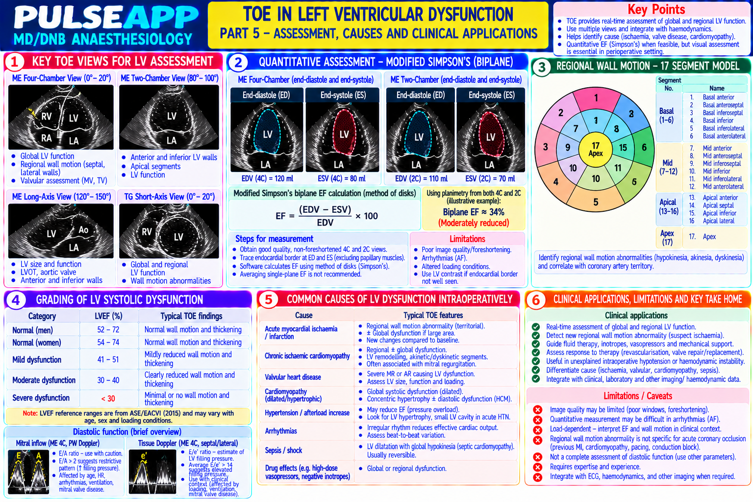
Describe the five standard TOE views used in the perioperative period. Outline the 5E (Effusion, Ejection, Equalities, Exits, Expected motion) focused cardiac assessment. Describe how TOE diagnoses: cardiac tamponade, massive PE (D-sign), LV dysfunction, and air embolism during surgery.

description Clinical Response
⚙ Core Concept
TOE is the gold-standard real-time haemodynamic monitor for the anaesthetised cardiac surgery patient — providing resolution and proximity to the heart that transthoracic echocardiography cannot achieve in the intubated patient. A focused TOE using the five standard perioperative views and the 5E framework can rapidly diagnose life-threatening haemodynamic instability in less than 5 minutes. (Shanewise JS — ASE/SCA guidelines; Perera P — 5E; Miller's Anaesthesia 9th Ed)
A. Five Standard Perioperative TOE Views3 marks

Probe Position View Structures Seen Primary Use and Angle Mid-Oesophageal Mid-oesophagus All four cardiac chambers simultaneously; Overall cardiac function; LV and RV size and function; valvular 4-Chamber (ME4C) (~30–35 cm); 0°; mitral valve; tricuspid valve; IAS; IVS assessment; ASD identification slight anteflexion Mid-Oesophageal Mid-oesophagus; LV and LA only; anterior and inferior LV walls; LV anterior and inferior wall motion; mitral valve anterior leaflet; LA 2-Chamber (ME2C) 90° mitral valve (2-chamber perspective) pathology Mid-Oesophageal Mid-oesophagus; LV outflow tract; aortic valve; proximal LVOT assessment; aortic valve morphology and stenosis/regurgitation; Long Axis 120–135° ascending aorta; posterior MV leaflet prosthetic aortic valve assessment (MELAX) Transgastric Short Transgastric (~45– LV cross-section at mid-papillary muscle Global LV function (all 16 segments simultaneously visible from a single Axis at Mid- 50 cm); 0°; neutral level; circular LV; anterior and posterior view); real-time ventricular wall motion; new regional wall motion Papillary Muscle position papillary muscles; RV (crescent-shaped, abnormalities = ischaemia; D-sign (septal flattening) for PE/RV strain (TG SAX) adjacent) Descending Aorta Mid-oesophagus; Descending thoracic aorta cross-section; Aortic atheroma (grading); aortic dissection; pleural effusion; identify Short Axis 0°; probe turned surrounding pleural space aorta for aortic cannula placement in cardiac surgery leftward (counterclockwise)

B. 5E Focused TOE Assessment for Haemodynamic Instability2 marks

Effusion: pericardial effusion? Diastolic collapse of RV free wall = tamponade; anechoic fluid in the pericardial sac; measure dimension; respiratory variation of mitral inflow (pulsus paradoxus equivalent on Doppler)

Ejection: LV ejection fraction estimated by visual assessment of wall motion in ME4C and TG SAX; normal (>55%), mildly reduced (40–55%), moderately reduced (30–40%), severely reduced (<30% — walls barely move); hyperdynamic (walls touch in systole = EF >70%) Equalities (RV:LV ratio): normal RV:LV area ratio <0.6 in ME4C; D-sign in TG SAX (interventricular septal flattening — septal bowing into the LV forming a "D" shape when RV pressure exceeds LV) = RV pressure overload from massive PE, RV infarction, or pulmonary hypertension

Exits (IVC collapsibility and valvular assessment): IVC diameter >2.5 cm non-collapsing with respiration = elevated RA pressure = cardiogenic shock or obstructive shock; IVC <2 cm collapsing = low CVP = hypovolaemia; aortic and mitral valve structural assessment

Expected motion (wall motion assessment): regional wall motion abnormalities (hypokinesis, akinesis, dyskinesis) indicate myocardial ischaemia; new RWMA = acute coronary syndrome; global LV dysfunction = cardiomyopathy

C. Specific Diagnoses2 marks

Emergency TOE Finding Cardiac Pericardial effusion (anechoic fluid) + right atrial/RV diastolic free wall collapse (the low-pressure right heart chambers collapse when pericardial pressure tamponade exceeds diastolic filling pressure); swinging heart; respiratory variation in MV inflow >25% (pulsus paradoxus equivalent); dilated IVC (>2.5 cm, noncollapsing) Massive PE Acute RV dilation (RV:LV >1.0 in ME4C); McConnell's sign — regional RV wall motion abnormality: RV free wall akinesis with preserved RV apical function (unique to acute PE); D-sign in TG SAX (septal bowing into LV from acute RV pressure overload); LV underfilling (compressed by dilated RV); occasionally: direct thrombus visualisation in the pulmonary artery (MELAX or modified short axis views) LV Reduced EF visually (<30%); dilated LV; global hypokinesis; elevated LVEDP inferred from dilated LA; mitral regurgitation from papillary muscle dysfunction dysfunction; may coexist with new RWMA (MI as the cause) (cardiogenic shock) Air Characteristic "snowstorm" or "swirling" echobrightness in the right heart (microbubbles of air appear as hyperechoic masses in the RA and RV); small embolism amounts detectable before any haemodynamic compromise; TOE is the MOST SENSITIVE monitor for VAE; in sitting-position neurosurgery: even trivial (VAE) air entrainment is visible before the precordial Doppler signal changes

🎤 Viva Corner
Q. On post-CPB TOE, the TG SAX shows a D-sign (interventricular septal bowing into the LV) and the RV is visually larger than the LV. What does this indicate and how do you manage it?
A D-sign on TG SAX (the interventricular septum bows into the LV forming a D-shape when cross-sectioned) combined with RV dilation larger than the LV on the ME4C indicates acute right ventricular pressure overload — the RV is operating at a pressure level exceeding the normal pulmonary artery systolic pressure, causing the septum to bow away from the high-pressure RV toward the lower-pressure LV. In the post-CPB context, the causes are: (1) Pulmonary hypertension — from pre-existing RV dysfunction, protamine-induced pulmonary vasoconstriction (a recognised reaction to protamine reversal of heparin), air in the pulmonary vasculature, or embolic phenomena during bypass; (2) Acute RV myocardial ischaemia — particularly relevant after procedures involving the right coronary artery (CABG — right coronary graft may be kinked or incomplete); (3) Inadequate myocardial protection during bypass — RV is most vulnerable to hypothermic cardioplegia (the right heart faces the anterior chest in the open chest and may rewarm preferentially). Management depends on the cause: assess with Doppler — tricuspid regurgitation jet velocity gives the estimated PA systolic pressure; akinesis of the RV free wall with preserved apex (McConnell sign) suggests acute RV ischaemia; pale appearance of the RV surface suggests ischaemia. Pharmacological management: milrinone (PDE-III inhibitor — reduces PVR and improves RV contractility simultaneously); inhaled NO (10–40 ppm — selective pulmonary vasodilator, reduces PVR without affecting SVR; the most targeted therapy for pulmonary hypertension in the post-CPB setting); vasopressin (maintains SVR to keep systemic BP up without worsening pulmonary hypertension, unlike noradrenaline which also raises PVR through some α₁ activity in the pulmonary vasculature); adrenaline for acute RV failure (β₁ inotropy + α₁ for systemic support). If protamine-induced pulmonary hypertension is suspected (occurred within 10 minutes of protamine administration) → the pulmonary vasoconstriction is usually transient (15–30 minutes) and responds to supportive treatment with inhaled NO and waiting.
★ Examiner's Pearl
The five standard views (ME4C at 0°; ME2C at 90°; MELAX at 120–135°; TG SAX at 0° transgastric; Descending aorta short axis) with the angle and key structures for each must be reproduced. D-sign (septal bowing into LV in TG SAX) = RV pressure overload = massive PE or pulmonary hypertension. McConnell's sign (RV free wall akinesis + preserved RV apex) = specific for acute massive PE. Air embolism: TOE is the MOST SENSITIVE monitor (snowstorm appearance in the right heart — detects air before any other monitor).
Shanewise JS et al. ASE/SCA guidelines for performing a comprehensive intraoperative multiplane TOE examination (Anesth Analg 1999;89:870-884). Perera P et al. 5E — focused cardiac ultrasound (Emerg Med Clin North Am 2010;28:29-56). Hahn RT et al. ASE comprehensive echocardiography guidelines 2019. Miller RD et al. Miller's Anaesthesia, 9th Ed.
lock Protected Clinical Document • Pulse App • Screenshots Strictly Prohibited
QUESTION 128 person Asked by .
bookmark_add

Describe the cell-based model of haemostasis (replacing the cascade model). Outline the pathophysiology of disseminated intravascular coagulation (DIC) — triggers, consumptive coagulopathy, and the paradox of simultaneous thrombosis and haemorrhage. Describe management including TEG/ROTEM-guided blood product use and recombinant Factor VIIa.

collections Question Diagrams & Reference Images (1)
Question Reference Diagram
zoom_in View Image
description Clinical Response
⚙ Core Concept
DIC is the consumptive coagulopathy of systemic activation of coagulation — occurring in obstetric emergencies, sepsis, severe trauma, malignancy, and transfusion reactions — in which simultaneous activation of clotting (microvascular thrombosis → organ failure) and depletion of clotting factors and platelets (haemorrhage) creates a life-threatening paradox. Management requires treating the trigger and replacing consumed components. (Levi M — Lancet 2010; Gando S — Nature Rev; Hoffman M — cell-based model; Miller's Anaesthesia 9th Ed)
A. Cell-Based Model of Haemostasis2 marks

The traditional "coagulation cascade" model (intrinsic + extrinsic pathways → common pathway → thrombin → fibrin) is a laboratory construct that does not accurately represent in vivo clotting; the cell-based model (Hoffman M, 2003) describes coagulation in three overlapping phases occurring on cell surfaces:

Phase 1 — Initiation: occurs on tissue factor (TF)-bearing cells (fibroblasts, monocytes); vascular injury exposes TF → TF binds factor VIIa (pre-existing in plasma) → TF-VIIa complex activates factor X → Xa + Va → small initial thrombin burst (insufficient for full clot formation but enough to prime the system); factor IX is also activated by TF-VIIa

Phase 2 — Amplification: the small thrombin burst activates platelets (via PAR-1 receptors on the platelet surface) → activated platelets expose negatively charged phospholipid surface (phosphatidylserine) → coagulation complexes assemble on the platelet surface → cascade amplifies; thrombin activates factors

V, VIII, and XI

Phase 3 — Propagation: on the activated platelet surface: intrinsic tenase (IXa + VIIIa) → massive factor Xa generation → prothrombinase (Xa + Va) → massive thrombin burst → cleaves fibrinogen → fibrin; thrombin activates Factor XIII → cross-links fibrin → stable clot; THIS is the clinically important thrombin burst that forms the actual clot

Why this matters clinically: Factor VIIa (extrinsic initiation) and Factor VIII/IX (intrinsic amplification) are both essential; patients with haemophilia A (Factor VIII deficiency) have intact initiation but fail amplification → bleeding; recombinant FVIIa (NovoSeven) works by overwhelming the TF-VIIa pathway to generate enough thrombin even without Factor VIII/IX

B. DIC — Pathophysiology2 marks

Triggers: obstetric emergencies (amniotic fluid embolism, placental abruption, HELLP syndrome — massive TF release from placental and amniotic fluid); sepsis (LPS and cytokines upregulate TF on monocytes and endothelium); severe trauma (tissue injury → TF exposure); acute haemolytic transfusion reaction; malignancy (many tumour cells constitutively express TF); burns

Pathophysiology: systemic TF upregulation → continuous low-grade thrombin generation throughout the circulation → microvascular thrombi (fibrin deposits in small vessels → end-organ ischaemia: renal failure, cerebral dysfunction, hepatic failure) → CONSUMPTION of: fibrinogen (→ <1 g/L in severe DIC); platelets (→ <50×10⁹/L); clotting factors (V, VIII, XIII) → secondary fibrinolysis activation (plasmin dissolves the microthrombi but also destroys circulating fibrinogen and activates FDPs/D-dimers) → SIMULTANEOUS thrombosis and haemorrhage

Laboratory diagnosis: ↑ PT, ↑ APTT, ↑ TT (all clotting tests prolonged from factor consumption); ↓ fibrinogen (<1.5 g/L — most specific marker of DIC); ↑ Ddimers (fibrin degradation products — most sensitive marker); ↓ platelets (thrombocytopenia); fragmented red cells (schistocytes) on blood film — microangiopathic haemolytic anaemia from RBCs caught in fibrin strands

C. Management3 marks

Treat the underlying trigger: the most important intervention — DIC will not resolve until the trigger is removed; in obstetric DIC → deliver the fetus and placenta; in septic DIC → antibiotics and source control; in haemolytic transfusion reaction DIC → stop the transfusion and treat the haemolysis Blood product replacement (guided by TEG/ROTEM or conventional coagulation tests): Fibrinogen concentrate 4–6 g IV or cryoprecipitate 10 units (when fibrinogen <1.5 g/L or FIBTEM MCF <10 mm) — the most urgently needed replacement; fibrinogen is the first clotting factor to be critically depleted in DIC FFP 10–15 mL/kg (factor replacement when PT/APTT >1.5× normal and there is active bleeding) Platelets (when <50×10⁹/L with active bleeding; or <20×10⁹/L even without active bleeding) Vitamin K 10 mg IV if concurrent warfarin or hepatic failure is contributing to factor deficiency

Recombinant Factor VIIa (rFVIIa / NovoSeven): 90–120 mcg/kg IV; bypasses the intrinsic pathway defects in DIC by overwhelming the extrinsic TF-VIIa pathway with supraphysiological concentrations of FVIIa → generates thrombin even in the absence of Factors VIII, IX, V; use: licensed for haemophilia with inhibitors; off-label for life-threatening haemorrhage refractory to all other measures (obstetric haemorrhage, major trauma); NOT recommended without adequate fibrinogen (>1 g/L) and platelets (>50×10⁹/L) — rFVIIa cannot work without substrate

Heparin in DIC: historically discussed as a treatment for the thrombotic component of DIC; not routinely recommended for acute haemorrhagic DIC; considered only in DIC predominantly manifesting as thrombosis (e.g., thrombotic DIC in purpura fulminans, malignancy-associated DIC) after very careful riskbenefit assessment

🎤 Viva Corner
Q. Why does DIC cause BOTH bleeding AND thrombosis simultaneously — and which should you treat first?
DIC creates both bleeding and thrombosis simultaneously through a single pathological process — systemic, uncontrolled thrombin generation throughout the circulation. The thrombin causes: THROMBOSIS — microvascular fibrin deposition in small vessels throughout the organs (kidney, liver, brain, adrenal glands) → microvascular occlusion → end-organ ischaemia and failure; AND BLEEDING — because the same uncontrolled thrombin generation rapidly CONSUMES all the available clotting factors and platelets throughout the circulation; fibrinogen is cleaved by thrombin into fibrin; Factor V, VIII, and XIII are consumed; platelets are activated and aggregated into microthrombi → platelet count falls; the coagulation cascade has been so completely overwhelmed that there is nothing left to form a clot at sites of vascular injury — hence paradoxical haemorrhage. Secondary fibrinolysis amplifies the haemorrhage: the body's endogenous fibrinolytic system (plasmin) is activated to try to dissolve the pathological microthrombi → but plasmin also degrades circulating fibrinogen and formed clots → fibrin degradation products (FDPs) accumulate → FDPs themselves inhibit platelet aggregation and fibrin polymerisation → worsening haemostatic failure. In terms of treatment priority: you must always treat BOTH arms simultaneously, but the sequence depends on which is more immediately life-threatening. In clinical practice: active haemorrhage is the immediate killer in acute DIC — haemorrhage that cannot clot is visible and immediately threatening; microthrombi are silent and cause organ dysfunction over hours to days; therefore: blood product replacement (fibrinogen, FFP, platelets) to restore haemostatic competence takes priority to stop the active haemorrhage; simultaneously, treat the underlying trigger which will stop the thrombin generation that is driving both the thrombosis and the consumption. Do NOT give heparin for acute haemorrhagic DIC (even though theoretically it would address the thrombotic arm — it will catastrophically worsen the already-present haemorrhage).
★ Examiner's Pearl
Cell-based model three phases: initiation (TF-bearing cells → small thrombin burst); amplification (thrombin activates platelets → phospholipid surface assembly); propagation (platelet surface tenase and prothrombinase → massive thrombin burst → fibrin) — the three phases and the cell-substrate transitions between them must be described in sequence. DIC laboratory diagnosis: ↑ PT + ↑ APTT + ↑ D-dimers + ↓ fibrinogen + ↓ platelets — fibrinogen is the MOST SPECIFIC (first depleted) and D-dimers MOST SENSITIVE. rFVIIa requirements before use: fibrinogen >1 g/L AND platelets >50×10⁹/L (no substrate = no thrombin generation even with massive FVIIa).
Levi M. Disseminated intravascular coagulation (Crit Care Med 2007;35:2191-2195). Gando S et al. DIC (Nat Rev Dis Primers 2016;2:16037). Hoffman M. A cell-based model of haemostasis (Thromb Haemost 2003;85:958-965). Miller RD et al. Miller's Anaesthesia, 9th Ed.
lock Protected Clinical Document • Pulse App • Screenshots Strictly Prohibited
QUESTION 129 person Asked by .
bookmark_add

Describe the AAGBI pre-use anaesthesia machine check (Montague checklist). Explain the variable bypass vaporiser design (Tec 5/Mk 6) including the splitting ratio, temperature compensation mechanism, and back-bar. Describe the pin index safety system, O₂ failsafe devices, and the hypoxic guard system.

collections Question Diagrams & Reference Images (1)
Question Reference Diagram
zoom_in View Image
description Clinical Response
⚙ Core Concept
The anaesthesia machine is the most complex piece of equipment routinely used in clinical medicine — its correct function is fundamental to patient safety. Equipment-related anaesthetic adverse events, including hypoxic gas mixtures, vaporiser malfunctions, and circuit leaks, are preventable through meticulous pre-use checks. The AAGBI checklist (Montague 2012) provides a standardised framework that must be completed before every operating list. (AAGBI — Checking Anaesthetic Equipment 2012; Dorsch JA — Understanding Anaesthesia Equipment; Miller's Anaesthesia 9th Ed)
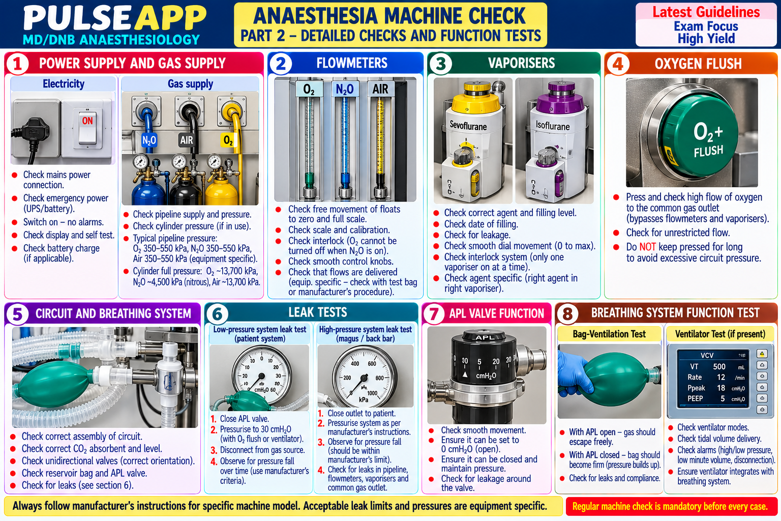
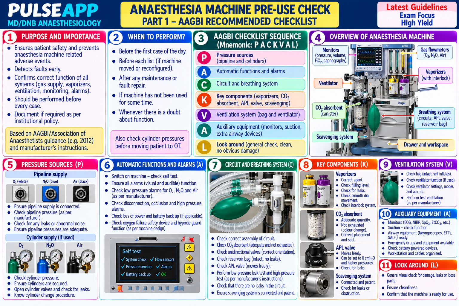
A. AAGBI Pre-Use Anaesthesia Machine Check (Montague Checklist 2012)3 marks

Step 1 — Self and Environment: confirm own identity, site, patient; check theatre environment and emergency equipment available

Step 2 — Electrical supply: connect mains power; check all monitoring equipment powered on and functional; check backup battery charge

Step 3 — Gas supplies: pipeline O₂, N₂O, and air pressure (400 kPa pipeline = 60 psi); check cylinder O₂ reserve cylinder is full (turn on, check pressure, turn off); check N₂O cylinder if applicable

Step 4 — Flowmeters: flowmeter tubes intact and correctly calibrated; bobbins spin freely; set to minimum at rest; check integrity of the rotameter assembly (no cracks)

Step 5 — Vaporisers: adequately filled; seated correctly on the back-bar (Selectatec® locking mechanism engaged); inter-lock functioning (cannot select two vaporisers simultaneously); not contaminated with wrong agent; vaporiser dial function at zero

Step 6 — Breathing system: assemble the circle breathing system; connect to machine; perform a leak test (close APL valve, occlude patient end → squeeze bag → confirm pressure maintained at ≥30 cmH₂O for ≥10 seconds without gas escape); confirm CO₂ absorber is functional (colour indicator fresh; weight appropriate)

Step 7 — Ventilator: switch on; test function; confirm appropriate settings (volume-control or pressure-control mode as required); test the disconnection alarm (pull the ventilator hose)

Step 8 — Monitoring: confirm SpO₂, ETCO₂, airway pressure, and agent monitoring are functioning and calibrated

Step 9 — Ancillary equipment: suction working; airway equipment available and checked; drugs prepared and labelled

Step 10 — Documentation: sign the machine check log; document time and initials

B. Variable Bypass Vaporiser — Physics and Design3 marks

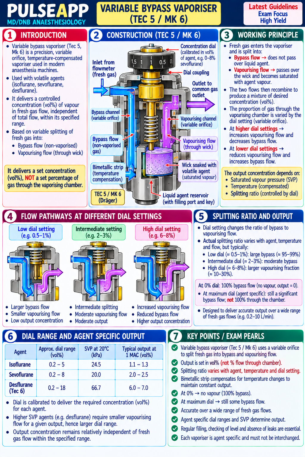
Principle of variable bypass (Tec 5, Tec 6, Tec 7 — Datex-Ohmeda; Mk 6 — GE): fresh gas flow (FGF) entering the vaporiser is split into two streams: a BYPASS FLOW (the larger portion — passes through the bypass chamber without contacting liquid anaesthetic) and a VAPORISING CHAMBER FLOW (the smaller portion — passes over or through the liquid anaesthetic where it becomes saturated with vapour); the two streams rejoin downstream; the output concentration is determined by the ratio of bypass flow to vaporising chamber flow (the "splitting ratio")

Splitting ratio: the fraction of total FGF directed through the vaporising chamber is controlled by the concentration dial; at the dial setting of 2% sevoflurane: the splitting ratio is adjusted so that the small saturated stream, when mixed with the large bypass stream, produces 2% sevoflurane in the output; the saturation vapour pressure (SVP) of the agent and the dial setting determine the mathematical splitting ratio required

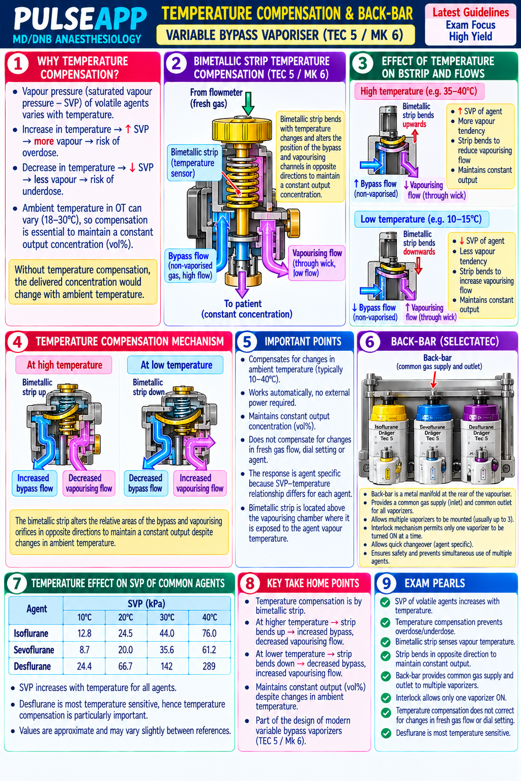
Temperature compensation: as temperature falls, SVP falls → less vapour enters the vaporising chamber per unit of FGF → output concentration would fall; the vaporiser compensates with a bimetallic strip or temperature-sensitive valve — as temperature falls, the valve opens WIDER to allow a HIGHER proportion of FGF through the vaporising chamber, compensating for the lower SVP and maintaining the set output concentration constant; conversely at higher temperatures → the valve partially closes → maintains output

Flow compensation: at very high FGF rates (>15 L/min), the vaporising chamber may not achieve full saturation → the output concentration may be slightly lower than set; at very low FGF (<0.5 L/min), the output may be slightly higher than set (less dilution of the saturated vapour with bypass gas); Tec 5/6 vaporisers are calibrated for FGF 0.5–15 L/min

Agent-specific vaporisers: each vaporiser is calibrated for a specific volatile agent (SVP and MAC are agent-specific); filling the wrong agent into a vaporiser → catastrophic: e.g., desflurane (SVP 669 mmHg at 20°C) mistakenly filled into a sevoflurane vaporiser (SVP 157 mmHg) would result in delivery of a hypnotic overdose; colour-coding and agent-specific keyed fillers (Quik-Fil, Saf-T-Fill) prevent this

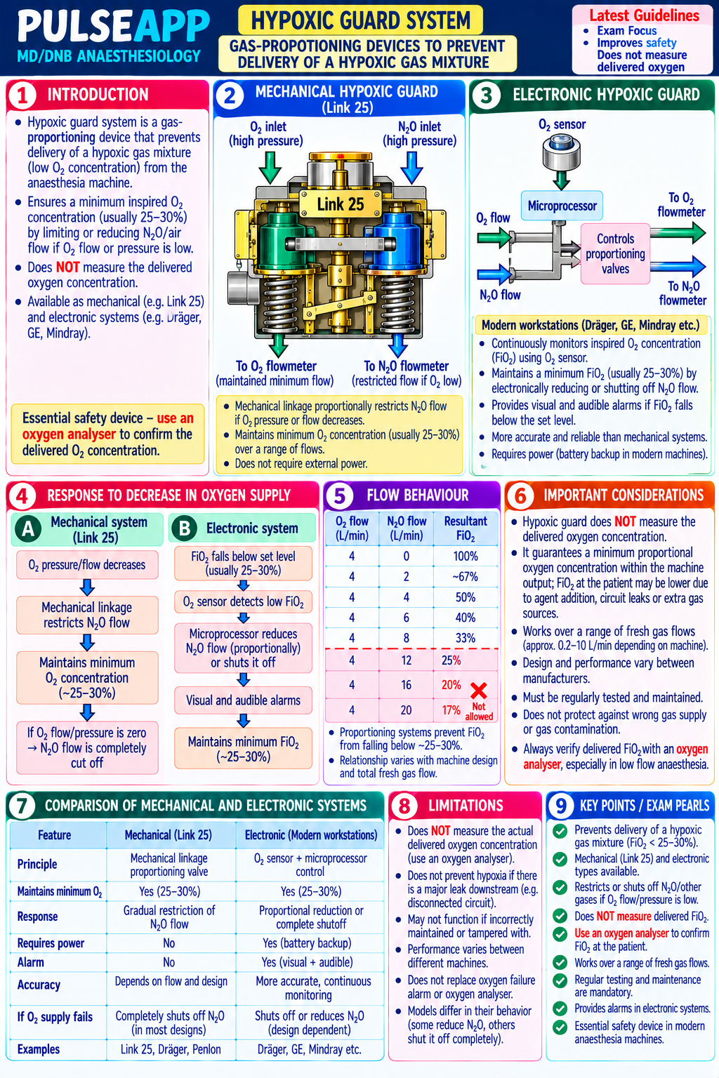
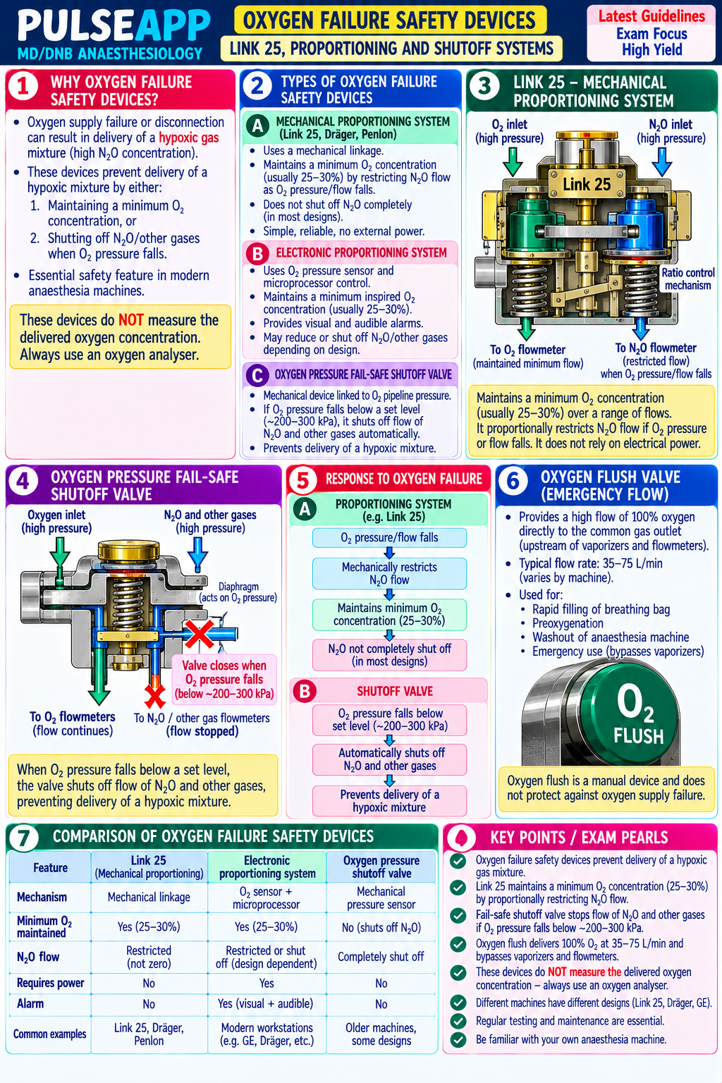
C. Safety Systems — Pin Index, O₂ Failsafe, Hypoxic Guard4 marks

Safety System Design Prevents Pin Index Safety A unique arrangement of pins on the yoke (cylinder valve interface) for each gas cylinder; two pins project from Wrong gas cylinder being

System (PISS) the yoke in positions unique to each gas (O₂: positions 2,5; N₂O: 3,5; Air: 1,5); the cylinder valve has connected to the wrong gas corresponding holes; a cylinder can ONLY be connected to the correct yoke (O₂ cylinder cannot be connected to supply outlet; prevents the N₂O yoke) accidental substitution of N₂O for O₂ O₂ Failsafe When pipeline O₂ pressure falls below a threshold (approximately 200 kPa / 30 psi): an alarm sounds Delivery of a hypoxic gas System immediately; ALL gas flows to the flowmeters are automatically cut off (not just N₂O — ALL gases); the design mixture to the patient if the O₂ ensures that the patient cannot receive a hypoxic gas mixture if O₂ supply fails; on older machines: a pressure pipeline fails; also prevents regulator valve closes when O₂ pressure falls → blocks N₂O flow; on modern machines: electronic flowmeter N₂O only delivery without O₂ control automatically stops all flows; a reserve O₂ cylinder is mounted on the machine for this scenario Hypoxic Guard (O₂ A mechanical or electronic linkage between the O₂ and N₂O flowmeter controls that prevents the total gas mixture Accidental delivery of a Ratio delivered from containing less than a minimum O₂ concentration (typically 25%); on mechanical systems hypoxic gas mixture through

Monitor/Controller) (Ohmeda Link-25): a chain-and-sprocket mechanism links the O₂ and N₂O needle valves — increasing N₂O flow operator error on the automatically increases O₂ flow proportionally; on electronic systems: the O₂ concentration in the outflow is flowmeters (e.g., turning N₂O continuously measured and N₂O flow is automatically reduced if FiO₂ falls below 25% to maximum without proportionally increasing O₂) O₂ analyser An oxygen sensor (paramagnetic O₂ analyser — uses the paramagnetic properties of O₂ molecules) placed on Delivery of a hypoxic gas (paramagnetic) the inspiratory limb of the breathing circuit measures the inspired O₂ concentration continuously; if FiO₂ falls mixture to the patient despite below 21% (or a set alarm threshold) → alarm sounds immediately all upstream safety systems functioning — the final safety layer before the patient

🎤 Viva Corner
Q. During a case, the oxygen failure alarm sounds. What is your immediate response?
The O₂ failure alarm indicates that O₂ pipeline pressure has fallen below the safe threshold (approximately 200 kPa). My immediate actions in sequence: First: maintain the patient's airway manually — if the patient is intubated and on the ventilator, switch immediately to manual ventilation with the breathing bag (do not continue machine ventilation without confirmed gas supply); if the machine has automatically cut gas flow (as designed), the patient must be hand-ventilated immediately. Second: check the reserve O₂ cylinder on the back of the anaesthetic machine — every anaesthetic machine has a reserve O₂ cylinder; open the reserve cylinder immediately (turn the cylinder key anticlockwise); this restores O₂ supply to the machine; confirm the O₂ analyser shows adequate FiO₂ on the reserve supply. Third: if the reserve cylinder is also empty or unavailable: use the self-inflating bag (Ambu bag) with a free-standing O₂ cylinder from the emergency trolley; this is completely independent of the anaesthetic machine and pipeline supply. Fourth: alert the theatre team and call the medical gas engineer: "O₂ pipeline failure — engineer needed urgently"; theatre coordinator should check if the pipeline failure affects other theatres (hospital-wide O₂ failure — a major incident). Fifth: do NOT continue elective surgery if O₂ supply is uncertain — wake the patient and close the surgical field if the procedure can be safely paused; continue only if the surgery is immediately life-saving and O₂ can be maintained from an alternative source (cylinders). Prevention lesson: the pre-use checklist specifically requires turning on and checking the O₂ reserve cylinder pressure before each list — a full reserve cylinder should always be present before starting any anaesthetic.
★ Examiner's Pearl
Pin Index System: O₂ (positions 2,5); N₂O (positions 3,5); Air (positions 1,5) — the specific pin index positions for the three common gases must be reproduced. O₂ failsafe: triggers at <200 kPa (30 psi) pipeline pressure → ALL gas flows cut off (not just N₂O) → alarm. Temperature compensation in the vaporiser (bimetallic strip opens wider as temperature falls to compensate for lower SVP → maintains constant output) is the specific mechanism for temperature-related accuracy maintenance. Hypoxic guard: maintains minimum FiO₂ 25% in the gas mixture by mechanically linking N₂O and O₂ flowmeter controls.
AAGBI — Checking Anaesthetic Equipment 2012. Dorsch JA, Dorsch SE. Understanding Anaesthesia Equipment, 5th Ed. Sykes MK, Vickers MD, Hull CJ. Principles of Measurement and Monitoring in Anaesthesia and Intensive Care. Miller RD et al. Miller's Anaesthesia, 9th Ed.
lock Protected Clinical Document • Pulse App • Screenshots Strictly Prohibited
QUESTION 130 person Asked by .
bookmark_add

Describe validated pain assessment tools for acute and chronic pain including NRS, VAS, BPS, CPOT, and the DN4 neuropathic pain questionnaire. Outline the structure of an Acute Pain Service (APS). Describe the pathophysiology and risk factors for chronic post-surgical pain (CPSP) and the evidence-based prevention strategies including gabapentinoids and ketamine.

collections Question Diagrams & Reference Images (1)
Question Reference Diagram
zoom_in View Image
description Clinical Response
⚙ Core Concept
Acute pain is among the most common undertreated medical conditions — and inadequate management of acute post-operative pain is a major risk factor for the development of chronic post-surgical pain (CPSP), which affects 10–30% of patients after major surgery. CPSP represents a transition from acute nociception through central sensitisation to persistent chronic pain, and is at least partially preventable through early multimodal analgesia targeting the sensitisation pathways. (Kehlet H — CPSP Lancet 2006; IASP definition 2020; Fletcher D; Macintyre PE — acute pain management; Miller's Anaesthesia 9th Ed)
A. Pain Assessment Tools3 marks

Tool Patient Type Scale Details NRS (Numeric Communicative; 0–10 (0 = no Patient verbally rates pain; simple and quick; most widely used; 0–3 = mild; 4–6 = moderate; ≥7 = severe; Rating Scale) post-operative; pain; 10 = worst clinically meaningful change = ≥2 points; NRS ≤3 is the target for adequate analgesia adults; paediatric pain imaginable) ≥8 yrs VAS (Visual Communicative; 100 mm Patient marks a point on the line; ruler measures distance from left end = pain score in mm; more sensitive Analogue adults; requires horizontal line to change than NRS for research purposes; less practical at bedside Scale) literacy/visual (left = no pain, ability right = worst pain) Faces Pain Children 4–12 Six faces from Child points to the face that matches their pain; validated across cultures; revised version (FPS-R) uses Scale (FPS-R) years; adults with happy to crying faces without tears for better intercultural validity communication (scored 0–10) difficulty BPS Non- 3–12 (3 = no Three domains: Facial expression (1–4), upper limb position (1–4), compliance with mechanical ventilation (Behavioural communicative pain; 12 = (1–4); BPS ≥6 = clinically significant pain requiring treatment Pain Scale) adult ICU patients maximum pain) on ventilation CPOT Non- 0–8 Four domains: facial expression (0–2), body movements (0–2), muscle tension (0–2), compliance with (Critical-Care communicative ICU ventilator or vocalization (0–2); CPOT ≥3 = unacceptable pain level; validated in medical and surgical ICU Pain patients (intubated patients; PADIS 2018 recommends CPOT or BPS for pain assessment in non-communicative ICU patients Observation and non-intubated) Tool) DN4 (Douleur Chronic pain; 0–10 10 yes/no questions (7 symptoms, 3 clinical examination findings); score ≥4/10 = neuropathic pain likely; Neuropathique neuropathic pain validated for identifying neuropathic component of chronic pain (diabetic neuropathy, post-herpetic 4) screening neuralgia, CPSP)

B. Acute Pain Service (APS) Structure2 marks

An Acute Pain Service (APS) is a multi-disciplinary specialist team providing: consultant anaesthesiologist leadership; specialist pain nurses (pain sisters/APS nurses) who conduct daily ward rounds, troubleshoot analgesic failures, and educate nursing staff; dedicated pharmacist; physiotherapy input for rehabilitation pain management; psychology support for chronic pain patients admitted acutely; the APS is responsible for: managing all patient-controlled analgesia (PCA) infusions and epidural catheters in the hospital; developing and updating acute pain management protocols; monitoring adverse events (respiratory depression, PONV, inadequate analgesia); education of ward nursing staff in pain assessment and opioid safety

APS functions: daily review of all patients with epidurals, PCAs, or difficult acute pain management; 24-hour on-call support (pain nurse and pain anaesthesiologist available); adverse event monitoring and reporting; protocol development (multimodal analgesia pathways, procedure-specific protocols); training and education; quality improvement (NRS tracking, opioid consumption, adverse event rates)

C. Chronic Post-Surgical Pain (CPSP) — Definition, Pathophysiology & Risk Factors3 marks

IASP 2020 definition: CPSP is chronic pain that develops after a surgical procedure; it must: persist beyond the normal healing time (at least 3–6 months post-surgery); be distinct from pre-existing pain; be located in the surgical area or referred from it; significantly affect quality of life or daily function

Incidence: 10–50% of patients after major surgery develop some degree of CPSP; severe CPSP (significantly impacting function): 2–10%; highest incidence: thoracotomy (30–50% CPSP), limb amputation (phantom pain 50–70%), mastectomy (20–30%), inguinal hernia repair (10–12%); lowest after laparoscopic procedures

Pathophysiology — three phases:

Peripheral sensitisation: surgical tissue injury → prostaglandins, bradykinin, substance P, NGF released at the wound → sensitise peripheral nociceptors (threshold ↓, response ↑) → ↑ afferent firing

Central sensitisation: prolonged intense C-fibre activity activates NMDA receptors in the dorsal horn → glutamate-mediated windup → increased excitability of dorsal horn neurons → expansion of pain receptive fields → allodynia and hyperalgesia extend beyond the wound

Neuroplastic changes: persistent dorsal horn sensitisation → structural and functional changes in the CNS → chronic pain pathways established; this is the point at which pain transitions from a protective acute response to a maladaptive chronic condition

Risk factors: pre-operative: pre-existing pain at the surgical site (most powerful predictor); psychological factors (depression, anxiety, catastrophising — measured by Pain Catastrophizing Scale); genetic predisposition (COMT gene variants); younger age; female sex; intraoperative: nerve injury (direct or traction) during surgery; poor surgical technique; high-dose remifentanil infusion (OIH); post-operative: poorly controlled acute post-operative pain (NRS >6 in the first 24 hours is the most modifiable perioperative risk factor for CPSP)

D. CPSP Prevention Strategies2 marks

Strategy Evidence Gabapentinoids Block α₂δ Ca²⁺ channel subunits → reduce presynaptic glutamate release → reduce central sensitisation → blunt windup in the dorsal horn; pre(pregabalin, operative pregabalin 150 mg + continued post-operatively reduces CPSP incidence in thoracotomy, breast surgery, and total hip/knee replacement gabapentin) pre- (multiple RCTs and meta-analyses); PROSPECT guidelines recommend peri-operative gabapentinoids for specific procedures with high CPSP risk operatively Ketamine (sub- NMDA antagonism → prevents central sensitisation directly; multiple RCTs (Lavand'homme 2005; Himmelseher 2005): intraoperative ketamine 0.2– anaesthetic) 0.5 mg/kg/hr reduces acute post-operative pain scores AND reduces CPSP incidence at 3 and 6 months after thoracotomy and colorectal surgery; effect is independent of its acute analgesic action — it specifically reduces the dorsal horn sensitisation that drives CPSP development Regional Blocks afferent nociceptive traffic to the dorsal horn throughout surgery → prevents the initial sensitisation trigger; well-designed neuraxial or anaesthesia peripheral nerve blocks that provide complete analgesia from incision → reduces acute pain AND reduces CPSP at 3–6 months in multiple procedures; thoracic epidural analgesia for thoracotomy dramatically reduces the 30–50% CPSP incidence

Minimising Surgical technique modification: nerve-sparing approaches (nerve-sparing radical prostatectomy; nerve identification and preservation during inguinal surgical nerve hernia repair — ilioinguinal, iliohypogastric nerve preservation reduces inguinal CPSP from 12% to <5%) injury Psychological Pre-operative cognitive behavioural therapy (CBT) targeting pain catastrophising reduces CPSP in patients with high pre-operative catastrophising pre-conditioning scores; mindfulness-based approaches; pain education (explaining the biology of pain transition to chronic pain)

🎤 Viva Corner
Q. What is the most important modifiable perioperative risk factor for CPSP development and how do you target it?
The most important modifiable perioperative risk factor for chronic post-surgical pain is poorly controlled acute post-operative pain — specifically, high pain intensity in the first 24–48 hours after surgery (NRS ≥7). The association is both strong and mechanistically sound: acute nociceptive pain from surgical tissue injury drives ongoing C-fibre afferent activity into the dorsal horn; sustained high-frequency C-fibre input activates spinal NMDA receptors → glutamate-mediated wind-up → central sensitisation; if this sensitisation process is not interrupted, it can persist beyond the healing period and become the structural and functional basis of chronic pain (the nociceptive stimulus is removed — the wound heals — but the sensitised dorsal horn and brain circuitry continue to generate pain). Targeting this: (1) the most effective strategy is PRE-EMPTIVE and PREVENTIVE analgesia — beginning multimodal analgesia BEFORE surgery (gabapentinoids; NSAIDs; ketamine), performing regional nerve blocks BEFORE incision so the dorsal horn receives no nociceptive input from the very first surgical incision, and maintaining excellent analgesia throughout surgery and the entire post-operative period; (2) the goal is to keep NRS ≤3/10 in the first 24–48 hours — this is the threshold below which central sensitisation is significantly less likely to establish; (3) specifically: ketamine 0.1–0.2 mg/kg/hr intraoperatively directly targets the NMDA sensitisation mechanism; pregabalin 75–150 mg pre-operatively targets the presynaptic Ca²⁺ channels that potentiate glutamate release; a well-placed regional block (thoracic epidural for thoracotomy; brachial plexus block for shoulder surgery; TAP block for abdominal surgery) eliminates the afferent nociceptive drive entirely during and after surgery; opioids treat the acute pain but do NOT prevent CPSP (remifentanil high-dose may actually worsen CPSP risk through OIH); (4) the APS (acute pain service) daily review in the first 48–72 hours post-operatively catches undertreated acute pain before it has the opportunity to transition to central sensitisation. The bottom line: excellent acute pain management is not just about patient comfort — it is a direct intervention to prevent chronic pain.
★ Examiner's Pearl
IASP 2020 CPSP definition (pain persisting ≥3–6 months post-surgery, at the surgical site, causing significant functional impairment) is the current definition. High acute post-operative pain (NRS ≥7 in first 24h) = the most important MODIFIABLE risk factor for CPSP. Three prevention strategies with mechanisms: gabapentinoids (α₂δ Ca²⁺ block → ↓ glutamate → ↓ sensitisation), ketamine (NMDA antagonism → prevents dorsal horn wind-up), regional analgesia (eliminates afferent drive). CPOT and BPS for ICU pain assessment in non-communicative patients — PADIS 2018 recommendation.
Kehlet H et al. Persistent post-surgical pain — risk factors and prevention (Lancet 2006;367:1618-1625). IASP definition of chronic pain and CPSP 2020 (Treede RD et al. Pain 2019;160:19-27). Fletcher D et al. Chronic postsurgical pain — a pharmacological prevention (BJA 2012;108:690-700). Macintyre PE, Schug SA. Acute Pain Management: A Practical Guide, 4th Ed. Miller RD et al. Miller's Anaesthesia, 9th Ed.

Showing 121130 of 337 questions

account_tree

Subcategory Tree

Explore Anesthesia subcategories

folder_special Anesthesia
Main
lock

Category Subscription

Subscribe to Anesthesia to unlock this module and all nested subcategories.

  • check_circle Access Anesthesia & all subcategories
  • check_circle Detailed, Peer-Reviewed Answers
  • check_circle High-yield visual aids & imaging
Get Category Subscription arrow_forward
Secure 256-bit SSL Connection

Anatomical Models

Explore high-fidelity 3D visualizations included in premium modules.

Case Reviews

Real-world clinical scenarios narrated by senior consultants.

Pulse App