Anesthesia | Eklavya Medical
vaccines
medical_services Main Specialty Domain lock Subscription Required

Anesthesia

Clinical anesthesiology, local and general anesthesia delivery systems, monitoring, and perioperative care.

verified Verified Medical Faculty menu_book 337 Q&A Modules
lock Protected Clinical Document • Pulse App • Screenshots Strictly Prohibited
QUESTION 91 person Asked by .
bookmark_add

Describe femoral triangle anatomy (NAVY). Contrast the femoral nerve block (complete quadriceps paralysis) with the adductor canal block (quadriceps-sparing). Explain anatomically why the ACB spares the quadriceps. State doses and the PROSPECT recommendation for TKR analgesia.

collections Question Diagrams & Reference Images (5)
Question Reference Diagram
zoom_in View Image
Question Reference Diagram
zoom_in View Image
Question Reference Diagram
zoom_in View Image
Question Reference Diagram
zoom_in View Image
Question Reference Diagram
zoom_in View Image
description Clinical Response
⚙ Core Concept
The shift from femoral nerve block to adductor canal block for TKR analgesia is the most clinically important recent development in lower limb regional anaesthesia — driven by ERAS-TKR evidence that early ambulation is equally as important as analgesia. The ACB provides equivalent medial knee analgesia while preserving quadriceps strength, enabling same-day physiotherapy and reducing falls risk. (Jaeger P — Anesthesiology 2013; Shah NA — J Arthroplasty 2014; PROSPECT Guidelines TKR 2022)
A. Femoral Triangle Anatomy — NAVY2 marks

Boundaries: superior (inguinal ligament), lateral (sartorius), medial (adductor longus); floor: iliopsoas laterally + pectineus medially

Contents lateral to medial — NAVY: Nerve (femoral nerve), Artery (femoral artery), Vein (femoral vein), Y-fronts (lymphatics/lymph nodes) Femoral nerve (L2–L4) lies just lateral and slightly deep to the femoral artery; divides immediately below the inguinal ligament into anterior division (purely sensory — medial and intermediate cutaneous nerves of the thigh) and posterior division (saphenous nerve + motor branches to all four quadriceps heads)

B. Adductor Canal Anatomy2 marks

A fascial tunnel in the middle third of the thigh; boundaries: roof = sartorius; lateral wall = vastus medialis; posteromedial wall = adductor longus then adductor magnus

Contents: saphenous nerve (PURELY SENSORY at this level — all motor branches to quadriceps have already branched off proximal to the canal); femoral artery; femoral vein; descending genicular artery

Key concept: the motor branches to the quadriceps (rectus femoris, vastus lateralis, vastus intermedius, vastus medialis) ALL originate within the femoral triangle or proximal thigh — ABOVE the adductor canal; the saphenous nerve within the canal is exclusively sensory

C. FNB vs ACB — The Mechanism of Quadriceps Sparing3 marks

Feature Femoral Nerve Block (FNB) Adductor Canal Block (ACB) Target Entire femoral nerve in femoral triangle (motor + sensory Saphenous nerve within the adductor canal (sensory only at this level) branches)

Quadriceps motor COMPLETE — all four quadriceps heads paralysed; patient PRESERVED — motor branches all originate proximal to the canal; block cannot extend knee → cannot weight-bear safely completely unaffected Analgesia quality Excellent anterior knee; partial posterior knee Equivalent anterior and medial knee (same saphenous nerve territory); with for TKR IPACK for posterior → comprehensive coverage

Falls risk High — quadriceps weakness → falls documented in multiple Very low — normal quadriceps strength maintained studies Rehabilitation Delayed until block resolves (12–18h); physiotherapy postponed Day 1 physiotherapy achievable; reduces hospital LOS by ~1 day PROSPECT 2022 NOT recommended (motor weakness/falls) RECOMMENDED as primary block for TKR + IPACK for posterior knee recommendation

D. Technique and Doses2 marks

ACB technique: supine with thigh externally rotated; high-frequency probe at mid-thigh level; identify sartorius (superficial, triangular shape), femoral artery; saphenous nerve appears as a small hyperechoic oval adjacent to the artery within the fascial compartment; inject 15–20 mL 0.25–0.375% ropivacaine or 0.25% bupivacaine; visualise spread within the canal on ultrasound IPACK block (Infiltration between Popliteal Artery and Capsule of the Knee): complements ACB by covering the posterior knee capsule — popliteal branches of the sciatic, obturator, and genicular nerves; 15–20 mL LA injected posterior to the knee between the capsule and popliteal artery; performed at end of surgery under ultrasound or arthroscopically

Fascia Iliaca Block: large volume (40 mL) deep to fascia iliaca → spreads to femoral nerve, LFCN, sometimes obturator; landmark technique; used for hip fracture analgesia in ED by non-anaesthesiologists; less reliable for surgical anaesthesia

E. Why the ACB Spares Quadriceps — The Detailed Explanation1 mark

When the femoral nerve enters the femoral triangle below the inguinal ligament, its posterior division immediately starts giving off motor branches to the quadriceps: nerve to rectus femoris, nerve to vastus lateralis, nerve to vastus intermedius, and nerve to vastus medialis — ALL of these motor branches originate within the femoral triangle (above or at the level of the inguinal ligament); by the time the saphenous nerve (the sensory continuation of the posterior femoral nerve division) reaches the adductor canal, it has already lost ALL its motor branches and is purely sensory; therefore, an ACB at mid-thigh level blocks ONLY sensory afferents without any motor component

🎤 Viva Corner
Q. A surgeon requests a femoral nerve block for TKR. You suggest an adductor canal block instead. How do you explain the difference?
The fundamental difference is where in the femoral nerve's course we place the block. The femoral nerve block is performed in the femoral triangle at the groin — at this level, the nerve is a mixed nerve carrying both motor fibres (to all four quadriceps muscles) and sensory fibres (to the knee and medial leg); blocking here produces excellent knee analgesia but also completely paralyses the quadriceps for 12–18 hours — the patient cannot extend the knee or bear weight safely, falls risk is documented, and early physiotherapy is impossible. The adductor canal block is performed at the mid-thigh level within the sartorius tunnel — at this point, all the motor branches to the quadriceps have already departed from the nerve trunk at the femoral triangle level; only the purely sensory saphenous nerve continues into the canal; blocking the saphenous nerve here provides equivalent medial and anterior knee analgesia with complete preservation of quadriceps strength; the patient can stand, walk, and actively participate in physiotherapy from day one. The PROSPECT 2022 guidelines specifically recommend the ACB over the FNB for TKR because of this rehabilitation advantage. With the ACB supplemented by an IPACK block for posterior knee coverage, we provide comprehensive TKR analgesia without any motor deficit.
★ Examiner's Pearl
NAVY mnemonic (Nerve/Artery/Vein/Y-fronts lateral to medial). The ACB quadriceps-sparing mechanism in full: all motor branches to quadriceps originate in the femoral triangle, proximal to the adductor canal; only the purely sensory saphenous nerve remains in the canal. PROSPECT 2022 recommendation: ACB + IPACK preferred; FNB not recommended (falls risk).
Jaeger P et al. ACB vs FNB for TKA (Anesthesiology 2013;118:409-415). PROSPECT Working Group. TKR pain management recommendations 2022. Grevstad U et al. Effect of ACB vs FNB on muscle strength (BJA 2015). Hadzic A. Regional Anesthesia Textbook, 3rd Ed.
lock Protected Clinical Document • Pulse App • Screenshots Strictly Prohibited
QUESTION 92 person Asked by .
bookmark_add

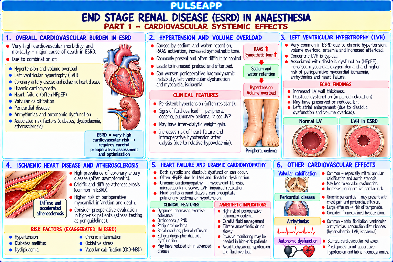
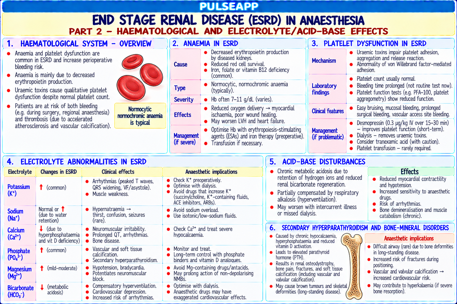
Describe systemic effects of ESRD on cardiovascular, haematological, and electrolyte systems. Outline drug pharmacokinetics (NMBs, opioids, NSAIDs). State AV fistula protection principles and the timing of surgery relative to dialysis.
description Clinical Response
⚙ Core Concept
ESRD creates a complex multi-system perioperative challenge. The most immediately dangerous anaesthetic considerations are: hyperkalaemia (avoid K⁺-containing fluids; avoid succinylcholine), altered drug kinetics (avoid morphine — M6G accumulation; prefer atracurium/cisatracurium — organindependent elimination), and AV fistula protection. (Miller's Anaesthesia 9th Ed; Stoelting — Coexisting Disease; Sear JW — BJA 2005; Hunter JM — NEJM 1995)
A. Systemic Effects Relevant to Anaesthesia3 marks

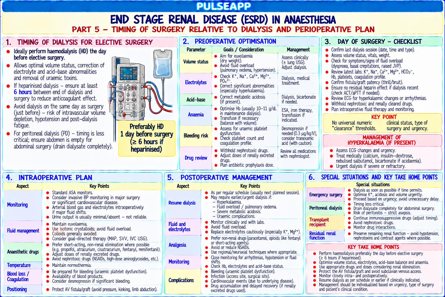
System ESRD Effect Clinical Action Cardiovascular Hypertension (80%); LVH; accelerated atherosclerosis; Pre-op echo (LV function); ECG; arterial line; careful fluid management; avoid rapid uraemic cardiomyopathy; pericardial effusion; arrhythmias boluses in diastolic dysfunction (hyperkalaemia, hypomagnesaemia) Haematological Normochromic normocytic anaemia (↓EPO); uraemic Pre-op: EPO + iron; target Hb ≥80–90 g/L; DDAVP 0.3 mcg/kg IV pre-op (releases vWF platelet dysfunction; thrombocytopenia (hypersplenism) → improves platelet adhesion); AVOID NSAIDs (worsen platelet dysfunction) Electrolytes Hyperkalaemia (K⁺ 5.5–7.5 mEq/L common); metabolic Check K⁺ DAY OF SURGERY; K⁺ >6.0 → postpone → emergency dialysis; AVOID acidosis; hypocalcaemia; hyperphosphataemia Hartmann's/Ringer's Lactate (contains K⁺ 4 mEq/L — FATAL in hyperkalaemic ESRD); use 0.9% NaCl ONLY; ECG monitoring throughout Fluid/Volume Fluid overloaded or depleted depending on dialysis; most Compare with dry weight; cautious fluid administration; echo for volume assessment patients should be dialysed 12–24h before elective surgery

B. Drug Pharmacokinetics in ESRD4 marks

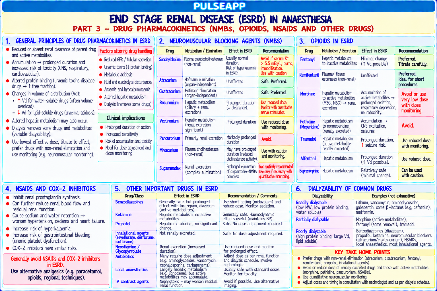
Drug Problem in ESRD Recommendation Morphine Morphine-6-glucuronide (M6G — active, renally cleared) accumulates → prolonged sedation AVOID morphine in ESRD; use fentanyl (hepatic and respiratory depression → inactive metabolites; safe) Fentanyl Hepatic metabolism to inactive norfentanyl; no renally-cleared active metabolites Safe — preferred opioid in ESRD; dose as normal with careful monitoring Succinylcholine K⁺ release 0.5–1.0 mEq/L → cardiac arrest if baseline K⁺ ≥5.5 mEq/L; also reduced AVOID if K⁺ >5.5 mEq/L; use rocuronium 1.2 pseudocholinesterase in severe uraemia mg/kg + sugammadex 16 mg/kg for CICO Atracurium / Organ-independent Hofmann elimination (pH/temperature-dependent chemical degradation) + PREFERRED NMBs in ESRD; cisatracurium

Cisatracurium ester hydrolysis — NOT renally cleared; predictable duration regardless of renal function preferred (3× more potent; less laudanosine production) Vecuronium 3-desacetyl-vecuronium (active metabolite) renally cleared → accumulates → prolonged block Avoid; if used, reduce doses and monitor TOF carefully Rocuronium Primarily hepatic elimination; some renal excretion; t½ modestly prolonged Generally safe with TOF monitoring; sugammadex reversal preferred for reliability NSAIDs COX inhibition → ↓ renal prostaglandins → ↓ GFR further; worsens uraemic platelet ABSOLUTELY AVOID ALL NSAIDs in ESRD; use dysfunction; risk of AKI paracetamol for baseline analgesia Midazolam 1-hydroxymidazolam glucuronide (active, renally cleared) accumulates → prolonged sedation Use cautiously; reduce dose; prefer shorter-acting alternatives for ICU sedation

C. Laudanosine — Atracurium Metabolite1 mark

Laudanosine is a byproduct of atracurium Hofmann elimination; a tertiary amine that crosses the BBB; CNS stimulant/potentially epileptogenic at high concentrations in animals; in humans, clinical doses of atracurium/cisatracurium produce laudanosine concentrations well below toxic thresholds; cisatracurium produces ~3× less laudanosine than atracurium per equivalent NMB dose (because it is more potent and lower doses are used) Clinically safe at recommended doses even in ESRD; prefer cisatracurium for prolonged ICU infusions in ESRD

D. AV Fistula Protection & Dialysis Timing2 marks

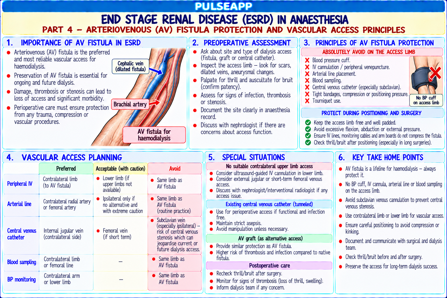
AV Fistula — absolute rules: NO BP cuff on fistula arm; NO IV cannula in fistula arm; NO arterial line in fistula arm; clearly label the fistula arm; pad to avoid positional compression; check for bruit/thrill pre- and post-operatively

Dialysis timing: perform dialysis 12–24 hours before elective surgery to normalise K⁺ (<5.5 mEq/L), optimise volume status (reach dry weight), and correct metabolic acidosis; avoid dialysis immediately before surgery (post-dialysis hypotension from volume depletion; heparin effect — withhold 4–6 hours)

Regional anaesthesia advantage: avoids systemic drugs with renally-altered kinetics; superior analgesia without NSAIDs; neuraxial is safe if platelet count and coagulation acceptable; check platelet count and recent coag profile in uraemic patients before neuraxial
🎤 Viva Corner
Q. Why is atracurium the preferred NMB in ESRD and what is laudanosine?
Atracurium is preferred in ESRD because it undergoes organ-independent elimination — it is broken down in the plasma itself through Hofmann elimination (spontaneous pH- and temperature-dependent chemical degradation at physiological pH 7.4 and 37°C) and non-specific ester hydrolysis; neither pathway requires the kidney or liver; therefore duration is predictable and NOT prolonged by renal failure, unlike vecuronium (renally-cleared active metabolite) or pancuronium (primarily renal elimination). Laudanosine is one of the breakdown products of Hofmann elimination. It is a tertiary amine that crosses the BBB and has CNS stimulant, potentially epileptogenic effects at high plasma concentrations in animal models. However, at clinical NMB doses in humans, even in ESRD patients where laudanosine has some renal accumulation, plasma laudanosine concentrations remain well below seizure thresholds. Cisatracurium (the R-cis, R-cis stereoisomer of atracurium) has the same Hofmann elimination mechanism but is 3× more potent, so lower doses are used → 3× less laudanosine production per unit of NMB effect. Cisatracurium is therefore the ideal NMB choice in ESRD, particularly for prolonged ICU infusions where laudanosine accumulation could theoretically become more significant.
★ Examiner's Pearl
AVOID Hartmann's/Ringer's Lactate in ESRD (contains K⁺ 4 mEq/L → fatal in hyperkalaemic patient) — use 0.9% NaCl only. AVOID morphine (M6G accumulation) → use fentanyl. AVOID succinylcholine if K⁺ >5.5. Atracurium/cisatracurium preferred (Hofmann elimination — organ-independent). Laudanosine: CNS stimulant metabolite, clinically safe at clinical doses, less with cisatracurium.
Sear JW. Kidney dysfunction in the postoperative period (BJA 2005;95:20-32). Hunter JM. New neuromuscular blocking drugs (NEJM 1995;332:1691-1699). Miller RD et al. Miller's Anaesthesia, 9th Ed. Stoelting RK. Anaesthesia and Coexisting Disease, 6th Ed.
lock Protected Clinical Document • Pulse App • Screenshots Strictly Prohibited
QUESTION 93 person Asked by .
bookmark_add

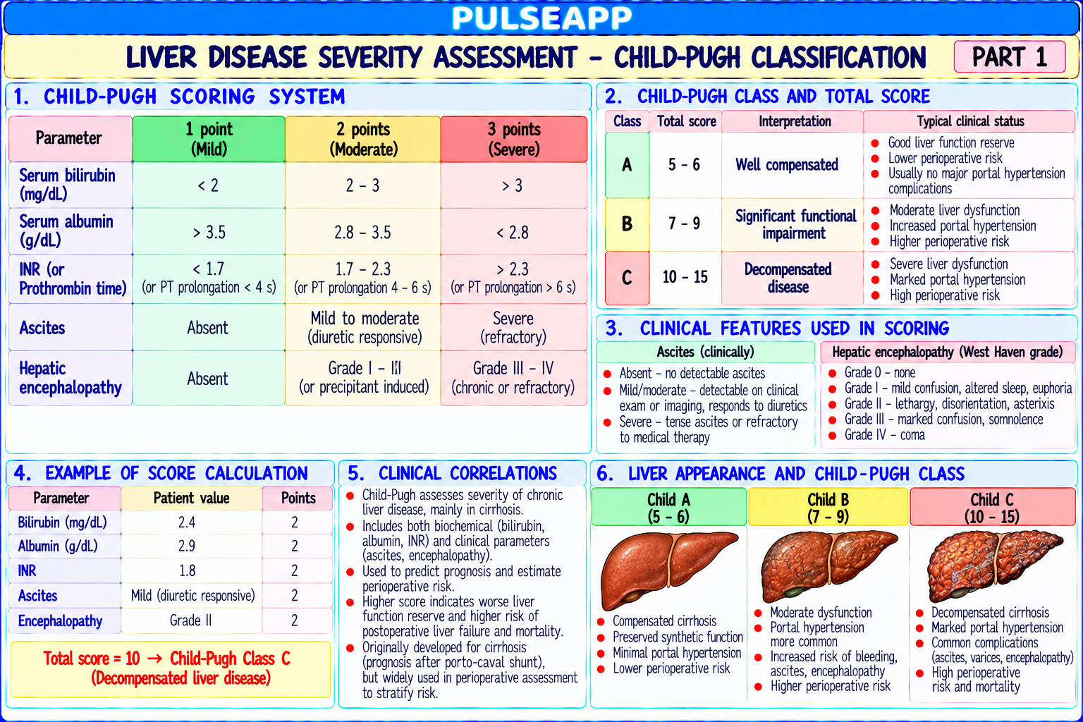
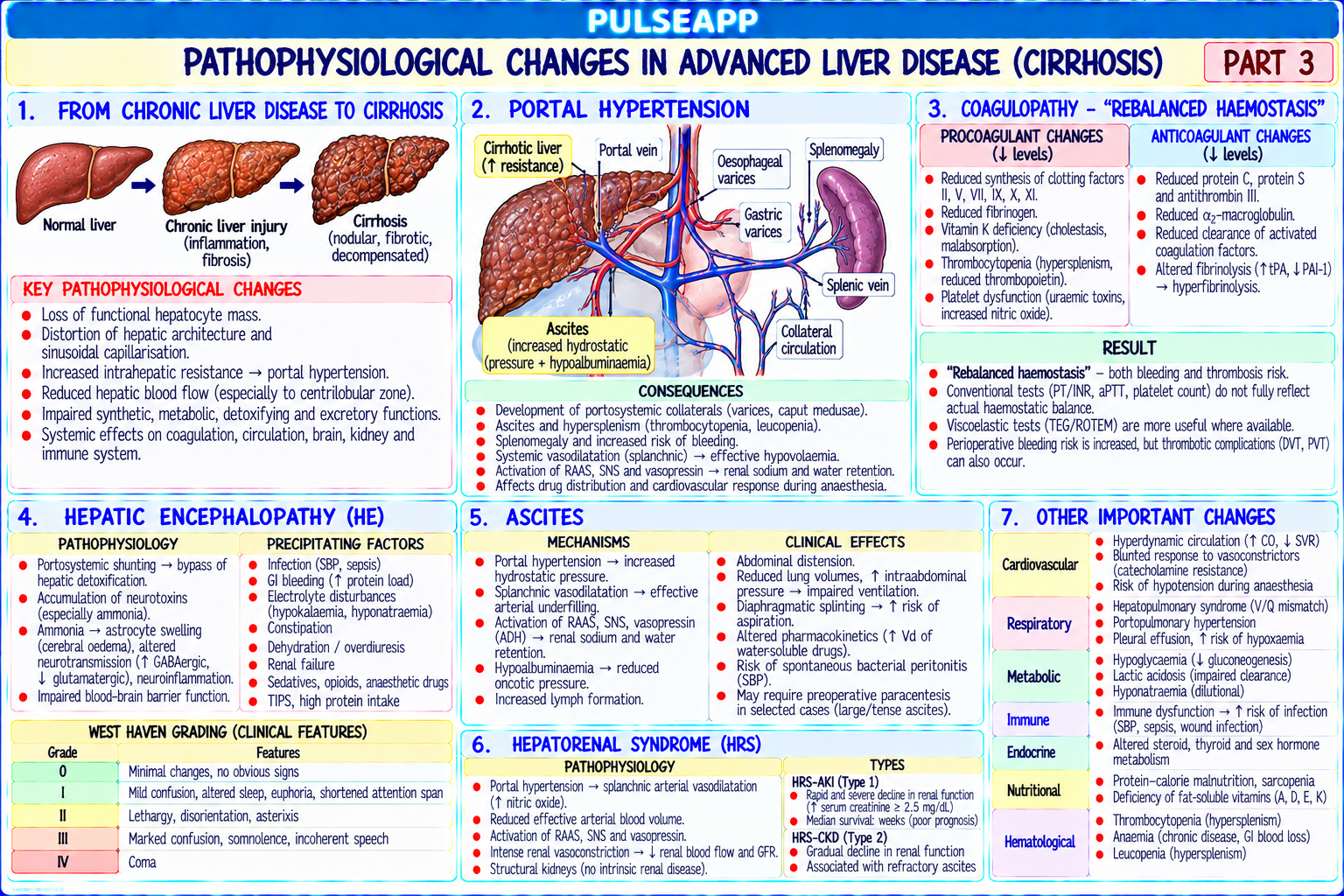
Classify liver disease severity using Child-Pugh and MELD scoring. Describe pathophysiological changes (coagulopathy, encephalopathy, hepatorenal syndrome). Outline drug pharmacokinetics in liver failure and the perioperative risk based on scoring.

collections Question Diagrams & Reference Images (1)
Question Reference Diagram
zoom_in View Image
description Clinical Response
⚙ Core Concept
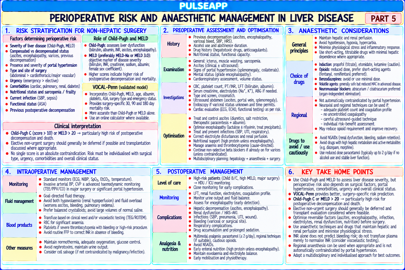
Liver disease profoundly alters every pharmacokinetic parameter and creates complex multi-organ dysfunction. Child-Pugh and MELD scores provide objective surgical mortality prediction. The key pharmacology insight: INR is unreliable in liver disease (rebalanced haemostasis — use TEG/ROTEM). The key drug principles: avoid benzodiazepines (precipitate encephalopathy); prefer atracurium/cisatracurium (organ-independent); prefer remifentanil (ester hydrolysis). (Friedman LS — Gastroenterology 1999; Wiesner RH — MELD 2003; Miller's Anaesthesia 9th Ed; Stravitz RT — Chest 2008)
A. Child-Pugh Score2 marks

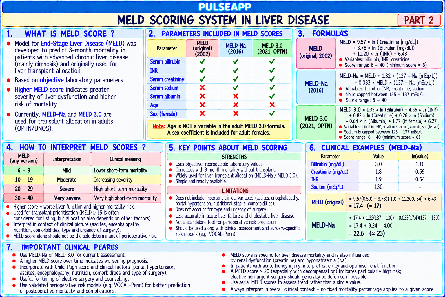
Parameter 1 Point 2 Points 3 Points Encephalopathy None Grade 1–2 Grade 3–4 Ascites Absent Mild Moderate-severe Bilirubin (μmol/L) <34 34–51 >51 Albumin (g/L) >35 28–35 <28 Prothrombin time (sec prolonged) <4 4–6 >6 Child-Pugh A (5–6 pts): 30-day surgical mortality ~10%; Child-Pugh B (7–9 pts): ~30%; Child-Pugh C (10–15 pts): ~76–82% — elective surgery generally contraindicated

MELD score: = 3.78 × ln(bilirubin mg/dL) + 11.2 × ln(INR) + 9.57 × ln(creatinine mg/dL) + 6.43; MELD <10 low risk; >15 high risk; >20 very high; >25 ≈50% mortality for major surgery; superior to Child-Pugh for predicting 90-day surgical mortality

B. Pathophysiological Changes3 marks

Derangement Mechanism Clinical Implication

Coagulopathy — ↓ synthesis of procoagulant factors (II, V, VII, IX, X) INR is unreliable for predicting bleeding risk in liver disease (rebalanced "rebalanced AND anticoagulant factors (Protein C, S, antithrombin); haemostasis — some patients with high INR bleed little; some with "normal" INR bleed haemostasis" thrombocytopenia (hypersplenism); primary fibrinolysis; massively from portal hypertension); use TEG/ROTEM (measures the full clot formation the INR ONLY measures procoagulant factor and fibrinolysis process in whole blood at physiological conditions); TEG-guided blood deficiency — misses the simultaneous anticoagulant product administration uses significantly less product than INR-guided with equivalent factor reduction haemostasis Hepatic Ammonia + gut-derived neuroactive substances + AVOID benzodiazepines (exquisite sensitivity; precipitates or worsens encephalopathy); encephalopathy astrocyte failure; GABA receptor dysfunction; cerebral avoid opioids where possible; lactulose pre-operatively; maintain haemodynamic stability oedema in acute liver failure (encephalopathy worsens with hypotension) Hyperdynamic Portal hypertension → ↑ NO/prostacyclin → splanchnic AVOID NSAIDs absolutely; maintain MAP ≥65 mmHg (vasopressors not fluids in fluidcirculation + vasodilation → low SVR, high CO; reduced effective overloaded cirrhosis); careful fluid balance; avoid hepatotoxic drugs and contrast without hepatorenal risk circulating volume despite high CO; renal adequate prehydration; HRS type 1 (acute renal failure) → terlipressin + albumin vasoconstriction → HRS risk from: NSAIDs, nephrotoxins, hypotension, paracentesis without albumin Hepatopulmonary Intrapulmonary vascular dilatation → arteriovenous Pre-op SpO₂ and ABG; supplemental O₂; echo to exclude portopulmonary hypertension syndrome shunting → hypoxaemia worsening on standing (PAP >35 mmHg) (orthodeoxia)

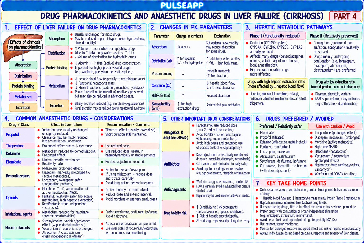
C. Drug Pharmacokinetics in Liver Failure3 marks

Drug Effect Recommendation Benzodiazepines Hepatic metabolism to active metabolites; prolonged t½; enhanced CNS AVOID in liver disease; can precipitate or worsen hepatic sensitivity in encephalopathy encephalopathy

Opioids Morphine: hepatic glucuronidation (relatively preserved) but M6G clearance Titrate carefully; prefer remifentanil (ester hydrolysis — organreduced; fentanyl: prolonged t½ with reduced clearance and protein binding independent, safe in liver failure); avoid high doses Atracurium / Hofmann elimination — organ-independent; safe in liver failure; duration First-choice NMBs in liver disease Cisatracurium NOT prolonged Rocuronium, Hepatic elimination → prolonged duration in severe liver disease (Vd Use cautiously; reduce repeat doses; TOF monitoring; sugammadex Vecuronium increases with ascites) for reversal Volatile agents Halothane → immune-mediated hepatic necrosis (20% subclinical hepatitis; AVOID halothane; sevoflurane preferred — least hepatotoxic volatile; — halothane rare fulminant failure) maintain hepatic blood flow (avoid hypotension and deep anaesthesia) Propofol Hepatic + extrahepatic metabolism; moderately reduced clearance; ↑ free Safe but reduce induction dose; titrate carefully; BIS monitoring during fraction (↓ albumin) TIVA

D. Perioperative Risk Summary & Drug Reversal2 marks

Child-Pugh A → acceptable risk for most elective surgery; Child-Pugh B → 30% mortality; careful MDT review, optimise first; Child-Pugh C → generally contraindicated for elective surgery; only life-saving surgery; consider liver transplantation evaluation Sugammadex preferred over neostigmine for NMB reversal in liver disease (neostigmine metabolised hepatically; unpredictable in severe disease; sugammadex reversal organ-independent)

🎤 Viva Corner
Q. Why is the INR unreliable for predicting bleeding risk in liver disease, and what should you use instead?
The INR was developed specifically to monitor warfarin anticoagulation — it measures extrinsic pathway (factors II, VII, IX, X) activity and was calibrated for warfarin patients who have deficient procoagulant factors while their anticoagulant factors (protein C, S) remain normal. In liver disease, BOTH procoagulant factors (deficient — causing elevated INR) AND anticoagulant factors (also deficient — protein C, protein S, antithrombin all reduced) are simultaneously impaired. The INR only detects the procoagulant deficiency and displays an elevated value suggesting bleeding tendency — but it completely fails to account for the simultaneous anticoagulant factor deficiency that would push in the opposite direction. This concept of "rebalanced haemostasis" means the actual bleeding risk may be much lower than the INR suggests; and conversely, some patients with "normal" INR bleed massively from variceal or portal hypertensive sources unrelated to clotting factors. TEG/ROTEM (viscoelastic haemostatic assays) assess the entire coagulation process from initial clot formation to fibrinolysis using whole blood at physiological conditions — measuring both procoagulant and anticoagulant contributions simultaneously. TEG-guided blood product administration in liver disease patients requires significantly less FFP and platelets than INR-guided administration, with equivalent surgical haemostasis, reducing transfusion complications and cost.
★ Examiner's Pearl
Child-Pugh table (5 parameters with 1/2/3 points; A=5–6/B=7–9/C=10–15; mortality 10%/30%/76%) must be reproduced. MELD components (bilirubin + INR + creatinine + 6.43; >20 = high surgical risk). INR unreliable → use TEG/ROTEM (rebalanced haemostasis — both procoagulant and anticoagulant factors deficient). Avoid benzodiazepines (encephalopathy); prefer atracurium (organ-independent); prefer remifentanil (ester hydrolysis).
Friedman LS. Risk of surgery in cirrhosis (Gastroenterology 1999;116:449). Wiesner RH et al. MELD and liver allocation (Gastroenterology 2003;124:91). Miller RD et al. Miller's Anaesthesia, 9th Ed. Stravitz RT. Critical management in acute liver failure (Chest 2008).
lock Protected Clinical Document • Pulse App • Screenshots Strictly Prohibited
QUESTION 94 person Asked by .
bookmark_add

Describe perioperative glucose dysregulation in diabetics. State the HbA1c postponement threshold. Outline perioperative medication management (metformin, SGLT2i, sulphonylureas, insulins). Describe the VRIII protocol with glucose target and management of hypoglycaemia and DKA.

description Clinical Response
⚙ Core Concept
Diabetes is the most common metabolic comorbidity in surgical patients (9–10%). Perioperative hyperglycaemia independently increases SSI risk, impairs wound healing, and worsens outcomes. Two specific dangers require awareness: HbA1c ≥69 mmol/mol (8.5%) mandates postponement; SGLT2 inhibitors cause euglycaemic DKA even with apparently normal blood glucose. Target: 6–10 mmol/L perioperatively. (NHS England/JBDS 2023; AAGBI Perioperative Diabetes 2015; Miller's Anaesthesia 9th Ed)
A. Perioperative Glucose Dysregulation2 marks

Surgical stress → ↑ cortisol, catecholamines, glucagon, growth hormone → all counter-regulatory (hyperglycaemic) → ↑ hepatic glucose production + ↑ insulin resistance; in T1DM the compensatory pancreatic response is absent → uncontrolled hyperglycaemia

Consequences of perioperative hyperglycaemia: SSI rate ↑ 2–3× for each 1 mmol/L above 10 mmol/L; ↓ neutrophil function; ↓ wound collagen synthesis; osmotic diuresis above 15–20 mmol/L; risk of HHS or DKA

B. Preoperative Assessment2 marks

Assessment Action HbA1c HbA1c ≥69 mmol/mol (8.5%) → POSTPONE elective surgery; refer diabetes team for optimisation; HbA1c 53–69 → proceed with caution + enhanced monitoring; HbA1c <53 (<7%) → well-controlled, proceed as planned Diabetic Autonomic neuropathy (↓ cardiovascular reflexes → orthostatic hypotension, silent MI, impaired tachycardia response to hypovolaemia); renal function complications (eGFR affects drug dosing); peripheral neuropathy (pre-existing deficit — document before regional anaesthesia)

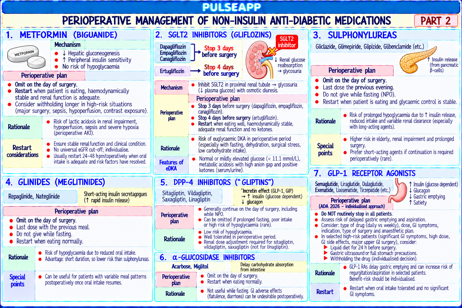
C. Perioperative Medication Management3 marks

Drug Morning of Surgery Rationale Metformin OMIT day of surgery Metformin + perioperative hypoperfusion → lactic acidosis; resume when eating and renal function confirmed (and 48h before contrast/major surgery) SGLT2 inhibitors OMIT ≥24–72 hours SGLT2i → euglycaemic DKA risk: blocks glucose reabsorption → glucosuria → shifts metabolism to fat → (empagliflozin, before surgery ketogenesis ↑; fasting + surgical stress + SGLT2i = DKA despite NORMAL blood glucose; check betadapagliflozin, hydroxybutyrate (ketones) if SGLT2i taken within 72h + acidosis present canagliflozin) Sulphonylureas OMIT morning dose Risk of hypoglycaemia; long half-life; check glucose hourly throughout day (gliclazide, glibenclamide) Long-acting insulin Give 80% of normal Do NOT omit completely in T1DM — always needs basal insulin to prevent ketogenesis; 80% reduces (glargine, detemir) dose the night before hyperglycaemia without hypoglycaemia risk Short/rapid-acting OMIT morning dose Only give prandial insulin when eating; if VRIII started → short-acting not separately required insulin

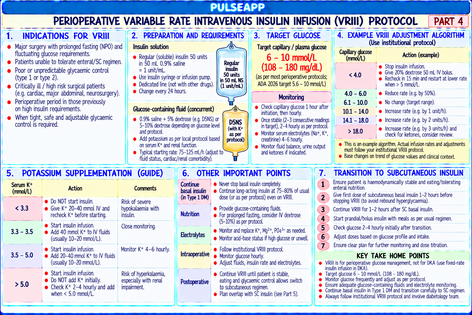
D. Variable Rate Insulin Infusion (VRIII)3 marks
✅ JBDS/NHS England 2023 VRIII Protocol
Indications: glucose persistently >12 mmol/L; T1DM for major surgery; T2DM on insulin + prolonged fasting (>1 meal missed); vomiting; unable to take oral medications >2 hours peri-operatively Target: 6–10 mmol/L (not too tight — avoids hypoglycaemia; not too loose — avoids SSI) Setup: 50 units Actrapid in 50 mL 0.9% NaCl (1 unit/mL); rate per VRIII table (glucose 4–7 → 1 unit/hr; 7–9 → 2 units/hr; 9–12 → 3 units/hr; 12–15 → 4 units/hr; >15 → call diabetes team); ALWAYS co-administer 5% dextrose / 0.45% NaCl with 0.15% KCl at 125 mL/hr (glucose substrate prevents hypoglycaemia); monitor K⁺ every 4–6 hours (insulin drives K⁺ into cells → hypokalaemia risk)
E. Hypoglycaemia & DKA Management —

Hypoglycaemia (<4 mmol/L): STOP VRIII; 150–200 mL 10% dextrose IV over 15 min; recheck at 15 min; once >6 mmol/L → restart VRIII at lower rate; do NOT stop basal insulin in T1DM (stops it → risks DKA)

DKA recognition: glucose >11 mmol/L (OR normal glucose if on SGLT2i = euglycaemic DKA) + ketones >3 mmol/L + pH <7.3, HCO₃⁻ <18 → start DKA protocol: fixed rate insulin 0.1 units/kg/hr + IV fluids + K⁺ replacement + hourly monitoring + endocrinology/diabetes team

🎤 Viva Corner
Q. Why must SGLT2 inhibitors be stopped 24–72 hours before surgery and what specific complication do they cause?
SGLT2 inhibitors must be stopped pre-operatively because they cause euglycaemic diabetic ketoacidosis (EDKA) — DKA with apparently NORMAL blood glucose. The mechanism: SGLT2i block glucose reabsorption in the renal proximal tubule → glucosuria → blood glucose appears normal or only mildly elevated (9–14 mmol/L) even as the body undergoes severe ketoacidosis. The reason for ketosis: glucosuria signals relative glucose deficiency → glucagon ↑ and insulin ↓ → fat metabolism predominates → fatty acid oxidation → ketone body production in the liver → ketoacidosis. Under normal circumstances this is counterbalanced, but perioperatively: fasting provides no carbohydrate → fat metabolism is the only fuel; surgical stress → catecholamines → further suppresses insulin → more ketogenesis; volume depletion from fasting → reduced renal ketone clearance; the SGLT2i from days before the surgery is still active (half-lives 12–24h or more). The danger: anaesthesia teams monitor blood glucose for diabetic safety; blood glucose appears acceptable at 11 mmol/L; the profound underlying acidosis (pH 7.0, HCO₃ 8 mmol/L) from ketoacidosis is missed until the patient is severely ill. By stopping SGLT2i 24–72h before surgery: the drug clears from the system; the ketogenic mechanism is removed before the perioperative fasting stress. If emergency surgery cannot wait: check beta-hydroxybutyrate (blood ketones) preoperatively; if >1.5 mmol/L → treat with insulin and dextrose before proceeding.
★ Examiner's Pearl
HbA1c ≥69 mmol/mol (8.5%) → postpone elective surgery. SGLT2i → euglycaemic DKA (DKA with normal glucose; check KETONES not just glucose; stop 24–72h pre-op). VRIII glucose target 6–10 mmol/L (not "tight control"). VRIII always co-administered with dextrose substrate (5% dextrose/0.45% NaCl) and K⁺ monitoring (hypokalaemia from insulin).
NHS England/JBDS. Management of adults with diabetes undergoing surgery 2023. AAGBI. Perioperative management of diabetes 2015 (Anaesthesia 2015;70:1427). Turchin A et al. SGLT2 and perioperative DKA (JAMA 2023). Miller RD et al. Miller's Anaesthesia, 9th Ed.
lock Protected Clinical Document • Pulse App • Screenshots Strictly Prohibited
QUESTION 95 person Asked by .
bookmark_add

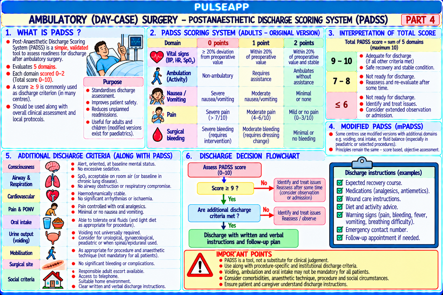
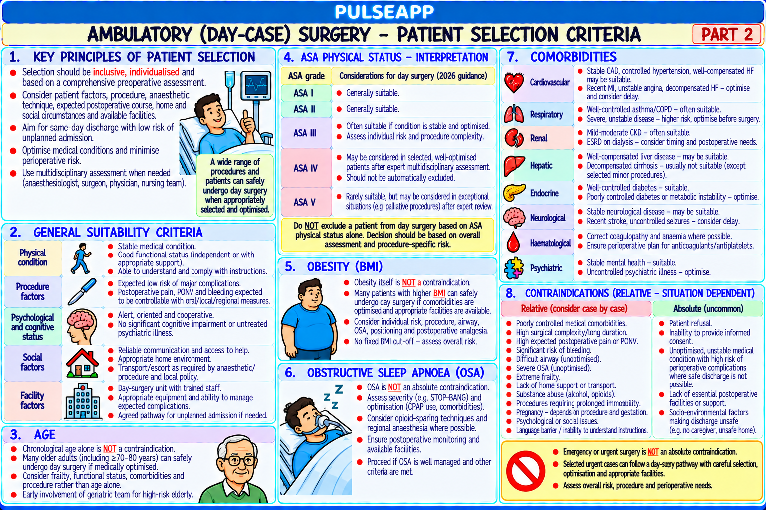
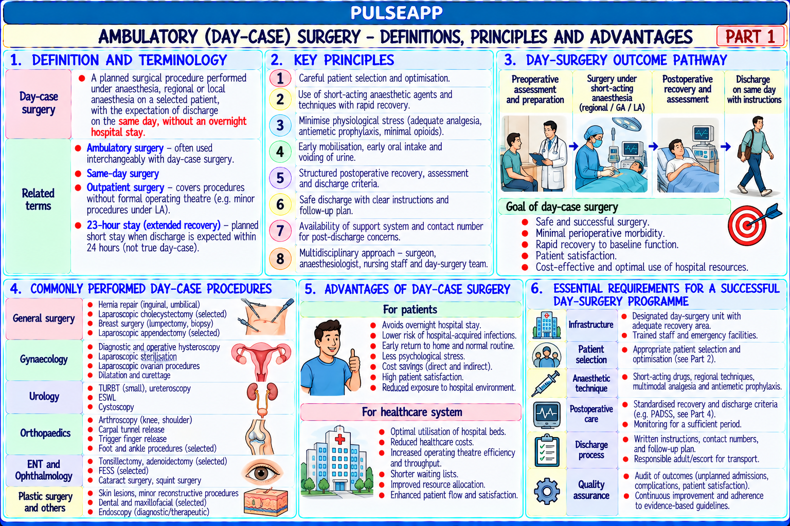
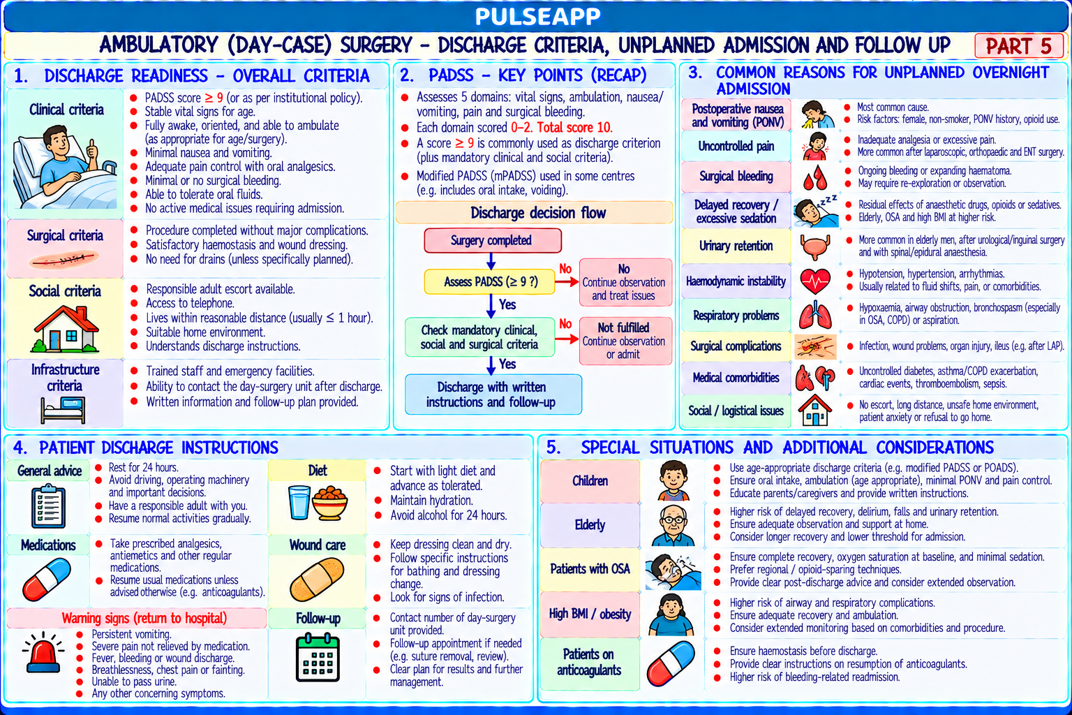
Define day-case surgery and patient selection criteria. Describe anaesthetic technique modifications for ambulatory surgery and the PADSS discharge scoring system. Outline the most common reasons for unplanned overnight admission.

description Clinical Response
⚙ Core Concept
Day-case surgery accounts for 70–80% of elective surgery in developed healthcare systems. The anaesthetic technique must achieve three goals: rapid recovery, adequate home analgesia, and prevention of PONV — the #1 cause of unplanned admission (35–40%). The PADSS (Post-Anaesthesia Discharge Scoring System) provides objective discharge criteria. (Chung F — J Clin Anesth 1995; BADS Directory 2019; Verma R — BJA 2011)
A. Patient Selection Criteria2 marks

Criterion Eligible Requires Overnight Admission ASA ASA I, II; selected well-optimised ASA III ASA IV; unstable ASA III; recent decompensation BMI BMI up to 35–40; BADS 2019: BMI alone should NOT exclude patients; assess BMI >40 with severe OSA + major surgery; obesity individually hypoventilation syndrome OSA Mild-moderate OSA on CPAP + minor surgery; brings own CPAP device Severe OSA (AHI >30) with opioid requirement post-op Social Responsible adult to drive home and stay 24h; within 60–90 min of hospital; telephone Lives alone; remote from medical care; unable to understand access; able to follow instructions discharge instructions Procedure Expected duration <3 hours; pain controllable with oral analgesia; manageable blood loss Major surgery; significant expected blood loss; complex wound management

B. Anaesthetic Technique for Day-Case Surgery3 marks

Element Day-Surgery Recommendation Rationale Induction Propofol 2–2.5 mg/kg IV — preferred Antiemetic properties; pleasant emergence; faster return to street fitness vs thiopentone Maintenance Propofol TIVA (propofol + remifentanil TCI) OR sevoflurane; TIVA TIVA: lower PONV (propofol antiemetic); no volatile emissions; sevoflurane preferred for Apfel ≥3 suitable for shorter cases; desflurane: fastest recovery for long cases but environmental concerns NMB Short-acting NMBs; sugammadex reversal (faster and more reliable than Residual NMB delays discharge; neostigmine → nausea adds to PONV neostigmine); ideally avoid NMB (LMA + spontaneous breathing for burden suitable cases) Analgesia Regional where possible; paracetamol 1 g IV at induction + ketorolac 15 Opioids → PONV + sedation at home → most common cause of failed day mg IV; wound infiltration bupivacaine 0.25%; prescribe 3–5 days surgery; adequate multimodal analgesia enables discharge paracetamol + NSAID for home PONV Minimum 2 antiemetics for Apfel ≥2: ondansetron 4 mg at end + PONV is #1 cause of unplanned admission; every Apfel point = +20% risk; prophylaxis dexamethasone 4–8 mg at induction; consider TIVA for Apfel ≥3; maximum prevention is cost-effective prescribe rescue antiemetics for home Airway LMA preferred over ETT where safe — reduces sore throat, coughing, Smooth emergence without coughing prevents PONV and cardiovascular laryngospasm; i-gel or ProSeal for controlled ventilation stress; less stimulating extubation with LMA

C. PADSS — Post-Anaesthesia Discharge Scoring System3 marks

Parameter Score 2 Score 1 Score 0 Vital signs ≤20% change from pre-op baseline 20–40% change >40% change Ambulation Steady gait; no dizziness Requires assistance Unable to ambulate Nausea/Vomiting Minimal; oral treatment sufficient Moderate; parenteral medication needed Severe; continues despite treatment Pain Minimal; controlled with oral analgesics; acceptable to patient Moderate; requires parenteral analgesia Severe; not controlled Surgical bleeding Minimal; no dressing change Moderate; 1–2 dressing changes Severe; >2 dressing changes

Discharge criterion: PADSS ≥9 out of 10 required; developed and validated by Chung et al. (J Clin Anesth 1995) specifically for ambulatory surgery

D. Causes of Unplanned Overnight Admission2 marks

PONV (35–40% of failures): most common; prevent with Apfel-stratified multimodal prophylaxis; prescribe rescue antiemetics for home; discharge only when PONV PADSS score ≥1 Inadequate pain control (25–30%): pain too severe for home management; multimodal analgesia pre-discharge; VAS ≤3/10 at discharge; written pain management plan; adequate take-home analgesia

Surgical complications: haemorrhage requiring return to theatre; urinary retention (particularly after spinal with opioids — ensure void before discharge); wound concerns

Residual anaesthetic effects: prolonged sedation, dizziness, residual NMB; avoid long-acting benzodiazepines; quantitative NMB monitoring

🎤 Viva Corner
Q. Can you do day-case laparoscopic hernia repair in a 72-year-old, BMI 38, well-controlled hypertension, mild COPD (FEV1 72%)?
Yes — this patient is an appropriate candidate for day-case laparoscopic inguinal hernia repair, provided all comorbidities are confirmed as well-optimised. Working through the selection criteria: this patient is ASA II–III; ASA III patients are eligible when comorbidities are stable. BMI 38 — BADS 2019 specifically states BMI should NOT be used as a single exclusion criterion; BMI 38 in the context of a laparoscopic rather than open procedure, with normal respiratory reserve, is acceptable at most day surgery units. COPD FEV1 72% is GOLD II (moderate) — unless the patient has daytime hypoxaemia, CO₂ retention, or is on home O₂, this represents adequate reserve for a 1–2 hour laparoscopic procedure. Pre-operative confirmation needed: BP well-controlled (SBP <160 consistently); SpO₂ ≥94% on room air; no active respiratory exacerbation in the past 6 weeks; inhaler compliance confirmed; responsible adult at home; within 60 minutes of hospital; telephone access. Anaesthetic technique: TIVA with propofol-remifentanil preferred (lowest PONV risk for a likely Apfel score 2–3 patient); ondansetron 4 mg + dexamethasone 4 mg; multimodal analgesia (paracetamol + ketorolac + port-site bupivacaine infiltration); LMA with controlled ventilation; no NMB if possible. PADSS ≥9 before discharge; take-home: paracetamol 1 g QID + ibuprofen 400 mg TID for 5 days; rescue antiemetic (metoclopramide 10 mg) at home.
★ Examiner's Pearl
PADSS table (5 parameters; each 0–2; discharge requires ≥9/10) — must be reproduced. PONV as #1 cause of failed day surgery (35–40% of unplanned admissions). BADS 2019 position on BMI: BMI alone should NOT exclude — assess individually (corrects the outdated "BMI 35 hard cutoff"). TIVA preference for day surgery (antiemetic + lower PONV vs volatile).
Chung F et al. PADSS for home readiness after ambulatory surgery (J Clin Anesth 1995;7:500-506). British Association of Day Surgery (BADS). Directory 2019. Verma R et al. Day surgery: speciality or service? (BJA 2011). White PF. Ambulatory anaesthesia, 3rd Ed. Miller RD et al. Miller's Anaesthesia, 9th Ed.
lock Protected Clinical Document • Pulse App • Screenshots Strictly Prohibited
QUESTION 96 person Asked by .
bookmark_add

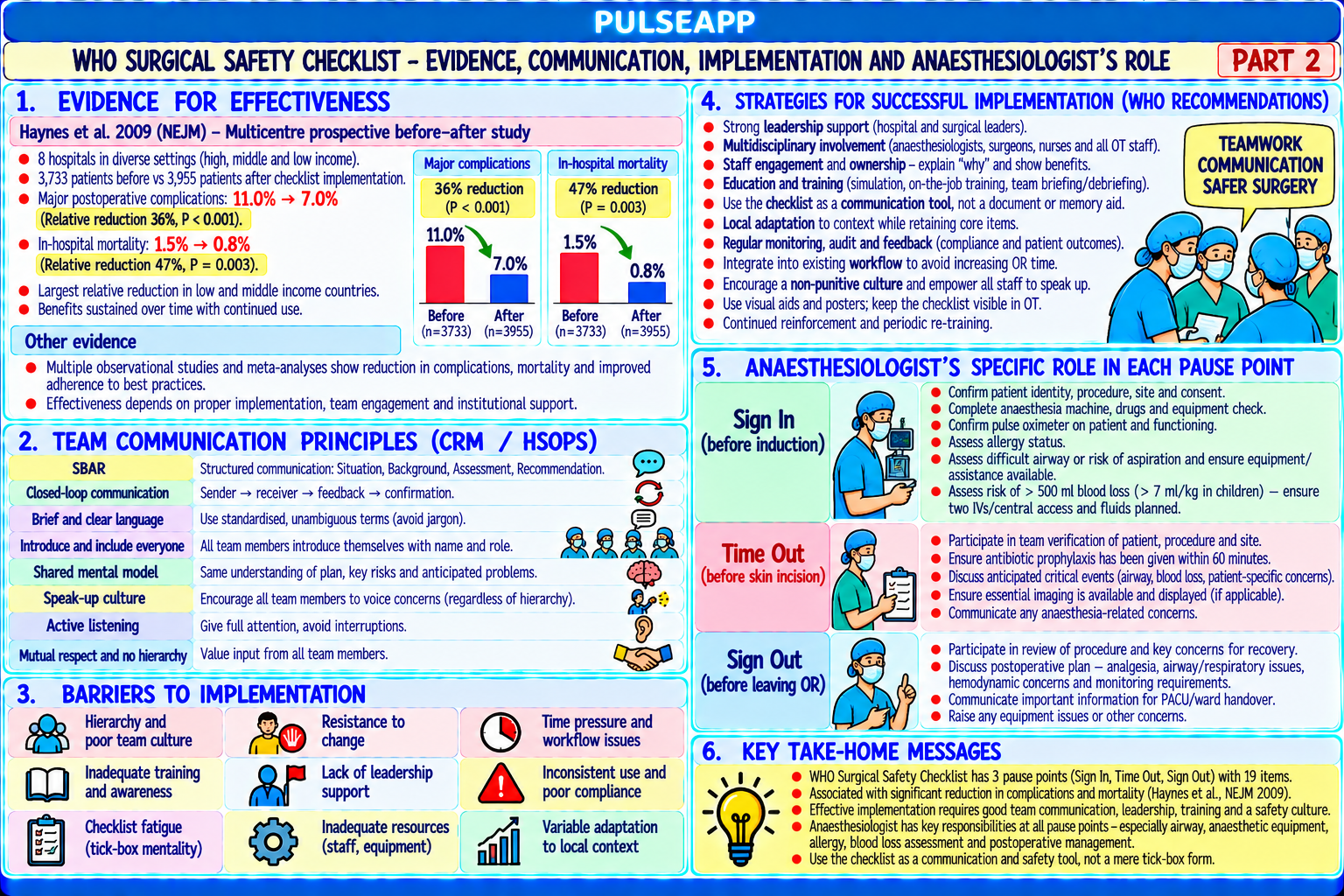
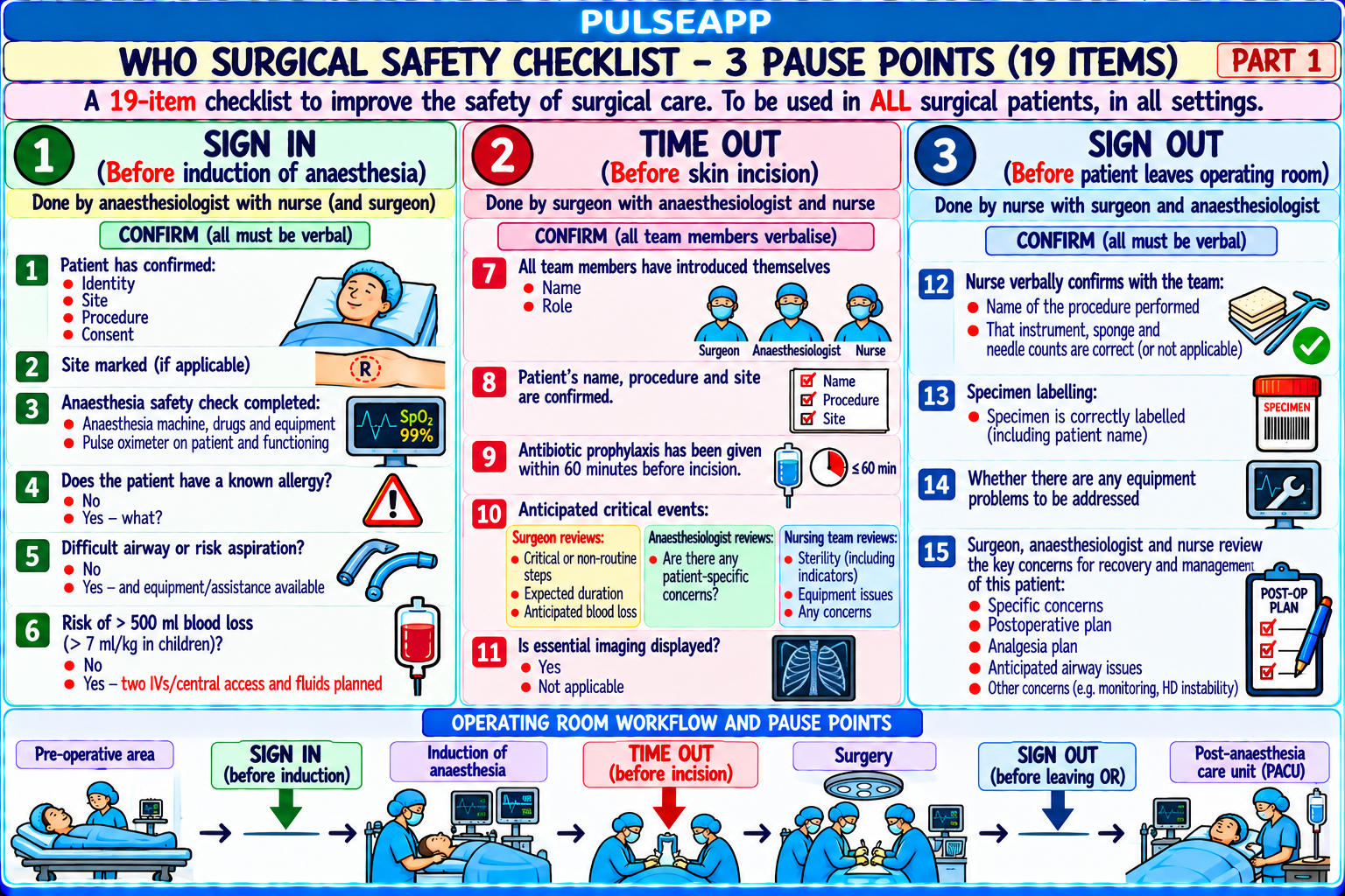
Describe the three WHO SSC pause points (Sign In, Time Out, Sign Out) with specific items at each. Cite the evidence for effectiveness. Discuss team communication principles, barriers to implementation, and the anaesthesiologist's specific role.

collections Question Diagrams & Reference Images (1)
Question Reference Diagram
zoom_in View Image
description Clinical Response
⚙ Core Concept
The WHO Surgical Safety Checklist (2008) is the single most evidence-based intervention for reducing surgical mortality globally — Haynes (NEJM 2009) demonstrated a 36% reduction in complications and 47% reduction in mortality across 8 hospitals worldwide. Its 19-item, three-pause-point structure addresses the most common causes of preventable surgical harm: wrong site surgery, retained instruments, allergic reactions, and team communication failures. (Haynes AB — NEJM 2009; de Vries EN — Lancet 2010; WHO Safe Surgery Saves Lives 2008)
A. The Three Pause Points4 marks

Stage When Leader Key Items Sign Before anaesthesia induction Anaesthesiologist 1. Patient identity confirmed verbally (name + DOB against wristband + In notes) 2. Procedure and site confirmed (including marking) 3. Consent signed and present 4. Pulse oximeter functioning and applied 5. Known allergy? → state allergen 6. Difficult airway/aspiration risk? → equipment ready 7. Blood loss risk ≥500 mL? → IV access, cross-match, blood products ordered Time Before surgical incision (patient draped, all Surgeon or team leader; any 1. All team members introduce themselves and roles Out team assembled) member may initiate 2. Surgeon + anaesthesiologist + nurse verbally confirm: patient name, procedure, site 3. Surgeon: anticipated critical steps, expected duration and blood loss 4. Anaesthesiologist: patient-specific concerns (allergy, airway, cardiac risk) 5. Nursing team: equipment sterility confirmed, essential equipment available 6. Antibiotic prophylaxis given within 60 min of incision if required 7. Imaging confirmed displayed and correct patient Sign Before patient leaves the operating room Nursing team (scrub/circulating) 1. Procedure recorded by name Out 2. Instrument, sponge, and needle count completed and documented 3. Specimen labelling confirmed (patient name, DOB, procedure, surgeon on label) 4. Equipment problems to address 5. Key concerns for recovery: surgeon and anaesthesiologist verbally handover to recovery team

B. Evidence for Effectiveness2 marks

Haynes AB et al. (NEJM 2009): 8 hospitals, 8 countries, n=7,688 surgical patients; WHO SSC reduced: overall complications 11.0% → 7.0% (36% relative reduction); in-hospital mortality 1.5% → 0.8% (47% reduction); SSI 6.2% → 3.4% (45% reduction); benefit was significant in all income settings de Vries EN et al. (Lancet 2010): SURPASS trial, Netherlands; 11-step checklist across multiple perioperative time points; complications 15.4% → 10.6%; mortality 1.5% → 0.8% — independently confirmed Haynes findings

Implementation challenges: despite strong evidence, compliance <50% in many audits; primary barriers: tick-box exercise without engagement; time pressure (OR schedule); hierarchical culture (junior reluctant to pause senior surgeon); checklist fatigue

C. Team Communication — The Core Mechanism2 marks

Aviation research and human factors science (Reason — Swiss cheese model, 1990): most high-consequence errors occur from team communication breakdowns, not individual failures; the SSC embeds structured communication into the surgical routine

TeamSTEPPS core strategies: SBAR (Situation-Background-Assessment-Recommendation) for structured handover; closed-loop communication (receiver explicitly confirms the message); call-out and check-back; the Time Out team introduction step specifically breaks down hierarchical barriers and creates a culture where any team member can halt the procedure

Challenging authority: the SSC gives every team member — including the most junior nurse — specific authorisation to raise a safety concern; the ABCDE framework for challenging decisions: Assert concern clearly; call Supervisor; call Expert help; Explain to the team; Document concerns

D. Anaesthesiologist's Specific Role2 marks

Leads the Sign In — the most complex section, requiring knowledge of both the patient's medical history and the anaesthetic plan; confirms all anaestheticspecific safety items (allergy, airway, aspiration, blood loss risk) Contributes specific expertise at all three pause points; at Time Out: states patient-specific anaesthetic concerns: "this patient is allergic to penicillin — please ensure a non-penicillin antibiotic is used"; "this patient has known difficult airway — video laryngoscope is in the room"; "this patient is on anticoagulation — the surgeon should anticipate increased bleeding" Has a duty to halt the procedure if the checklist reveals an unresolved safety concern — patient identity discrepancy, unsigned consent, absent blood crossmatch in a high-blood-loss case, unavailable specialised equipment; escalate through the chain of command if the initial concern is dismissed

🎤 Viva Corner
Q. During Time Out before laparotomy, the nurse notices the surgical site marking is absent and imaging shows a right-sided lesion but the patient is positioned with the left side up. What do you do?
This is a wrong-site surgery near-miss — the Time Out has functioned exactly as intended by detecting this discrepancy before the first incision. My immediate response: call a stop confidently and clearly: "Stop — we need to resolve this discrepancy before we can proceed." This is not optional. Every team member has authority to pause the procedure when a safety concern is identified. Systematic verification: confirm patient identity and procedure from the signed consent form (should specify the procedure and laterality); review the imaging directly in theatre — radiologist's report must be available with correct patient ID confirmed; check if the surgical site marking was performed preoperatively (NPSA requirement — marking should be done before anaesthesia with patient's verbal confirmation while awake); call the responsible surgeon to verify the correct side from imaging, consent, and operative notes before any incision. Do NOT proceed until the correct side has been unambiguously confirmed from multiple independent sources. Document everything: the discrepancy identified, the pause, the confirmation process, and who verified the correct side. Report through the hospital incident reporting system (near-miss prevents a never-event) — mandatory even though the wrong-site surgery was prevented. The checklist worked. This is the system functioning as designed.
★ Examiner's Pearl
Three pause points with leader (Sign In/anaesthesiologist → Time Out/surgeon → Sign Out/nursing team) and specific items at each must be reproduced completely. Haynes NEJM 2009 specific numbers: complications 11%→7% (36%); mortality 1.5%→0.8% (47%). The anaesthesiologist's Sign In items (allergy, airway, aspiration, blood loss) are the most specifically tested practical content.
Haynes AB et al. A surgical safety checklist (NEJM 2009;360:491-499). de Vries EN et al. SURPASS trial (NEJM 2010;363:1928-1937). WHO. Surgical Safety Checklist 2009. Gawande A. The Checklist Manifesto. TeamSTEPPS 2.0 AHRQ.
lock Protected Clinical Document • Pulse App • Screenshots Strictly Prohibited
QUESTION 97 person Asked by .
bookmark_add

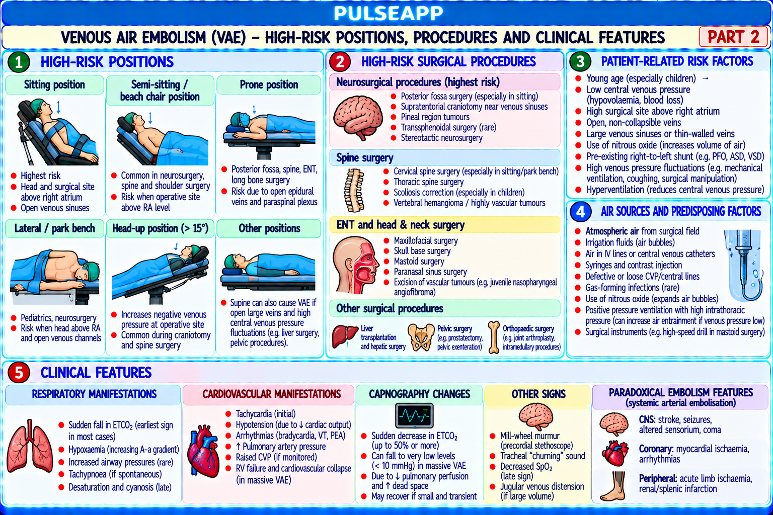
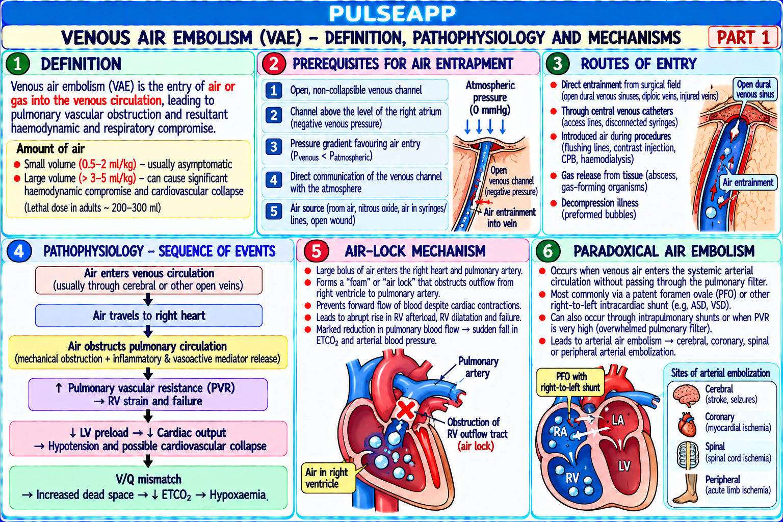
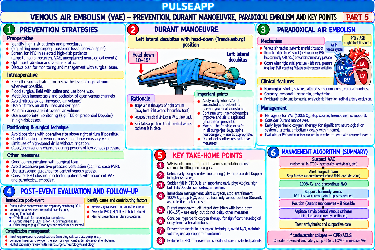
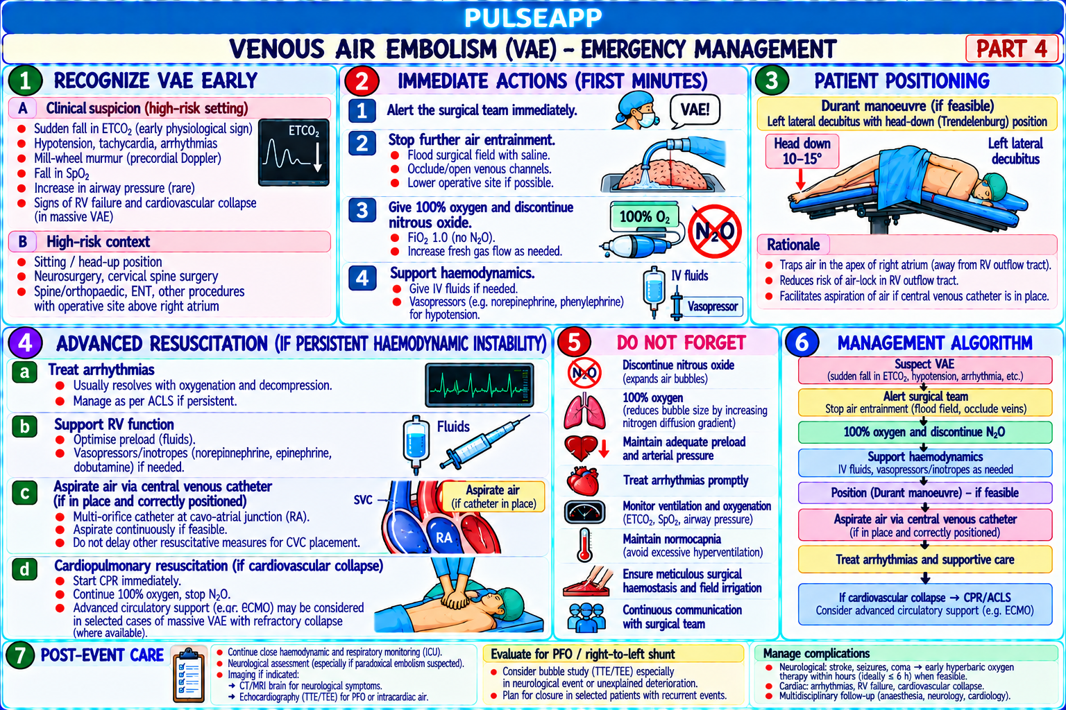
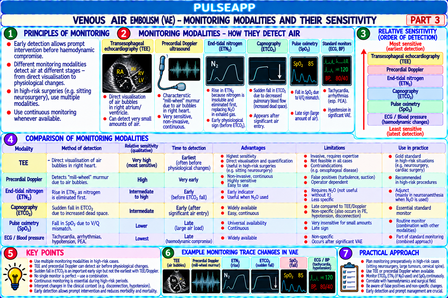
Define VAE and describe the pathophysiology including the air-lock mechanism and paradoxical air embolism. Compare monitoring modalities by sensitivity. List high-risk positions and procedures. Outline the stepwise emergency management including the Durant manoeuvre.

collections Question Diagrams & Reference Images (1)
Question Reference Diagram
zoom_in View Image
description Clinical Response
⚙ Core Concept
Venous air embolism — air entering the venous system and travelling to the right heart — causes cardiovascular collapse in proportion to the rate and volume of air entrainment. The sitting position for neurosurgery carries up to 45% VAE incidence by Doppler. Management depends on early detection (precordial Doppler is the standard monitoring), flooding the field, stopping N₂O, and positioning the patient to dislodge the air lock (Durant's manoeuvre). (Mirski MA — Crit Care Med 2007; Porter JM — BJA 1999; Miller's Anaesthesia 9th Ed)
A. Pathophysiology2 marks

Air enters a venous sinus or vessel held open by surrounding anatomy (bone, surgical retractors, tethered dura) where the venous pressure is below atmospheric → negative pressure gradient drives air into the venous system → right atrium → right ventricle → pulmonary vasculature

Air-lock mechanism: large amounts (2–5 mL/kg at rapid rate) → air accumulates in the right ventricle → impedes RV ejection → acute cor pulmonale → ↓ CO → cardiovascular collapse; simultaneously pulmonary arteriolar obstruction → ↑ dead space → rising then falling ETCO₂ as cardiac output falls

Paradoxical air embolism (PAE): patent foramen ovale (PFO — present in 25–30% of population) → right atrial pressure elevated by VAE → reverses atrial pressure gradient → air crosses PFO from right to left atrium → systemic arterial circulation → coronary artery (MI) or cerebral (stroke, seizures) air embolism; PAE can occur even with relatively small VAE amounts

B. Monitoring by Sensitivity3 marks

Monitor Sensitivity Details

Transoesophageal Most sensitive: Visualises air bubbles directly in right atrium and ventricle; detects paradoxical embolism across PFO; impractical for echo (TOE) detects 0.01–0.02 routine use — requires expertise, limits positioning mL/kg

Precordial Very sensitive: Placed over the right precordium (right sternal border, 2nd–4th ICS); characteristic churning "mill-wheel" quality change in Doppler detects 0.05–0.1 Doppler signal; STANDARD MONITOR for sitting-position neurosurgery; non-invasive; cannot quantify volume mL/kg

ETCO₂ Moderate: detects Initially ↑ CO₂ then ↓ as CO falls; non-specific; part of routine monitoring; the first bedside indicator in most cases (capnography) >0.5 mL/kg

Pulmonary artery Sensitive: detects 0.25 Direct PA pressure rise; allows air aspiration via PA catheter; invasive — not routine catheter mL/kg CVP / CVC Moderate CVP rises with right heart obstruction; allows air aspiration via right atrial catheter

Oesophageal Least sensitive — Mill-wheel murmur only with massive embolism; delayed detection; backup only stethoscope detects only very large VAE

C. High-Risk Positions and Procedures2 marks

Setting VAE Incidence Mechanism Sitting position Up to 45% by Doppler; 1–2% Surgical site far above heart → cranial venous sinuses at sub-atmospheric pressure → nonneurosurgery clinically significant collapsible (held open by bone) → air entrainment Posterior fossa surgery 15–25% by Doppler Same mechanism; dural venous sinuses (prone/sitting) Total hip arthroplasty Varies; BCIS related Intramedullary pressurisation during cementing → fat + air embolism (Bone Cement Implantation

Syndrome)

Laparoscopic surgery Rare CO₂ embolism if trocar in CO₂ embolism: 25× more soluble than air → less severe; sudden ETCO₂ fall + CVS collapse vein

CVC insertion/removal Air embolism during Prevent: Trendelenburg + Valsalva during removal; immediately occlude on removal disconnection Caesarean section 30–50% by TOE but almost all Open uterine veins during closure; vast majority are clinically irrelevant small amounts subclinical

D. Emergency Management — Stepwise Protocol3 marks
⚠ VAE Emergency — Immediate Actions
1. Notify the surgeon immediately — flood the surgical field with saline or wet patties (seals open venous sinuses; stops further air entrainment) 2. Discontinue N₂O immediately — N₂O is 25× more soluble than N₂ → rapidly enters the air embolism → expands it dramatically; switch to 100% O₂ (also helps absorb the embolism) 3. Compress jugular veins bilaterally — manual bilateral neck compression raises jugular venous pressure → reduces the negative pressure gradient driving air entrainment 4. Aspirate via the right atrial catheter — vigorously aspirate through CVC positioned at the right atrial level (Bunegin-Albin multi-orifice catheter placed pre-operatively is ideal); 5–20 mL frothy bloody aspirate confirms intracardiac air; continue until air-free blood returns 5. Durant's manoeuvre — left lateral decubitus + Trendelenburg positioning; moves the air lock from the right ventricular OUTFLOW TRACT (where it impedes ejection) to the right ventricular APEX → allows some continuation of cardiac output; also positions air near the CVC tip for aspiration 6. Supportive resuscitation — IV fluid bolus; vasopressors for BP; CPR if cardiac arrest (chest compressions may physically break up the air lock) 7. Lower the surgical site — lower the patient's head below heart level if anatomically feasible → reverses the pressure gradient driving air entrainment
🎤 Viva Corner
Q. During sitting-position craniotomy, precordial Doppler changes quality and ETCO₂ falls from 35 to 22 mmHg. SpO₂ is 90%. List management steps in order. Significant VAE in the sitting position. Management in order: Step 1 — inform the surgeon immediately: "Stop — significant air embolism detected; please flood the field with saline now." All surgical activity stops. Field flooded with wet patties. Step 2 — discontinue N₂O immediately; switch to 100% FiO₂. Step 3 — compress jugular veins bilaterally (assistant compresses both sides simultaneously). Step 4 — aspirate vigorously via the pre-placed right atrial catheter; 10 mL syringe; continue until air-free blood returns. Step 5 — check haemodynamics: if hypotension → IV fluid bolus 500 mL + phenylephrine 100 mcg IV or noradrenaline infusion. Step 6 — if cardiovascular compromise worsening → Durant's manoeuvre: left lateral decubitus + Trendelenburg; moves air from RVOT to RV apex allowing some cardiac ejection. Step 7 — ask surgeon to lower the surgical site if anatomically feasible. Step 8 — monitor for paradoxical embolism (new ECG changes, arrhythmia, neurological changes). Step 9 — if cardiac arrest: CPR (chest compressions may disrupt the air lock). Step 10 — post-event: TOE if available to confirm clearance; document and report; screen for PFO post-operatively; advise against sitting position for future procedures.
★ Examiner's Pearl
Monitoring sensitivity ranking (TOE 0.01 mL/kg most sensitive → precordial Doppler 0.05 mL/kg standard monitor → ETCO₂ moderate → oesophageal stethoscope least sensitive) with specific detection thresholds. Durant's manoeuvre: left lateral + Trendelenburg — moves air from RV outflow tract to RV apex → allows RV ejection — state the specific mechanism. N₂O discontinuation rationale: N₂O 25× more soluble than N₂ → rapidly enters and expands the air embolism.
Mirski MA et al. Venous air embolism (Crit Care Med 2007;35:1439-1448). Porter JM et al. Sitting position in neurosurgery (BJA 1999;82:117-128). Miller RD et al. Miller's Anaesthesia, 9th Ed, Chapter 70. Palmon SC et al. VAE review (J Clin Anesth 1997). Shaikh N et al. Acute management of VAE (J Emerg Trauma Shock 2009).
lock Protected Clinical Document • Pulse App • Screenshots Strictly Prohibited
QUESTION 98 person Asked by .
bookmark_add

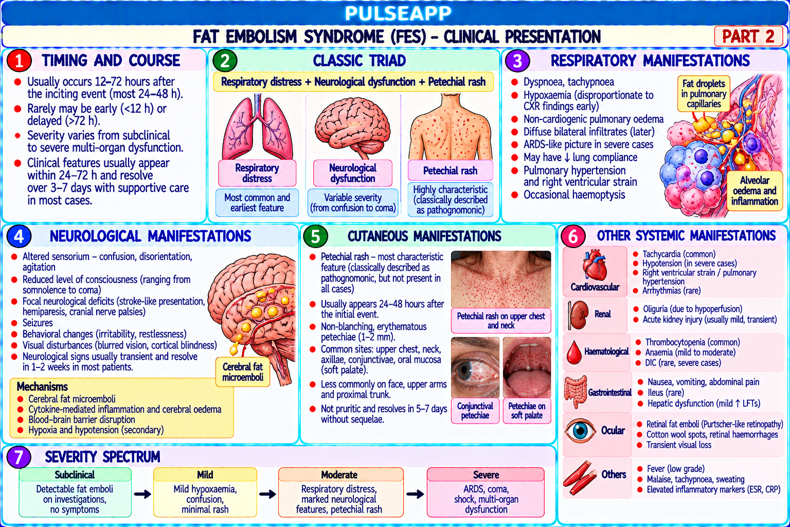
Define fat embolism syndrome (FES). Describe the two proposed pathophysiological mechanisms. State the Gurd diagnostic criteria (major and minor). Outline the clinical presentation including the pathognomonic petechial rash, and describe the management in the ICU.

collections Question Diagrams & Reference Images (1)
Question Reference Diagram
zoom_in View Image
description Clinical Response
⚙ Core Concept
Fat embolism syndrome (FES) is a serious complication of long bone fractures (femur, tibia, pelvis) occurring 12–72 hours after injury — characterised by the classic triad of respiratory distress, neurological dysfunction, and petechial rash. It is distinct from simple fat embolism (mere presence of fat globules in the bloodstream, which is almost universal after major fractures). FES develops when the embolised fat globules cause systemic inflammatory injury, particularly in the lungs (ARDS pattern) and brain. (Gurd AR — J Bone Joint Surg 1970; Mellor A — BJA 2001; Lindeque BG; Miller's Anaesthesia 9th Ed)
A. Definition and Incidence1 mark

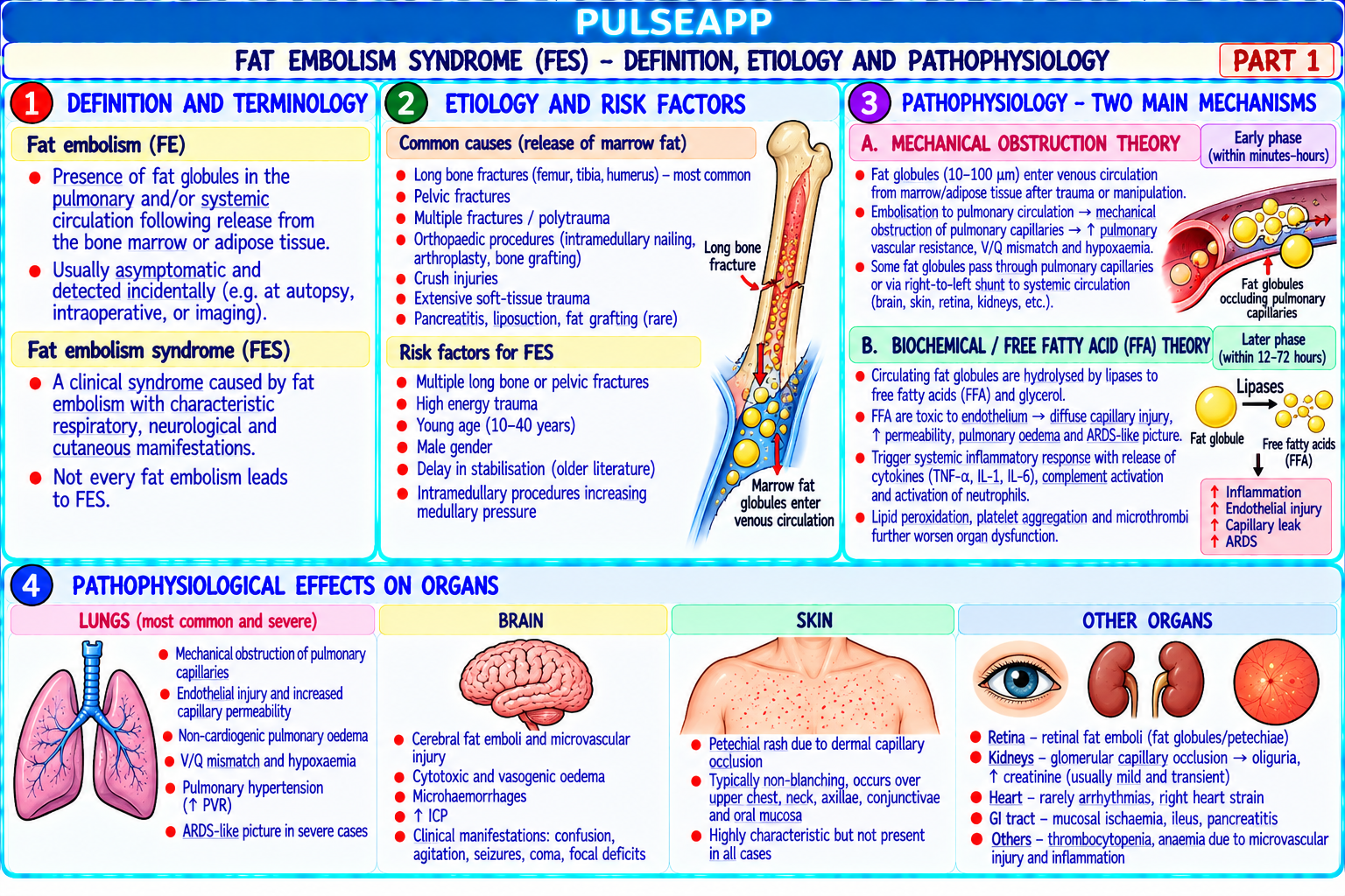
FES is a clinical syndrome caused by embolisation of fat globules from the bone marrow into the systemic circulation following long bone or pelvic fractures (or other triggers: liposuction, total hip arthroplasty, sickle cell crisis, pancreatitis); incidence after closed femoral shaft fracture: 0.5–2% clinical FES; incidence of fat emboli in the bloodstream (without syndrome): nearly 100%; the syndrome requires an inflammatory amplification beyond simple fat embolisation

B. Pathophysiology — Two Proposed Mechanisms2 marks

Mechanism Details Mechanical Disruption of bone marrow fat cells → fat droplets enter disrupted marrow sinusoids → enter the venous system → travel to the pulmonary vasculature (embolic) → mechanical obstruction of pulmonary arterioles → impaired gas exchange; fat droplets large enough to traverse the pulmonary capillaries or cross theory through a PFO → systemic embolisation to brain, skin, retina, kidneys Biochemical Fat droplets in the bloodstream undergo hydrolysis by lipases (plasma and tissue lipases) → release of FREE FATTY ACIDS (FFAs); FFAs are directly (inflammatory) toxic to: pulmonary endothelium (causing capillary leak → non-cardiogenic pulmonary oedema → ARDS); cerebral vasculature (cerebral oedema); theory — the platelet membranes (platelet aggregation → thrombocytopenia, DIC); the systemic inflammatory response amplifies this injury through cytokine release; primary this biochemical theory explains why ARDS onset is delayed (12–72 hours — time for lipases to generate sufficient FFAs) rather than immediate as mechanism would be expected from mechanical obstruction alone for FES

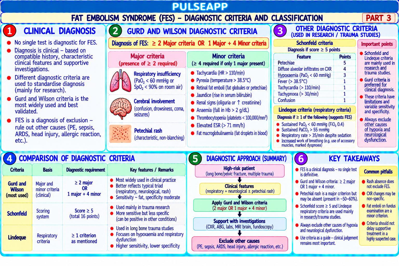
C. Gurd Diagnostic Criteria (1970, modified)3 marks
✅ Gurd Criteria — FES Diagnosis Requires: 1 Major + 4 Minor OR 1 Major + Fat Macroglobulinaemia
Major Criteria (1 required) Minor Criteria (4 required) Respiratory insufficiency: PaO₂ <60 mmHg on FiO₂ 0.4; or required mechanical ventilation; bilateral Tachycardia >120 bpm; fever >38.5°C; urinary fat pulmonary infiltrates on CXR (ARDS pattern) globules or fat macroglobulinaemia in urine Cerebral involvement: neurological deterioration (confusion, agitation, coma) that is NOT explained by Thrombocytopenia (platelets <150,000 — often head injury or hypoxia alone; cerebral fat emboli visible on MRI (diffuse white matter lesions, "starfield" sudden drop of >50% from baseline); raised ESR pattern on DWI) (>71 mm/hr) Pathognomonic: petechial rash — a specific distribution: axillae, conjunctivae, neck, anterior chest, and Anaemia (acute fall in Hb >20 g/L unexplained by base of neck; caused by fat emboli to dermal capillaries; appears 24–36 hours after injury; TRANSIENT bleeding alone); renal fat globules; retinal fat emboli (fades in hours); present in only 50–60% of cases but HIGHLY SPECIFIC when present on ophthalmoscopy (Purtscher's retinopathy)
D. Clinical Presentation2 marks

Timing: characteristically presents 24–72 hours after injury; the lucid interval — the patient may initially be alert and oriented after the fracture, then gradually deteriorates with increasing confusion, agitation, and dyspnoea over the subsequent 24–48 hours

The classic triad: respiratory failure (most constant — present in 75–90% of FES; ARDS pattern; bilateral infiltrates; hypoxaemia refractory to supplemental O₂); neurological dysfunction (confusion, agitation, headache, seizures, coma — in 86%); petechial rash (in 50–60% — pathognomonic)

Other features: acute anaemia (haemolysis and marrow fat release); thrombocytopenia; fever; retinal fat emboli (Purtscher's retinopathy — white retinal patches visible on fundoscopy)

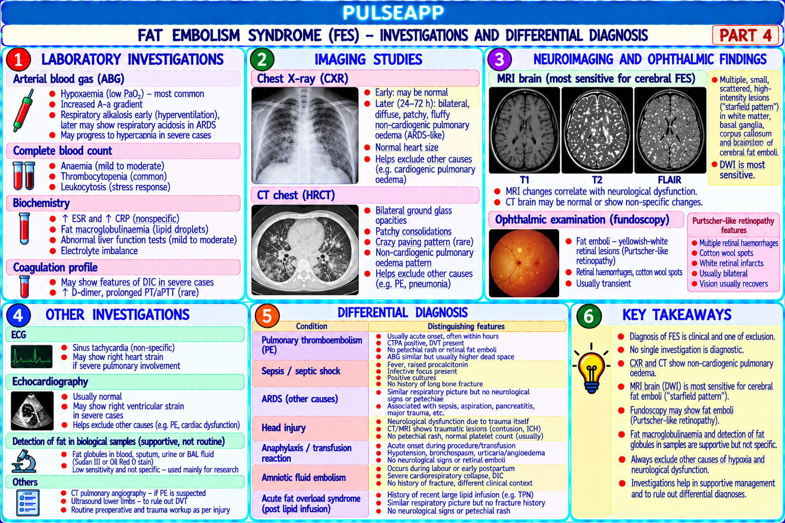
Investigations: ABG (hypoxaemia, ↓ PaO₂/FiO₂); CXR (bilateral infiltrates — "snowstorm" appearance); CT thorax (bilateral ground-glass opacities); MRI brain (DWI — "starfield" pattern of multiple small white matter diffusion restriction lesions — pathognomonic for cerebral fat embolism); FBC (thrombocytopenia, anaemia); serum lipase and urinary fat globules; urinalysis (lipiduria)

E. Management2 marks

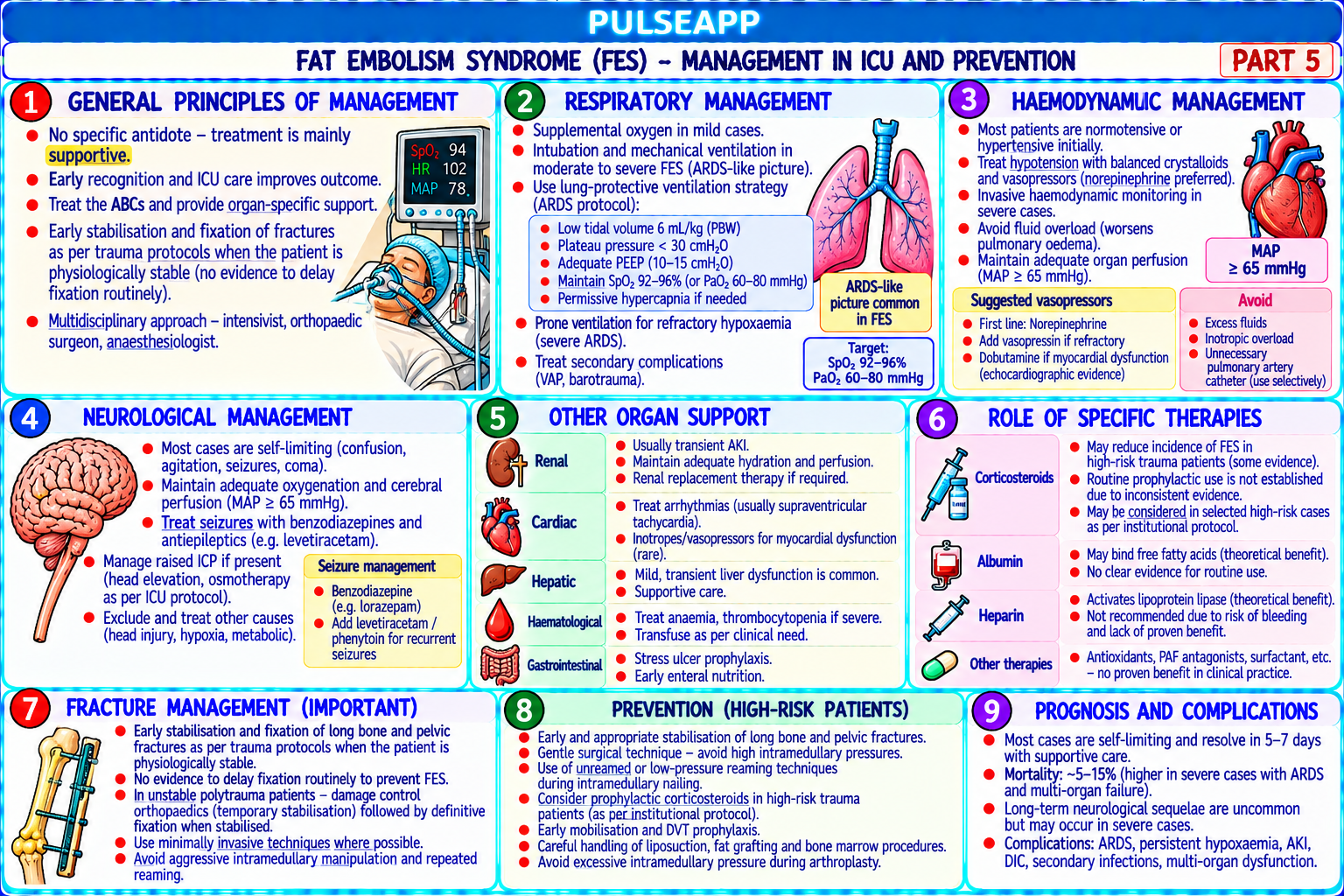
No specific antidote — treatment is primarily SUPPORTIVE; the syndrome is self-limiting if the patient survives the acute phase

Respiratory support: supplemental O₂; early CPAP/NIV for mild-moderate hypoxaemia; mechanical ventilation with lung-protective settings (6 mL/kg IBW, PEEP 8–12 cmH₂O, plateau ≤30 cmH₂O) for ARDS; prone positioning for severe ARDS (PaO₂/FiO₂ <150)

Haemodynamic support: IV fluids cautiously (avoiding fluid overload which worsens pulmonary oedema); vasopressors for hypotension; albumin may help bind and transport FFAs reducing free toxicity (theoretical)

Neurological: seizure control (benzodiazepines); maintain cerebral perfusion pressure; avoid hypoxia and hypotension (worsen cerebral injury); EEG monitoring for subclinical seizures

Early fracture fixation: definitive surgical fixation of the fracture reduces ongoing fat embolisation from the fracture site; early IMN (intramedullary nailing) is associated with lower rates of pulmonary complications than traction alone; however, reaming the intramedullary canal during nailing transiently increases fat embolisation — use of unreamed or solid nails, or reaming with suction, reduces this risk

Corticosteroids: prophylactic methylprednisolone (1.5 mg/kg every 8 hours for 3 doses) has been shown in some trials to reduce FES incidence in high-risk patients (multiple long bone fractures); not universally adopted; no evidence for therapeutic role once FES is established

🎤 Viva Corner
Q. Why does the petechial rash in FES occur specifically in the axillae, conjunctivae, and neck rather than on the limbs, and why is it transient?
The characteristic distribution of the petechial rash in FES — specifically in the axillae, conjunctivae, upper chest, and base of the neck — reflects the anatomical distribution of the non-dependent skin microcirculation that is perfused with blood at relatively low perfusion pressure. Fat globules in the systemic circulation after lung traversal or PFO passage enter the arterial circulation; these fat droplets, being less dense than blood, tend to float upward and preferentially embolise the nondependent (uppermost) capillary beds when the patient is in the semi-recumbent or sitting position — the axillae, conjunctivae, and neck are naturally the most nondependent areas of the skin in this position; gravity causes the lighter fat droplets to preferentially perfuse the skin capillaries in these areas. The axillary skin is characteristically thin and the capillaries are particularly superficial, making the petechial haemorrhages from capillary fat embolism visible as pinpoint 1–3 mm haemorrhagic spots. The transient nature of the rash — fading within hours of appearance — reflects the rapid metabolism and clearance of fat droplets from the skin capillaries once they are there; lipases in the skin and capillary endothelium rapidly hydrolyse the fat, the capillary plugs dissolve, and the petechiae fade as the capillary circulation is restored; this is unlike inflammatory petechiae (meningococcal purpura, vasculitis) which persist or enlarge. The transience means that if the rash is missed at initial examination, it may have disappeared by the time of the next assessment — making regular careful examination of the axillae and conjunctivae critical in the first 48 hours after major fractures in patients with unexplained hypoxaemia.
★ Examiner's Pearl
Gurd criteria: 1 major (respiratory/cerebral/petechiae) + 4 minor — must reproduce the major and minor categories. Petechial rash: specific distribution (axillae, conjunctivae, neck, anterior chest) + transient (24–36h after injury, fades in hours) + present in only 50–60% but HIGHLY SPECIFIC. Two pathophysiological mechanisms: mechanical (embolism) + biochemical (FFAs from lipase hydrolysis → capillary toxicity) — the biochemical mechanism explains the 24–72h delayed onset.
Gurd AR, Wilson RI. The fat embolism syndrome (J Bone Joint Surg Br 1974;56B:408-416). Mellor A, Soni N. Fat embolism (Anaesthesia 2001;56:145-154). Lindeque BG et al. Fat embolism syndrome (J Bone Joint Surg Am 1987;69A:128-141). Miller RD et al. Miller's Anaesthesia, 9th Ed.
lock Protected Clinical Document • Pulse App • Screenshots Strictly Prohibited
QUESTION 99 person Asked by .
bookmark_add

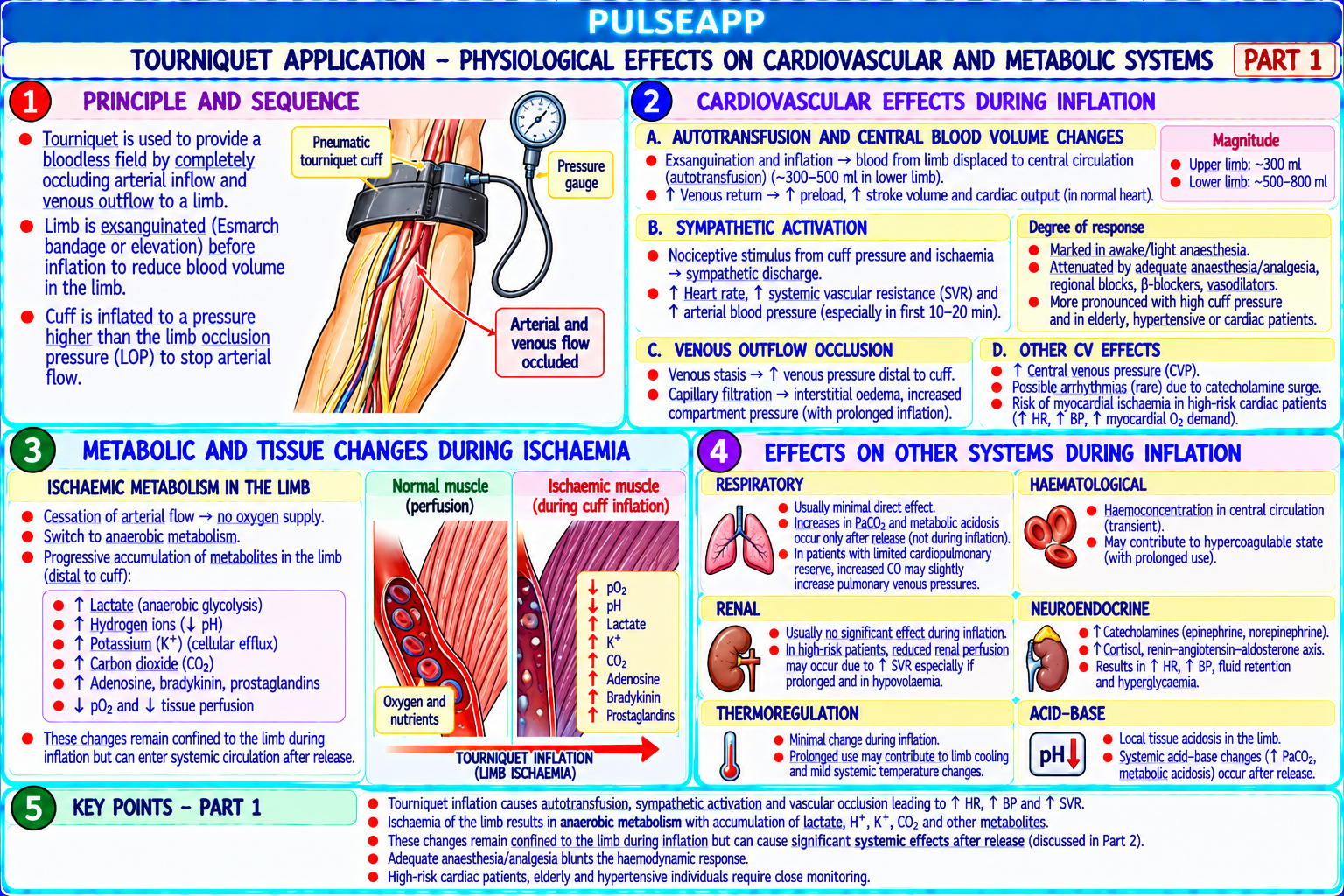
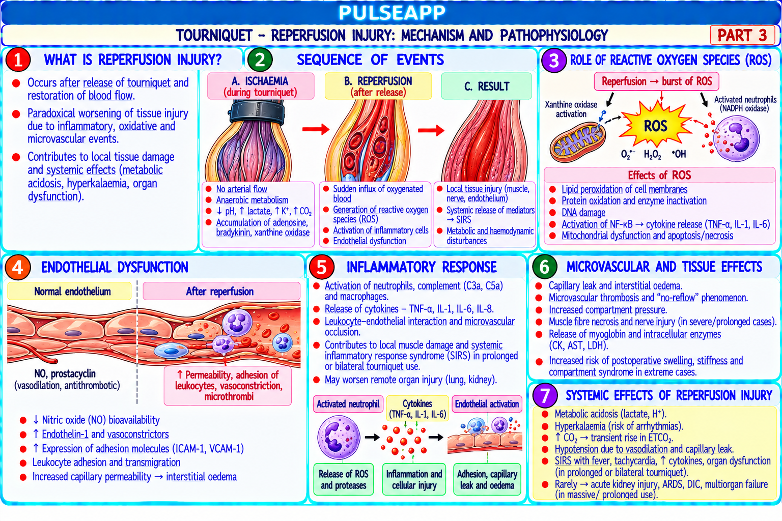
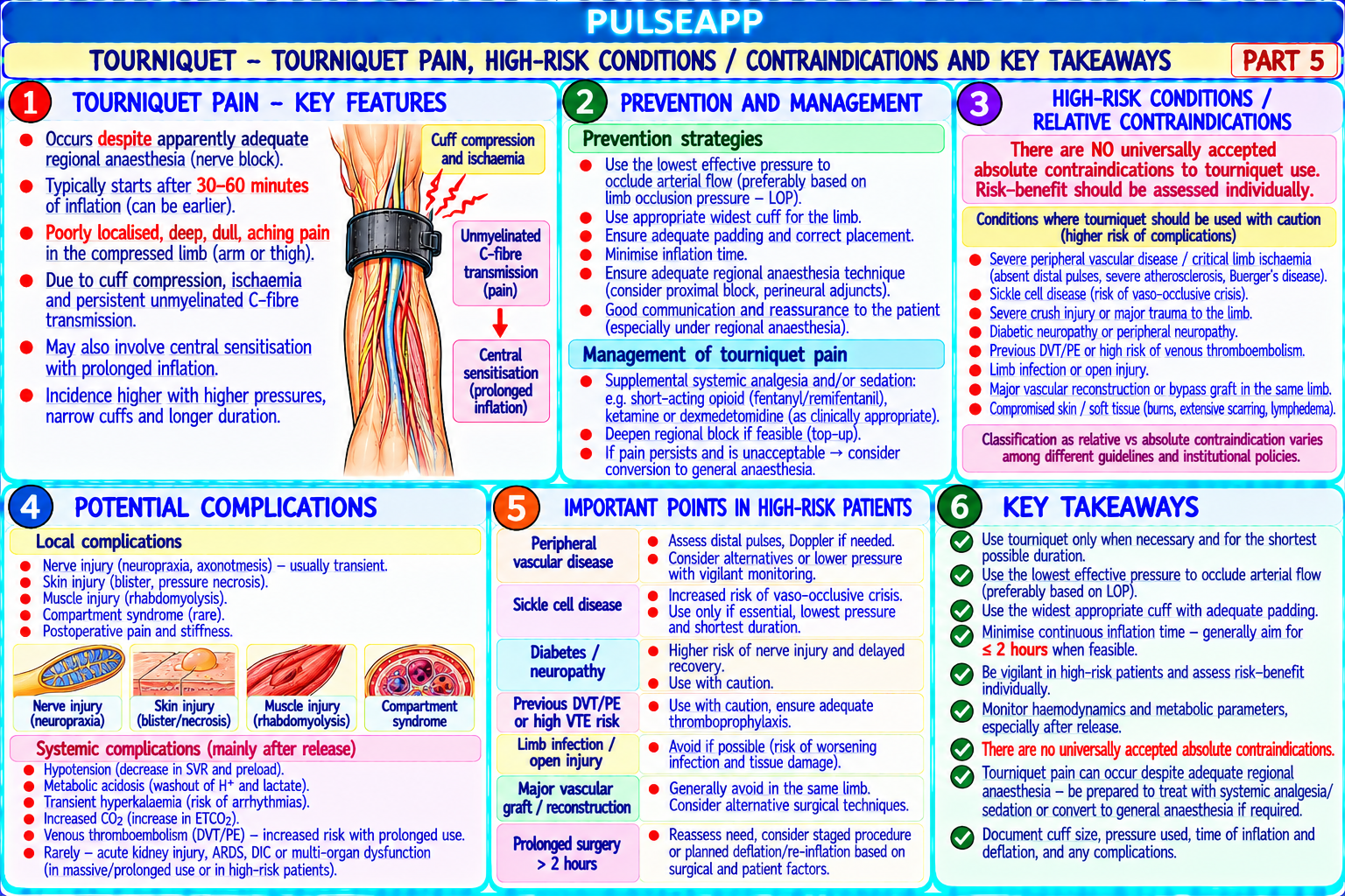
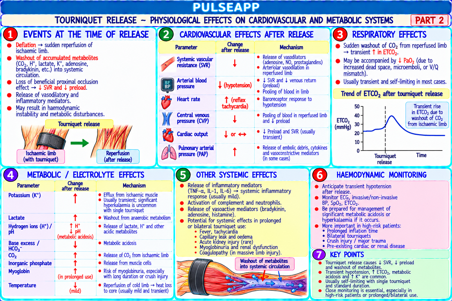
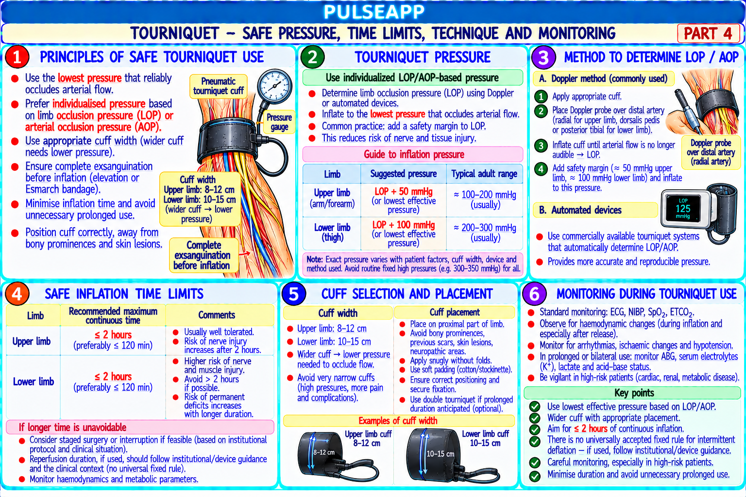
Describe the physiological effects of tourniquet application and release on the cardiovascular and metabolic systems. State safe tourniquet pressure, time limits, and the reperfusion injury mechanism. Discuss tourniquet pain under regional anaesthesia and absolute contraindications.

collections Question Diagrams & Reference Images (1)
Question Reference Diagram
zoom_in View Image
description Clinical Response
⚙ Core Concept
Pneumatic tourniquet use in orthopaedic surgery creates a predictable series of physiological perturbations: ischaemia of the limb during tourniquet inflation and reperfusion injury with systemic metabolite washout on release. Understanding these changes allows the anaesthesiologist to anticipate and manage the cardiovascular events — particularly the sudden haemodynamic changes at tourniquet release — and to optimise the management of tourniquet pain, which is a specific challenge under regional anaesthesia. (Odinsson A — BMJ 2006; Kam PC; Estebe JP — tourniquet pain; McLaren AC; Miller's Anaesthesia 9th Ed)
A. Tourniquet Application — Physiological Effects3 marks

System Effect During Inflation Mechanism Cardiovascular ↑ Circulating blood volume (+10–15% central volume); ↑ MAP; Exsanguination of the limb by Esmarch bandage before inflation + tourniquet inflation ↑ CVP; ↑ CO initially; ↑ HR → up to 1 L of blood redistributed from the limb to the central circulation → autotransfusion effect; raises preload, afterload, and myocardial work; may precipitate pulmonary oedema in patients with limited cardiac reserve

Metabolic — Complete ischaemia of the limb distal to the tourniquet → No arterial inflow; ischaemic reprogramming of cellular metabolism within 1–2 hours; limb anaerobic metabolism → lactic acid accumulation; K⁺ leaks safe limit is approximately 90–120 minutes before irreversible ischaemic damage from ischaemic cells; endothelial activation; neutrophil begins sequestration in capillaries; progressive tissue hypoxia Neurological Nerve injury from direct mechanical compression (at the Compression neuropraxia; ischaemic axonal damage; high cuff pressures + tourniquet edge) occurs at the periphery; ischaemia within the prolonged time → risk of permanent nerve injury; preserve by minimising pressure compressed zone; most nerve damage occurs at the (see below) tourniquet EDGE not the ischaemic limb distally

B. Safe Tourniquet Parameters2 marks

Parameter Safe Limit Rationale Inflation Systolic BP + 50–75 mmHg (or LOP × 1.2–1.4 where LOP = limb occlusion Lowest effective pressure reduces nerve injury and tissue damage; pressure — pressure); minimum pressure to occlude arterial flow only avoid arbitrary fixed pressures (e.g., 300 mmHg) which may be upper limb excessive Inflation Systolic BP + 75–100 mmHg; or LOP × 1.2–1.4; typically 250–300 mmHg for the Larger muscles and higher venous pressures in the lower limb pressure — thigh require higher tourniquet pressures for complete arterial occlusion lower limb Maximum 90–120 minutes maximum single inflation; if longer surgery anticipated: Beyond 90–120 minutes: progressive ischaemic myopathy, inflation time deflate for 10–15 minutes (reperfusion interval) then re-inflate for up to a further endothelial injury, nerve damage, and skin injury become 90 minutes irreversible Cuff width Widest cuff that fits the limb segment without wrapping beyond the surgical site Pressure per unit area is inversely proportional to cuff width; wider — wider cuffs achieve occlusion at lower pressures cuff = lower pressure needed = less nerve injury

C. Tourniquet Release — Reperfusion Injury2 marks

Tourniquet released → sudden re-perfusion of the ischaemic limb → massive washout of accumulated metabolites into the systemic circulation: lactic acid → metabolic acidosis (pH falls 0.1–0.2 units within minutes); K⁺ (2–4 mEq/L rise — risk of hyperkalaemic arrhythmia particularly in patients with pre-existing ↑ K⁺); CO₂ → ETCO₂ rises sharply (10–15 mmHg) within 2–3 minutes; hypotension (vasoactive metabolites + reactive hyperaemia in the reperfused limb acts as a "third space" drawing blood volume away from the central circulation)

Ischaemia-reperfusion injury mechanism: on re-oxygenation, xanthine oxidase in ischaemic endothelium generates reactive oxygen species (ROS) → oxidative damage to cell membranes → increased capillary permeability → oedema; neutrophils, sequestered in the ischaemic capillaries during tourniquet inflation, are activated by reperfusion mediators → massive ROS and protease release → amplification of local tissue injury; this explains why post-tourniquet limb swelling and pain can be disproportionate to the surgical injury alone

Anaesthetic management at tourniquet release: warn the surgeon to deflate gradually (slow release); increase minute ventilation to manage ETCO₂ rise; vasopressors for hypotension; monitor K⁺ if prolonged tourniquet time or pre-existing hyperkalaemia; monitor ECG for hyperkalaemia changes (peaked T waves, wide QRS)

D. Tourniquet Pain Under Regional Anaesthesia2 marks

Tourniquet pain (tourniquet tolerance) — pain or discomfort under a pneumatic tourniquet despite an apparently adequate regional anaesthetic block — develops in 30–60 minutes of inflation and becomes progressively more severe; affects 55–70% of patients with a functioning peripheral nerve block

Mechanism: the tourniquet compresses the skin and subcutaneous tissue at the cuff level; the pain fibres in this zone (C fibres) may escape block because: the cuff is often at the level of the sensory block margin (the most poorly blocked zone); C fibres are notoriously difficult to block reliably for prolonged periods; the slow onset of tourniquet pain suggests involvement of deep pain pathways (possibly spinal sensitisation) that are different from surgical pain pathways Management:

Supplement the regional block with IV analgesics: opioids (fentanyl 25–50 mcg IV), ketamine (0.25–0.5 mg/kg IV), or NSAIDs before tourniquet pain becomes established Sedation (midazolam, propofol sub-anaesthetic) can reduce the cortical processing of tourniquet pain TIVA with propofol-remifentanil can be added to maintain patient comfort while the regional block provides post-operative analgesia

If pain is intolerable: convert to GA

Double-cuff tourniquet: inflate the distal cuff first (over anaesthetised skin from the block); then inflate the proximal cuff (less discomfort as this is on the analgesia-free side); deflate the distal cuff; this technique delays but does not prevent tourniquet pain

E. Absolute Contraindications1 mark

Severe peripheral vascular disease (ischaemic limb — tourniquet-induced ischaemia in already-compromised tissue → limb-threatening ischaemia) Sickle cell disease (sickling may be precipitated in the ischaemic limb → vaso-occlusive crisis; if tourniquet essential → hydroxyurea pre-operatively, optimal oxygenation, avoid hypothermia) History of compartment syndrome of the relevant limb Severe crush injury of the limb (tourniquet on already-injured tissue) DVT in the limb (exsanguination before inflation → systemic embolisation of clot)

🎤 Viva Corner
Q. A patient undergoing total knee arthroplasty under spinal anaesthesia complains of escalating aching pain in the upper thigh at 75 minutes of tourniquet time. The sensory block is confirmed at T8. What is happening and how do you manage it?
This is tourniquet pain — pain arising from the pneumatic tourniquet cuff on the upper thigh that has developed despite an adequate sensory block. The spinal anaesthetic has provided complete surgical anaesthesia of the knee and lower limb, but the tourniquet cuff is positioned on the upper thigh at or near the cephalad margin of the spinal block; the C fibres from the skin and deep tissues at this level may not be completely blocked, and tourniquet pain characteristically develops through a different pathway from acute surgical pain — it involves a dull, aching, progressive pain that increases with time and is thought to involve slow C fibre polymodal nociceptors responding to the prolonged ischaemia and compression at the cuff level, with possible spinal sensitisation contributing as the pain intensifies. The 75-minute mark is typical — tourniquet pain usually begins at 40–60 minutes and peaks at 90 minutes. Management: first, confirm the sensory block level with ice or cold spray applied at the tourniquet site — if the cuff is on unanaesthetised skin (above the spinal block level), the patient has breakthrough pain from the unblocked zone. Options in order: IV supplemental analgesia — fentanyl 25–50 mcg IV (fastest and most effective first step); ketamine 0.25 mg/kg IV (NMDA antagonism specifically addresses the central sensitisation component of tourniquet pain); ketorolac 15–30 mg IV; if these provide only temporary relief — low-dose propofol infusion (0.5–1 mg/kg/hr) or midazolam 1–2 mg IV to provide sedation and reduce cortical pain processing; if pain persists and is intolerable — inform the surgeon that tourniquet tolerance has been reached; consider deflating the tourniquet (surgeon may accept increased bleeding for brief periods and reinflate); if absolutely necessary — convert to GA (laryngeal mask or intubation) with propofol-remifentanil which will eliminate tourniquet pain while the spinal continues to provide post-operative analgesia. At 75 minutes: the surgeon should also be preparing to complete the procedure expeditiously as the 90–120 minute safe time limit is approaching.
★ Examiner's Pearl
Safe tourniquet time (90–120 minutes single inflation; 10–15 minute reperfusion interval if longer surgery needed) and pressure formula (systolic BP + 50–75 mmHg for upper limb; +75–100 mmHg lower limb — use limb occlusion pressure not arbitrary fixed pressures) are the specific clinical parameters tested. Tourniquet release physiology: ETCO₂ rises 10–15 mmHg, pH falls 0.1–0.2, K⁺ rises 2–4 mEq/L — all within 2–3 minutes — the specific magnitude of each change is tested. Sickle cell disease as an absolute contraindication with the mechanism (ischaemia → sickling in the limb) is specifically tested.
Odinsson A, Finsen V. Tourniquet use and its complications (J Bone Joint Surg Br 2006;88:1090-1092). Estebe JP et al. Tourniquet pain under spinal anaesthesia (Reg Anesth 1995;20:142-147). Kam PC et al. Tourniquet and its complications (Anaesthesia 2001;56:534-545). McLaren AC et al. Safe tourniquet inflation (J Bone Joint Surg 1994;76A:605-611). Miller RD et al. Miller's Anaesthesia, 9th Ed.
lock Protected Clinical Document • Pulse App • Screenshots Strictly Prohibited
QUESTION 100 person Asked by .
bookmark_add

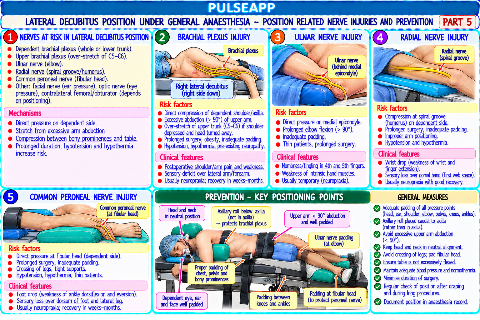
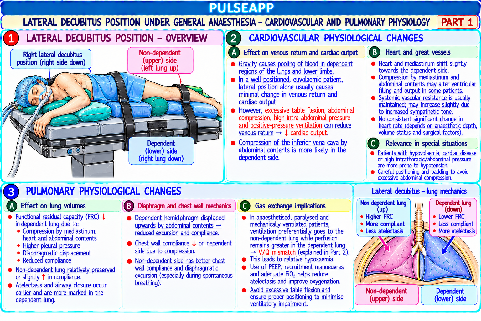
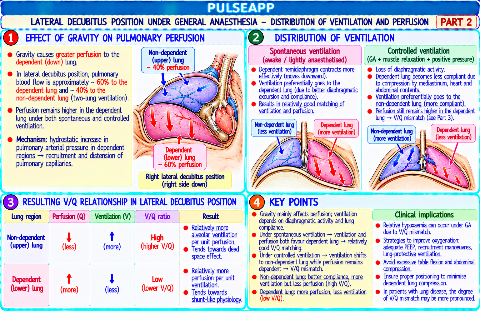
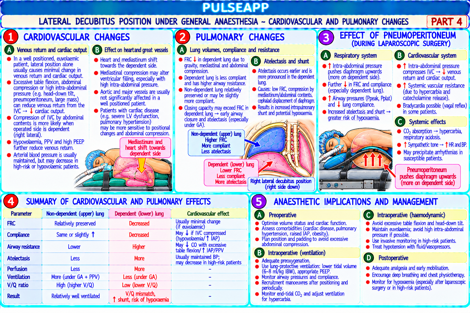
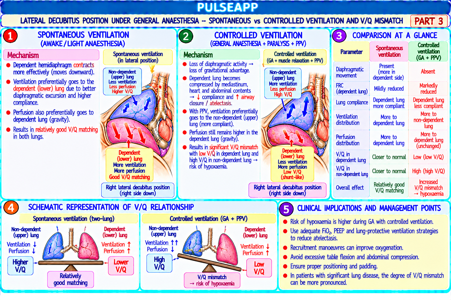
Describe the cardiovascular and pulmonary physiological changes that occur in the lateral decubitus position under general anaesthesia. Explain the V/Q mismatch and how spontaneous vs controlled ventilation alters perfusion distribution. List position-related nerve injuries specific to this position.

collections Question Diagrams & Reference Images (1)
Question Reference Diagram
zoom_in View Image
description Clinical Response
⚙ Core Concept
The lateral decubitus position creates a unique and complex pulmonary physiology challenge — in the awake patient, the dependent lung is better perfused (gravity) AND better ventilated (diaphragm mechanics favour the dependent side); under anaesthesia and paralysis, ventilation distributes to the NON-dependent lung (compliance is better) while perfusion remains greater to the dependent lung — creating a V/Q mismatch that is the fundamental physiological basis for one-lung ventilation techniques. (West JB — Respiratory Physiology; Benumof JL — Anesthesia for Thoracic Surgery; Miller's Anaesthesia 9th Ed)
A. Awake Lateral Decubitus — Physiology2 marks

Perfusion (Q): gravity causes blood to preferentially perfuse the dependent (lower) lung; pulmonary arterial pressure in the dependent lung is higher (hydrostatic gradient adds to zone 2 and zone 3 conditions → active blood flow); the dependent lung receives approximately 60% of total pulmonary blood flow

Ventilation (V): in the awake spontaneously breathing patient in the lateral position, the dependent hemidiaphragm is better positioned for contraction — it is higher (pushed up by mediastinal weight and abdominal pressure) and in a more efficient mechanical position (like a well-stretched muscle starting on the steep part of the length-tension curve); the dependent diaphragm moves more with each breath → dependent lung receives more ventilation (approximately 55–60% of tidal ventilation to the dependent lung)

Result (awake): V/Q matching is approximately preserved — the lung that gets more blood (dependent) also gets more ventilation → good oxygenation; this is the optimal physiological state

B. Anaesthesia + Muscle Relaxation — The V/Q Mismatch3 marks

Effect on ventilation: paralysis eliminates the active diaphragm advantage of the dependent side; the non-dependent (upper) lung has BETTER compliance under anaesthesia — it is less compressed, higher FRC, less atelectasis-prone; controlled ventilation distributes gas preferentially to the most compliant lung → the non-dependent lung receives more ventilation (the opposite of the awake state)

Effect on perfusion: gravity still preferentially perfuses the dependent (lower) lung → this does NOT change with anaesthesia; the dependent lung continues to receive ~60% of pulmonary blood flow

The critical mismatch: under anaesthesia, the non-dependent lung is WELL VENTILATED (high V) but receives less blood flow (low Q) → high V/Q ratio → dead space effect; the dependent lung is WELL PERFUSED (high Q) but receives less ventilation (low V due to compression, atelectasis, and splinting by mediastinal weight) → low V/Q ratio → shunt effect → hypoxaemia

Magnitude: FRC of the dependent lung decreases significantly (compression from mediastinal structures + abdominal pressure against the dependent diaphragm); closing capacity may exceed FRC in the dependent lung → small airway closure → atelectasis → intrapulmonary shunt

PEEP strategy for lateral decubitus: PEEP applied to the dependent lung helps recruit collapsed airways and restore dependent lung ventilation (reapproaches the awake V/Q relationship); PEEP titration to the dependent lung is the key ventilatory strategy during lateral decubitus under GA

C. Cardiovascular Effects2 marks

Venous return: the lateral position with a kidney bridge or flexed table can compress the IVC (depending on the flexion position) → ↓ venous return → ↓ CO; conversely, the Trendelenburg component (head-down) increases venous return; careful positioning with padding avoids abdominal compression

Dependent arm complications: the dependent arm may have impaired venous drainage, particularly with arm boards or in prolonged procedures — monitor the arm position carefully; avoid direct compression of the axillary vessels

Mediastinal shift: in open thorax (thoracotomy), the mediastinum may shift toward the operative (non-dependent) side during spontaneous breathing → paradoxical mediastinal movement → further impairs gas exchange

D. Position-Related Nerve Injuries3 marks

Nerve/Structure Mechanism Prevention

Brachial plexus Stretching of the brachial plexus from: dependent arm excessively abducted + Axillary roll placed BELOW the axilla (at the thoracic cage level, (dependent head turned away; axillary roll too high (pushing into the axilla rather than below not in the axilla itself); limit abduction to <90°; neutral head arm) it) → compresses the neurovascular bundle in the axillary region; dependent arm position; pad under the dependent shoulder; pillow between weight pulling on the shoulder knees; check arm position every 30 min Common Compression of the CPN as it winds around the head of the fibula; the lateral Generous padding (gel pad or foam) under the dependent knee; peroneal nerve aspect of the dependent knee presses against the table without adequate pillow between knees prevents the upper knee from compressing (dependent leg) padding the lower CPN; confirm the fibular head is well-padded before draping Lateral femoral Compression of the LFCN at the anterior superior iliac spine region when the Pad the dependent iliac crest; avoid kidney bridge position that cutaneous iliac crest is pressed against the table; particularly with kidney bridge positioning directly compresses the ASIS region nerve (LFCN) Eye (dependent Direct pressure on the dependent eye from inadequate head support → central Confirm eye position is NOT under pressure (headrest positioned eye) retinal artery occlusion (rare but devastating) → permanent blindness; also from behind the ear, not over the orbit); check eyes by lifting the head oedema from Trendelenburg component slightly immediately after positioning; document in anaesthetic chart

🎤 Viva Corner
Q. Why does the dependent lung become hypoxic under GA in the lateral position when it is better perfused, and how do you manage this intraoperatively?
The dependent lung becomes hypoxic because, while it continues to receive the majority of pulmonary blood flow (approximately 60% due to gravity — this does not change with anaesthesia), its ventilation is dramatically REDUCED under general anaesthesia and muscle relaxation compared to the awake state. In the awake patient, the dependent diaphragm actively contracts more effectively than the non-dependent side, distributing proportionally more ventilation to the dependent lung; this matches the greater perfusion and maintains good V/Q ratios. Under anaesthesia, paralysis eliminates the active diaphragm advantage; controlled positive pressure ventilation distributes gas preferentially to the most compliant region — which is the non-dependent (upper) lung, as it is less compressed, has a higher FRC, and contains less atelectasis; simultaneously, the dependent lung is compressed by the weight of the mediastinum (heart, great vessels) and by increased abdominal pressure against the dependent diaphragm, reducing its FRC below closing capacity in many patients → small airway closure → atelectasis → complete dependence on passive gas flow which is preferentially directed to the less-resistant non-dependent lung. The result: dependent lung has high Q but low V → low V/Q → intrapulmonary shunt → hypoxaemia. The dependent lung is both the most perfused AND the most atelectatic — these two factors conspire against oxygenation. Management: selective PEEP to the dependent lung (5–10 cmH₂O applied via the ventilator PEEP setting when the chest is closed; or CPAP to the dependent lung via a separate circuit when the chest is open during one-lung ventilation — CPAP 5 cmH₂O to the down lung recruits atelectatic units without wasting the one-lung ventilation strategy); recruitment manoeuvres to the dependent lung; ensure adequate FiO₂; avoid Trendelenburg if possible (worsens abdominal pressure on the dependent diaphragm); lateral tilt if the table allows (reduces mediastinal compression).
★ Examiner's Pearl
The key physiological comparison: awake lateral = dependent lung better perfused AND better ventilated (good V/Q match); anaesthesia + paralysis = dependent lung better perfused but LESS ventilated (V/Q mismatch → shunt → hypoxaemia). This reversal of ventilation distribution under anaesthesia is the fundamental concept tested. The axillary roll position (BELOW the axilla at thoracic level — NOT IN the axilla) is the specific positioning safety fact for brachial plexus injury prevention most tested.
West JB. West's Respiratory Physiology, 11th Ed. Benumof JL. Anesthesia for Thoracic Surgery, 2nd Ed. Brodsky JB. Positioning the surgical patient. Miller RD et al. Miller's Anaesthesia, 9th Ed, Chapter 41 (Patient Positioning).

Showing 91100 of 337 questions

account_tree

Subcategory Tree

Explore Anesthesia subcategories

folder_special Anesthesia
Main
lock

Category Subscription

Subscribe to Anesthesia to unlock this module and all nested subcategories.

  • check_circle Access Anesthesia & all subcategories
  • check_circle Detailed, Peer-Reviewed Answers
  • check_circle High-yield visual aids & imaging
Get Category Subscription arrow_forward
Secure 256-bit SSL Connection

Anatomical Models

Explore high-fidelity 3D visualizations included in premium modules.

Case Reviews

Real-world clinical scenarios narrated by senior consultants.

Pulse App