Anesthesia | Eklavya Medical
vaccines
medical_services Main Specialty Domain lock Subscription Required

Anesthesia

Clinical anesthesiology, local and general anesthesia delivery systems, monitoring, and perioperative care.

verified Verified Medical Faculty menu_book 337 Q&A Modules
lock Protected Clinical Document • Pulse App • Screenshots Strictly Prohibited
QUESTION 41 person Asked by .
bookmark_add

Define TIVA and explain the concept of Target-Controlled Infusion (TCI). Describe the pharmacokinetic models used, the propofol-remifentanil combination, advantages over inhalational anaesthesia, monitoring of depth of anaesthesia, and specific clinical indications for TIVA.
collections Question Diagrams & Reference Images (1)
Question Reference Diagram
zoom_in View Image
description Clinical Response
⚙ Core Concept
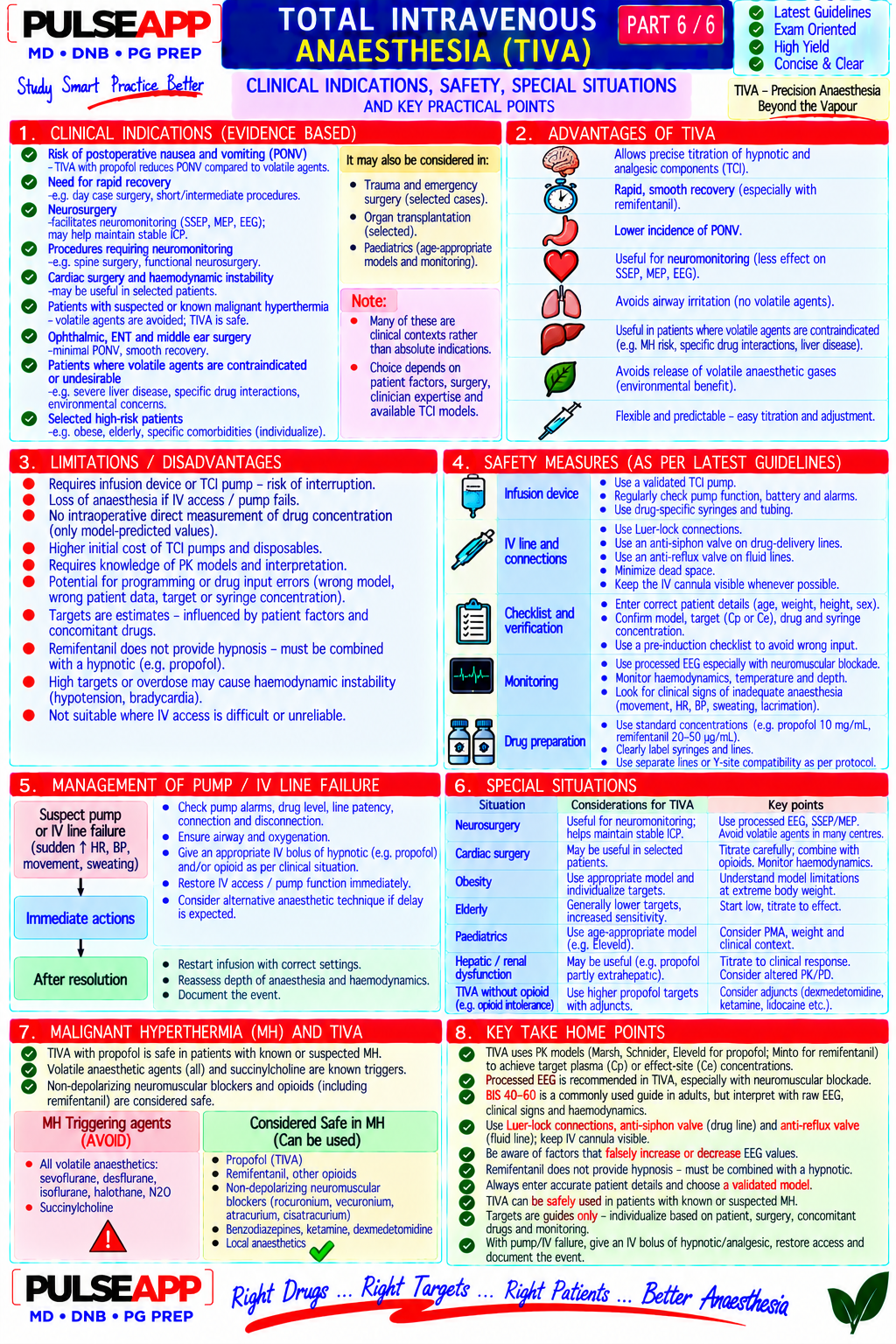
TIVA represents the complete administration of general anaesthesia using only intravenous drugs — eliminating inhaled anaesthetic agents entirely. With the propofol-remifentanil combination delivered via Target-Controlled Infusion (TCI), it is possible to achieve a reliably predicted plasma drug concentration at the bedside, titrate depth of anaesthesia with precision comparable to volatile agents, and guarantee rapid, predictable recovery regardless of case duration — a capability that revolutionised long-neurosurgical, thoracic, and day-case anaesthesia. (Miller's Anaesthesia 9th Ed; Absalom AR — TCI and TIVA; Struys MM; Shafer SL — Pharmacokinetics; Marsh B — propofol TCI model)
A. Definition and Components of TIVA1 mark

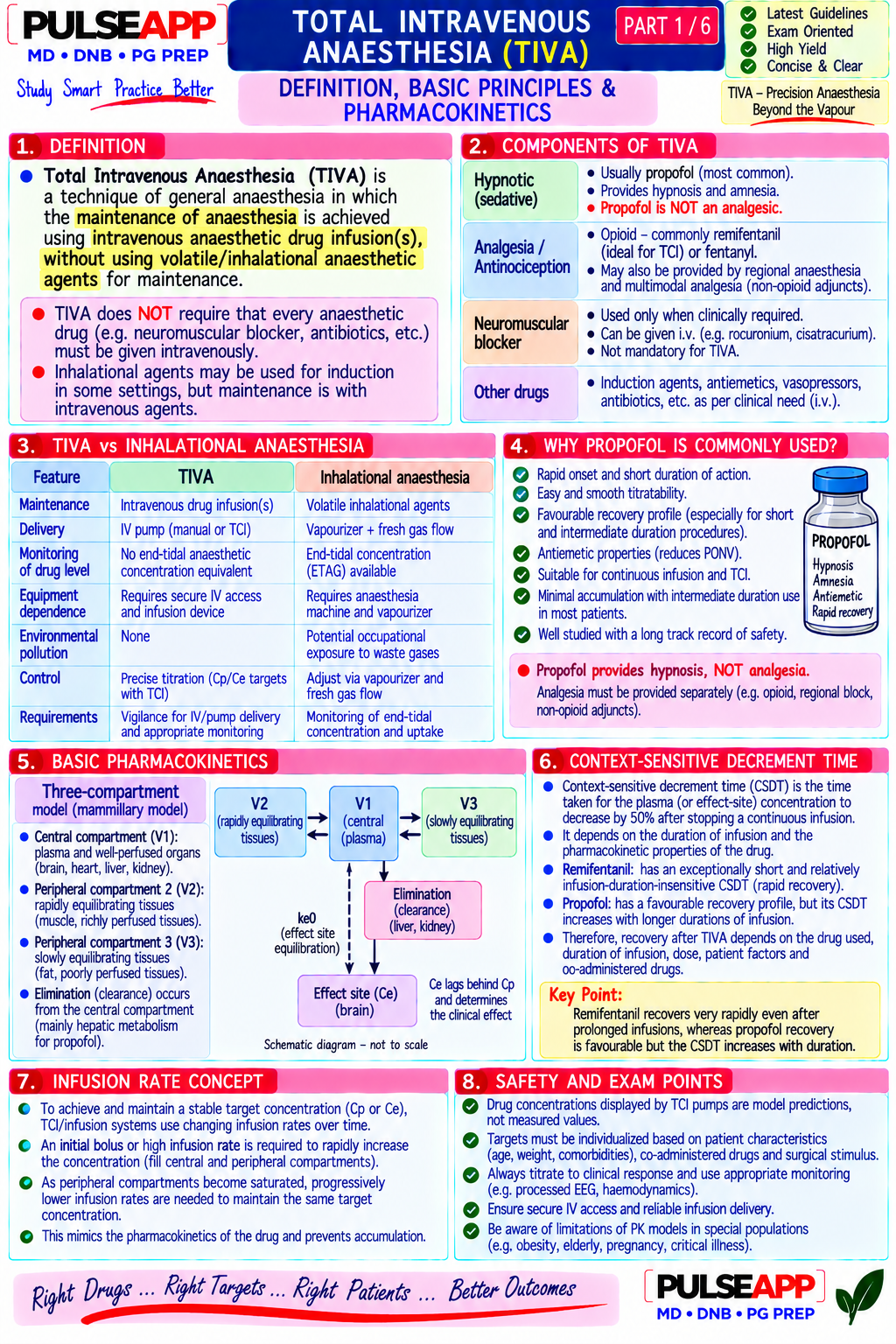
TIVA: induction AND maintenance of general anaesthesia using only intravenous drugs, without any inhalational anaesthetic agent; requires at least three pharmacological components:

Hypnotic agent (produces unconsciousness): propofol (almost universally); alternatives: thiopentone, ketamine, dexmedetomidine as adjuncts

Analgesic agent (blunts surgical pain response): remifentanil (preferred — ultra-short context-sensitive half-time, titrable); alternatives: fentanyl, alfentanil, ketamine Neuromuscular blocking agent (facilitates intubation and surgical relaxation, if required): rocuronium, vecuronium, cisatracurium

B. Target-Controlled Infusion (TCI) — Concept and Pharmacokinetics4 marks
✅ TCI — The Smart Syringe Pump
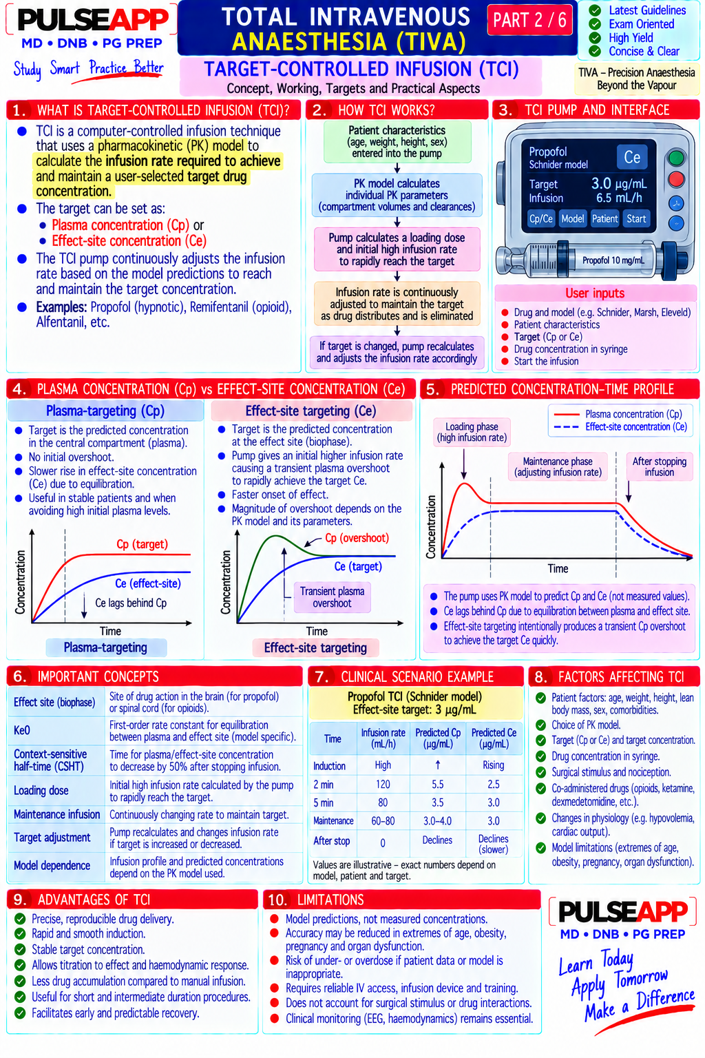
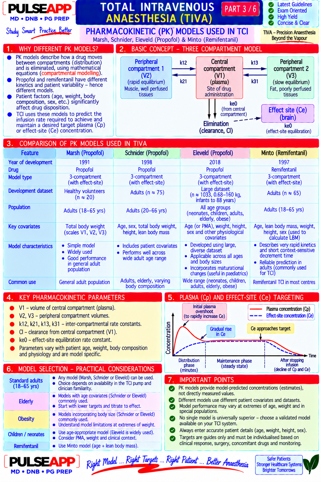
TCI is a drug delivery system in which the clinician specifies a TARGET PLASMA CONCENTRATION (Cp, in mcg/mL or ng/mL) rather than a dose rate (mL/hour). The pump's built-in pharmacokinetic model then continuously calculates the infusion rate required to achieve and maintain that target concentration, accounting for the patient's weight, age, height, sex, and the drug's known distribution and elimination kinetics. Pharmacokinetic Models for Propofol Model Basis Recommended Population Key Feature Marsh 3-compartment model; based on propofol PK data Adults (18–60 years); original Simpler; does not incorporate age effect; tends to slightly model from surgical patients; uses weight as the primary Diprifusor model; widely overpredict concentrations in elderly; the original TCI model covariate; validated for plasma-targeted TCI (Cpt) validated approved in most countries Schnider 3-compartment model incorporating weight, height, Adults; particularly better in Effect-site targeting allows faster induction (target the brain model age, lean body mass (LBM); validated for both elderly patients where age- concentration rather than plasma — effect compartment plasma-targeted and effect-site targeted TCI (Cet — correction reduces propofol equilibrates slightly later than plasma); incorporates age → effect compartment targeting) dose; preferred for effect-site TCI lower dose automatically for elderly Paedfusor Paediatric propofol PK model; accounts for age- Children 1–16 years Propofol TCI not licensed for <1 year; manual TIVA only in model dependent changes in volume of distribution and neonates/young infants clearance in children 1–16 years Three-Compartment Model — Basis of TCI The 3-compartment pharmacokinetic model divides the body into: Central compartment (V1 — blood, rapidly perfused organs = lungs, heart, liver, kidney) + Peripheral compartment 1 (V2 — rapidly equilibrating tissues = muscle) + Peripheral compartment 2 (V3 — slowly equilibrating tissues = fat). Drug moves between compartments according to rate constants (k12, k21, k13, k31); elimination occurs only from the central compartment (clearance CL). The TCI pump solves the differential equations describing these rate constants in real-time, adjusting infusion rate to match the target plasma concentration despite continuously changing distribution dynamics. Effect-Site (Ce) vs Plasma (Cp) Targeting Plasma targeting (Cpt): the pump targets a specific plasma concentration; the effect compartment (brain) equilibrates with plasma with a slight delay (ke0 — the rate of drug transfer from plasma to effect site); at induction, the effect compartment concentration rises after the plasma concentration Effect-site targeting (Cet): the pump targets the BRAIN concentration directly — it initially overshoots the plasma concentration (allowing rapid drug delivery to the brain) then reduces the infusion as the brain equilibrates with plasma; provides faster induction than plasma targeting for the same target brain concentration; more relevant to clinical drug effect
C. Propofol-Remifentanil TIVA — The Gold Standard Combination2 marks

Component Target Concentration Effect Rationale

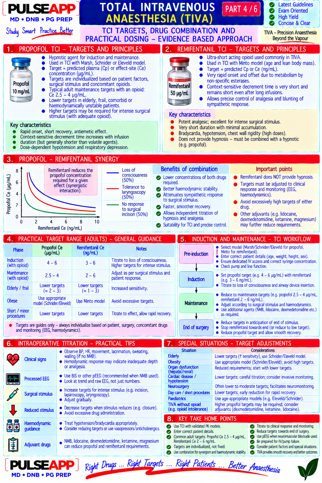
Propofol Induction: 4–6 mcg/mL Ce Unconsciousness, amnesia; Dose-dependent reduction of CMRO₂ and CBF (neuroprotective); lowest PONV (Schnider); Maintenance: 3–4 mild muscle relaxation at incidence of all agents; no HPV inhibition; environmentally clean; predictable recovery mcg/mL Ce (surgery); higher concentrations; with TCI

Sedation: 1–1.5 mcg/mL Ce antiemetic at subanaesthetic concentrations

Remifentanil Blunt intubation response: 4– Analgesia; suppression of Unique organ-independent ester hydrolysis metabolism → context-sensitive half-time = 8 ng/mL; Maintenance: 2–5 haemodynamic response to 3 min regardless of infusion duration; no drug accumulation; predictable emergence ng/mL (depending on surgical stimulation; reduces even after 12-hour infusion; CRITICAL: provide post-op analgesia (morphine/NSAIDs) stimulation); Emergence: propofol requirement by 30– before stopping remifentanil as its analgesic effect disappears within 5–10 min of reduce to 1–2 ng/mL 40% stopping

D. Depth of Anaesthesia Monitoring in TIVA1 mark

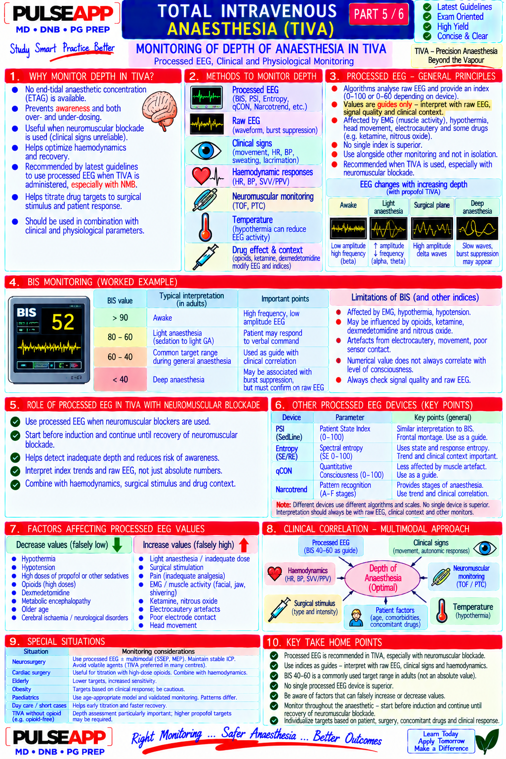
Unlike volatile anaesthesia (where ETCO₂ confirms drug delivery and MAC multiples correlate with depth), TIVA has no exhaled gas indicator of delivery or depth — processed EEG monitoring is essential

BIS (Bispectral Index): 0–100 scale; target 40–60 for surgical anaesthesia; values <40 suggest excessive depth (burst suppression); >60 → inadequate depth and awareness risk; specifically validated for propofol and volatile agents

Entropy (Spectral Entropy): State Entropy (SE) and Response Entropy (RE); similar utility to BIS; uses different mathematical processing of the EEG spectrum pEEG (Patient-State Index, Narcotrend): alternative processed EEG monitors; similar principle to BIS

Important limitation: all processed EEG monitors fail to reliably predict awareness in the concentration range where awareness most commonly occurs (0.2– 0.4 MAC equivalent of propofol); they provide a trend indicator, not an absolute guarantee of unconsciousness

E. Advantages of TIVA Over Inhalational Anaesthesia1 mark

Advantage Clinical Basis Lowest PONV incidence Propofol has intrinsic antiemetic properties (5-HT3 antagonism at sub-anaesthetic concentrations); absence of volatile agents (which are emetogenic); absence of N₂O; TIVA reduces PONV by ~25–30% vs inhalational techniques No HPV inhibition Propofol does not inhibit hypoxic pulmonary vasoconstriction (unlike volatile agents) → superior oxygenation during one-lung ventilation; preferred for thoracic anaesthesia No MH triggering Propofol and opioids are safe in MH-susceptible patients; TIVA is mandatory when volatile agents are contraindicated (MH, suspected MH susceptibility) No operating room No volatile agent exhaled into the OR environment; protects OR staff from chronic low-level anaesthetic exposure pollution Environmental No halogenated greenhouse gas emissions; environmentally superior to all volatile agent techniques sustainability Predictable recovery with Remifentanil's flat CSHT (3 min) means recovery time is independent of infusion duration; propofol's moderate CSHT rises slowly — propofol-remifentanil suitable for even very long cases (8+ hours) with predictable emergence Suitable for remote TIVA requires only a syringe pump and IV access — no anaesthetic machine, vaporizer, or gas supplies needed; ideal for MRI (no locations ferromagnetic components), remote sites, transport anaesthesia
🎤 Viva Corner
Q. You are running propofol TCI at Ce 3.5 mcg/mL (Schnider model) during neurosurgery. What does this specific number mean, and how was it determined?
The effect-site target concentration (Ce) of 3.5 mcg/mL means the TCI system is continuously adjusting the propofol infusion rate to achieve and maintain an estimated propofol concentration of 3.5 mcg/mL at the brain (effect compartment) as predicted by the Schnider pharmacokinetic model. The "estimated" is important — the TCI system calculates a predicted concentration based on the known population pharmacokinetics of propofol encoded in the Schnider model (which incorporates the patient's weight, height, age, and sex that I entered before starting). It does NOT measure the actual propofol plasma or brain concentration directly (real-time propofol concentration measurement is not clinically available). The value of 3.5 mcg/mL was chosen based on the clinical context: the Schnider model for adults predicts that an effect-site concentration of approximately 3–4 mcg/mL produces adequate anaesthesia for moderate surgical stimulation in most patients when combined with remifentanil at 3–5 ng/mL Ce; the combination of propofol and remifentanil is synergistic — less propofol is needed when remifentanil provides the analgesic component. The Schnider model specifically incorporates age into its parameters — an elderly patient of 75 years would receive lower initial doses from the same Ce target compared to a 35-year-old because the Schnider model adjusts the calculated infusion rate for age-related changes in volume of distribution and clearance. I monitor BIS continuously to verify that the estimated Ce corresponds to an appropriate depth of anaesthesia (target BIS 40–60); if BIS rises above 60 despite the Ce target, I increase the propofol target or check for pump malfunction or line disconnection.
Q. Why is it critical to provide post-operative analgesia BEFORE stopping remifentanil, and what happens if you forget?
Remifentanil is unique among opioids in having an essentially instantaneous elimination — its context-sensitive half-time is approximately 3 minutes regardless of infusion duration, because it is hydrolysed by non-specific plasma and tissue esterases ubiquitously distributed throughout the body (not dependent on hepatic or renal function). This means that when the remifentanil infusion is stopped, its plasma concentration falls by 50% within 3 minutes and to near-zero within 10–15 minutes. This is clinically extremely valuable during emergence — it allows precise, predictable moment-of-extubation control. However, this same property creates the "remifentanil analgesia cliff": remifentanil provides complete, profound analgesia at 3–5 ng/mL Ce during surgery, but this analgesia disappears completely within 10–15 minutes of stopping the infusion — leaving the patient with absolutely no residual opioid analgesia just as they are waking from surgery and becoming aware of their postoperative pain. If longer-acting analgesia (morphine, fentanyl, NSAIDs, paracetamol) has not been administered and had time to reach therapeutic effect before the remifentanil is stopped, the patient wakes into sudden, severe, completely unprovided-for acute pain — which triggers violent emergence agitation, hypertension, tachycardia, and sometimes self-extubation. The analgesia management strategy: administer morphine 0.1–0.15 mg/kg IV at least 30–45 minutes before planned end of surgery; give NSAIDs and paracetamol intraoperatively; ensure the pain score is assessed and analgesia confirmed effective in the recovery room before reducing remifentanil further; some anaesthesiologists maintain a very low background remifentanil infusion (0.05–0.1 mcg/kg/min) for the first 15–30 minutes in the recovery room while longer-acting analgesia takes effect — particularly useful for painful procedures like laparotomies and thoracotomies.
★ Examiner's Pearl
The three-compartment model explanation (central + two peripheral compartments; clearance only from central) is the fundamental PK concept that underpins TCI. Marsh vs Schnider differences (Schnider incorporates age and height → better for elderly; Schnider allows effect-site targeting) are tested as specific model comparisons. The remifentanil analgesia cliff — analgesia disappears in 3–5 minutes after stopping — with the clinical implication (must have long-acting analgesia established before stopping) is the single most important clinical safety fact about propofol-remifentanil TIVA.
Absalom AR, Glen JB. Pharmacokinetic and pharmacodynamic aspects of TCI (BJA 2009;103:i26-i37). Marsh B et al. Pharmacokinetic model driven infusion of propofol (BJA 1991;67:41-48). Schnider TW et al. The influence of age on propofol pharmacodynamics (Anesthesiology 1999;90:1502-1516). Struys MMRF et al. Performance of the Schnider and Marsh models (Anesthesiology 2007;107:213-221). Miller RD et al. Miller's Anaesthesia, 9th Ed, Chapter 30.
lock Protected Clinical Document • Pulse App • Screenshots Strictly Prohibited
QUESTION 42 person Asked by .
bookmark_add

Define Bone Cement Implantation Syndrome. Describe its grading, pathophysiology, risk factors, preventive measures during cemented arthroplasty, and anaesthetic management of the patient who develops sudden cardiovascular collapse during cemented hip replacement.

collections Question Diagrams & Reference Images (2)
Question Reference Diagram
zoom_in View Image
Question Reference Diagram
zoom_in View Image
description Clinical Response
⚙ Core Concept
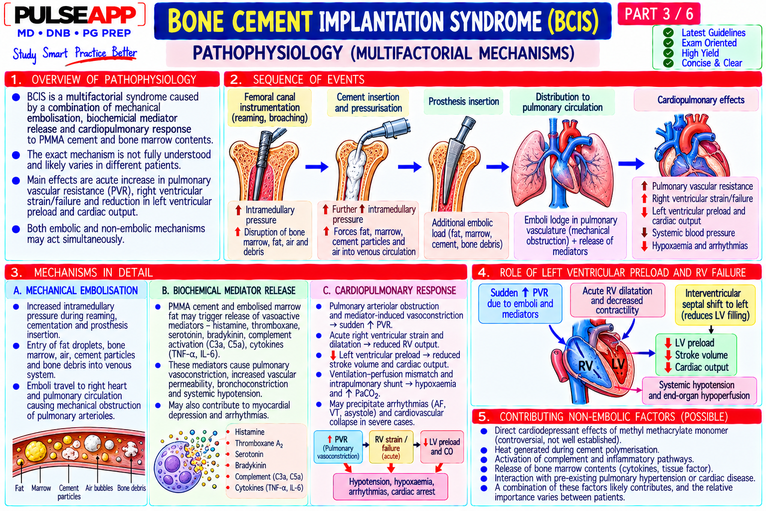
Bone Cement Implantation Syndrome (BCIS) is a potentially fatal cardiorespiratory complication occurring during or shortly after pressurisation of cement into the medullary canal and implantation of a prosthesis — characterised by hypoxia, hypotension, dysrhythmia, and in severe cases, cardiac arrest. It occurs in up to 30% of cemented hip arthroplasties in some series. Despite being a well-recognised complication since the 1970s, it remains responsible for approximately 18–34 deaths per 100,000 cemented hip replacements annually in UK data. Understanding the mechanism and having a prepared response protocol is a mandatory anaesthetic safety competency for all operating theatre practice. (Donaldson AJ et al. — BJA 2009; BCIS Consensus Statement; Staal MA; Miller's Anaesthesia 9th Ed)
A. Definition & Grading2 marks

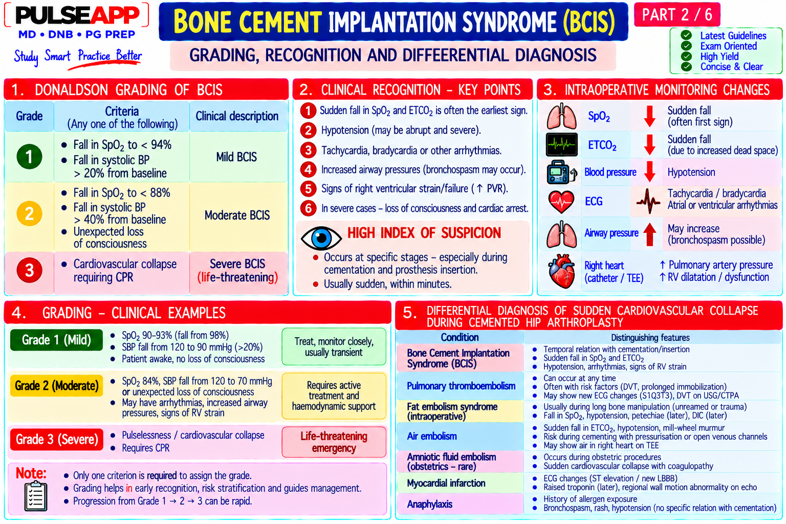
Definition: BCIS is defined as hypoxia (SpO₂ fall >10% from baseline), hypotension (systolic BP fall >20% from baseline), or both, occurring at the time of cementation, prosthesis insertion, reduction of the joint, or deflation of a tourniquet during orthopaedic surgery using PMMA bone cement Grade Clinical Features Incidence Mortality Grade 1 Moderate hypoxia (SpO₂ fall >10%) OR moderate hypotension (systolic BP Common — up to 30% Near-zero direct mortality (Mild) fall >20%) — not both; brief, self-limiting of cemented THRs Grade 2 Severe hypoxia (SpO₂ <94%) AND/OR severe hypotension (systolic BP fall ~5–10% Low but non-trivial; associated with increased (Moderate) >40%) AND/OR unexpected loss of consciousness postoperative complications Grade 3 Cardiovascular collapse requiring cardiopulmonary resuscitation — cardiac ~0.5–1% of cemented Very high — approaching 100% in full cardiac (Severe) arrest THRs arrest without immediate CPR

B. Pathophysiology3 marks
⚠ The Two Simultaneous Mechanisms of BCIS
Mechanism 1 — Embolisation into Pulmonary Vasculature Cement pressurisation into the femoral medullary canal → intramedullary pressure rises dramatically → fat, marrow elements, cement monomer (MMA), bone debris, and air are driven into the femoral medullary veins → enter the systemic venous circulation → reach the right heart and pulmonary vasculature → pulmonary embolism (mixture of fat, cement particles, air, marrow) → acute pulmonary hypertension → right heart failure → ↓ left heart filling → ↓ cardiac output → hypotension → hypoxia (V/Q mismatch from obstructed pulmonary circulation + shunting). Mechanism 2 — Direct Monomer Toxicity PMMA (polymethylmethacrylate) cement is mixed from powder + liquid monomer (methyl methacrylate, MMA); uncured MMA monomer is absorbed from the medullary canal into systemic circulation → direct cardiac myocyte depression (reduces contractility) → systemic vasodilation (direct smooth muscle relaxant effect on vascular endothelium) → produces additional hypotension independent of the embolic mechanism. MMA also causes direct pulmonary vasoconstriction (further worsening right heart afterload) and may trigger histamine release. Paradoxical Embolism via Patent Foramen Ovale In patients with PFO (25–30% of population): the acute pulmonary hypertension from cement embolisation raises right atrial pressure above left atrial pressure → right-to-left shunting through the PFO → embolic material (fat, air, cement particles) enters the systemic arterial circulation → cerebral embolism (stroke) or coronary embolism (MI) → explains why some BCIS patients develop stroke or myocardial ischaemia simultaneously with the haemodynamic collapse
C. Risk Factors2 marks

Risk Factor Specific Risk Factors Category Patient Advanced age (>70 years — greater intramedullary fat content, more fragile vasculature); male sex (larger intramedullary cavities); pre-existing factors cardiorespiratory disease (impaired ability to compensate for acute pulmonary embolism); PFO (paradoxical embolism risk); hypovolaemia at time of cementation; pathological bone (osteoporosis, Paget's, metastatic disease — more intramedullary blood and fat, more friable); pulmonary hypertension (reduced reserve for additional right ventricular afterload) Surgical Cemented implant (vs cementless — cementless arthroplasty does not cause BCIS); revision surgery (larger canals, more debris); long-stem prostheses factors (greater medullary surface area → more material driven into veins); inadequate canal cleaning before cementation (more marrow content → more embolic material); cement pressurisation technique; cement mixing (higher monomer content with improper mixing) Anaesthetic Hypovolaemia at time of cementation (reduced cardiac reserve for haemodynamic response); inadequate preloading; general anaesthesia reduces factors cardiovascular reserve vs regional; the haemodynamic challenge of BCIS may be tolerated better under spinal/epidural anaesthesia (better maintained cardiovascular reserve in spinal vs GA)

D. Prevention & Intraoperative Management3 marks

Preventive Measures

Communication: surgeon must verbally announce BEFORE cementation — "I am about to cement" — this is a mandatory communication point; the anaesthesiologist must be at full vigilance at the critical moment

Pre-cementation fluid loading: ensure the patient is euvolaemic or mildly hypervolaemic before cement is introduced; a small preloading fluid bolus (250–500 mL crystalloid) 5–10 minutes before cementation ensures adequate intravascular volume to buffer the haemodynamic response

FiO₂ to 1.0: increase to 100% O₂ immediately before cementation is announced; this maximises the oxygen reservoir in the FRC to buffer any hypoxic episode

ETCO₂ monitoring: a sudden fall in ETCO₂ is an early sign of massive embolism (reduced pulmonary blood flow → reduced CO₂ delivery to alveoli → ETCO₂ falls); warns the anaesthesiologist before SpO₂ falls

Surgical technique: thorough canal lavage (irrigation before cementation removes loose marrow and blood); use of cement restrictors (reduces the intramedullary volume receiving cement pressure); retrograde cementing technique (fills the canal from distal to proximal — minimises air entrapment); venting holes drilled by surgeon (releases intramedullary pressure) Management of Cardiovascular Collapse (Grade 3 BCIS)

⚠ Grade 3 BCIS — Immediate Management Protocol
1. Call for help immediately — cardiac arrest team 2. FiO₂ 1.0 and increase ventilation (for hypoxia and right heart offloading via hypocapnia) 3. IV fluid bolus 500–1000 mL crystalloid rapidly — restore intravascular volume 4. Vasopressors: ephedrine 6–12 mg IV (first-line — provides both alpha (vasoconstriction) and beta (cardiac stimulation) support; particularly appropriate for BCIS where both vasodilation and myocardial depression contribute); metaraminol 0.5–2 mg IV for pure vasopressor effect; adrenaline (epinephrine) 50–100 mcg IV boluses for cardiac arrest or profound collapse 5. CPR if cardiac arrest — standard ACLS; right heart failure specific: ensure adequate preload during CPR; consider the possibility of paradoxical embolism (cerebral/coronary involvement) 6. TOE if available — right heart distension from massive pulmonary embolism is directly visible; guide fluid therapy and confirm mechanism 7. Inform surgeon to stop further manipulation; surgeon may apply tourniquet pressure to femoral vessels if practical 8. Post-resuscitation: ICU admission; CT pulmonary angiogram when stable; anticoagulation after orthopaedic team review; investigate for PFO (bubble echocardiogram)
🎤 Viva Corner
Q. During a cemented total hip replacement, 90 seconds after the surgeon announces cement is in, the SpO₂ drops from 99% to 82% and BP falls from 130/80 to 65/40 mmHg with a concurrent fall in ETCO₂ from 35 to 12 mmHg. What is your diagnosis and step-by-step management?
This is Grade 3 Bone Cement Implantation Syndrome — the triad of sudden hypoxia (SpO₂ 82%), severe hypotension (BP 65/40), and dramatic ETCO₂ fall (35→12 mmHg) within 90 seconds of cementation is pathognomonic. The ETCO₂ fall to 12 mmHg indicates massively reduced pulmonary blood flow — consistent with acute pulmonary embolism from cement/fat/air embolism causing right heart outflow obstruction. Step-by-step management: First, call for help immediately (crash call + anaesthesia consultant + ODP). Increase FiO₂ to 1.0. Give a rapid fluid bolus 500 mL crystalloid (restore right heart preload against the increased RV afterload). Administer ephedrine 12 mg IV immediately — its combined alpha and beta effects support both BP and cardiac contractility; if no response within 60 seconds, escalate to adrenaline 50–100 mcg IV boluses. If available, connect TOE (transoeosphageal echocardiogram) — expect to see a dilated right ventricle with flattened interventricular septum (D-sign) from right heart pressure overload. Inform the surgeon to stop all surgical manipulation; if technically feasible, application of external pressure over the femoral vessels may slow venous drainage of embolic material. If the patient arrests: CPR immediately per ACLS; vasopressors (adrenaline 1 mg IV in cardiac arrest dose); continue CPR; if there is a witnessed surgical arrest from massive PE, ECMO or emergency cardiothoracic surgical consultation may be considered in appropriate centres. Post-event: move to ICU; investigate PFO status (bubble echo); CT pulmonary angiogram when stable; anticoagulation discussion with surgical team (competing haemorrhage risk from fresh hip arthroplasty); if paradoxical embolism suspected (new neurological signs), brain CT is urgent.
Q. A 78-year-old man with known COPD, pulmonary hypertension, and a previous MI is listed for cemented hemiarthroplasty for neck of femur fracture. How do you modify your perioperative management to reduce BCIS risk?
This patient has three major risk factors for severe BCIS: advanced age (78 years — greater intramedullary fat, reduced cardiovascular reserve), pre-existing pulmonary hypertension (his right ventricle is already operating near-maximally against elevated PVR — any additional acute pulmonary embolism will push him into acute right heart failure far more easily than a patient with normal PVR), and a previous MI (impaired left ventricular reserve). Perioperative modifications: First, discuss with the surgical team whether a cementless hemiarthroplasty implant is possible — modern cementless implants for NOF fractures are available; eliminating cement entirely eliminates BCIS risk; if cement is clinically necessary (bone quality too poor for press-fit cementless fixation), proceed with maximum precautions. Second, for anaesthetic technique: spinal anaesthesia is preferred over GA for this patient — regional anaesthesia maintains better sympathetic tone and cardiovascular reserve to compensate for BCIS haemodynamics; if GA required, use arterial line and invasive monitoring before induction. Third, precementation preparation: ensure euvolaemia or mild hypervolaemia before cement is introduced; have ephedrine drawn and ready at the bedside (12–24 mg in 10 mL syringe); FiO₂ 1.0 for at least 2 minutes before cementation. Fourth, pre-cementation communication: specifically discuss BCIS risk with the surgeon; agree on a verbal warning before cement is introduced; surgeon to use thorough canal lavage, venting holes, and retrograde cementing technique. Fifth, monitoring: arterial line in situ; ETCO₂ continuously monitored; watch for ETCO₂ fall as the first early warning sign of embolism. Sixth, preparation for severe BCIS: adrenaline 10 mcg/mL drawn and ready (not just ephedrine); crash call number posted clearly in theatre; TOE probe available if BCIS develops. If Grade 3 BCIS occurs, this patient will need ICU-level care and may need vasopressin or noradrenaline infusions for right heart support.
★ Examiner's Pearl
The three-grade classification (mild/moderate/severe) with specific thresholds (SpO₂ fall >10%, systolic BP fall >20/40%) is the most tested table structure in BCIS questions — reproduce it with the incidence figures (~30%/5–10%/0.5–1%). The two simultaneous mechanisms (embolism + MMA monomer toxicity) must both be stated — candidates who describe only the embolic mechanism miss the direct cardiac and vascular depressant effects of methyl methacrylate monomer. The precementation checklist (FiO₂ to 1.0, fluid preload, ephedrine drawn, surgeon communication) is the prevention protocol that must be reproduced in full.
Donaldson AJ et al. Bone cement implantation syndrome (BJA 2009;102:12-22). Staal MA et al. Cement restrictors and BCIS (Clin Orthop Relat Res 2004;423:152-155). Parvizi J et al. Mortality with total hip arthroplasty (J Arthroplasty 1999;14:122-125). Orsini EC et al. Cardiopulmonary function and pulmonary microemboli during arthroplasty using cemented prostheses (J Bone Joint Surg 1987). Miller RD et al. Miller's Anaesthesia, 9th Ed.
lock Protected Clinical Document • Pulse App • Screenshots Strictly Prohibited
QUESTION 43 person Asked by .
bookmark_add

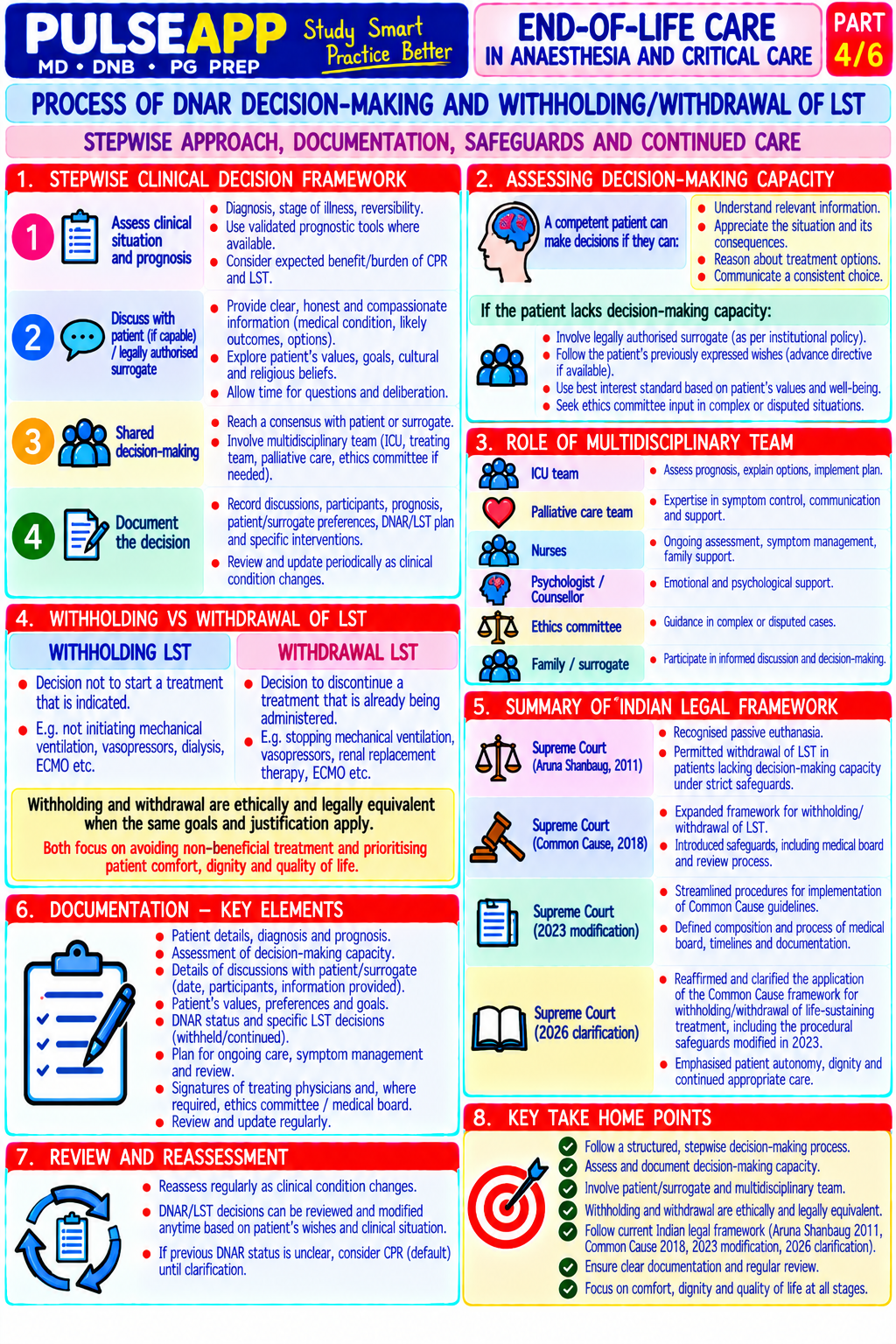
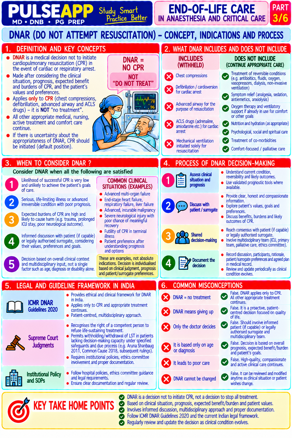
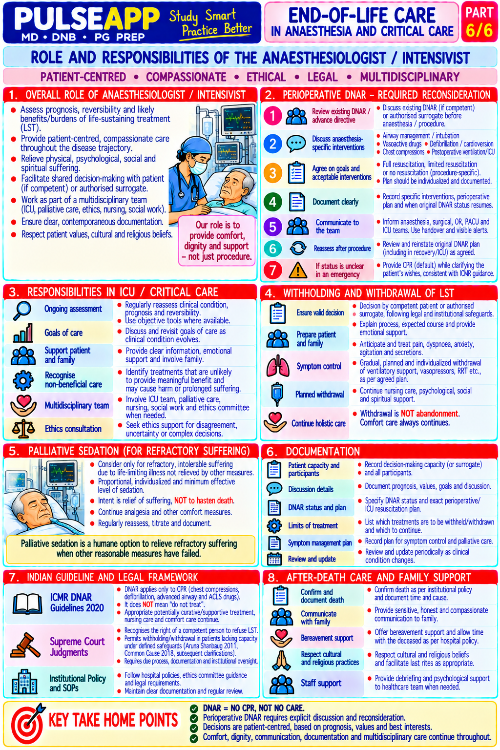
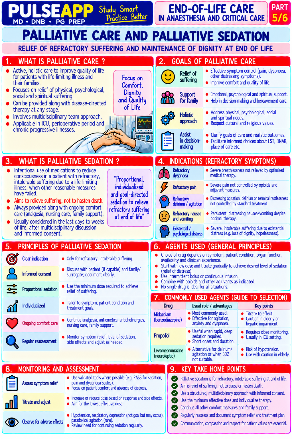
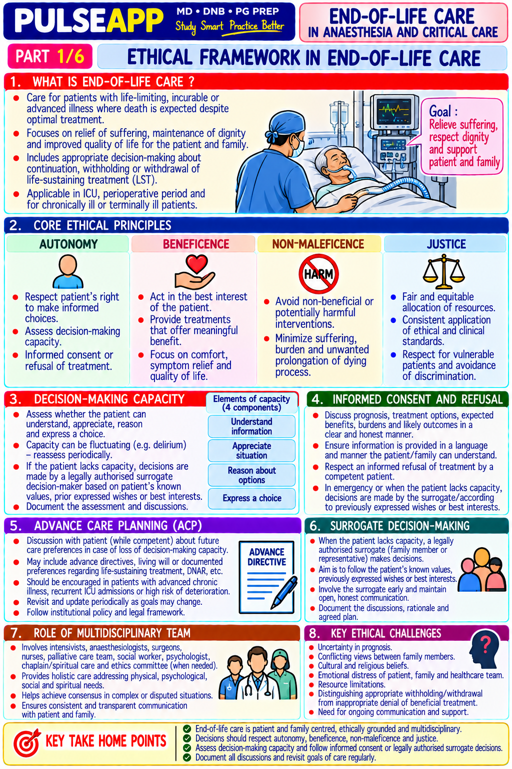
Discuss the ethical framework and practical approach to end-of-life counselling in anaesthesia and critical care. Describe DNR orders, withdrawal of life-sustaining treatment, palliative sedation, and the anaesthesiologist's specific responsibilities in end-of-life care.

collections Question Diagrams & Reference Images (2)
Question Reference Diagram
zoom_in View Image
Question Reference Diagram
zoom_in View Image
description Clinical Response
⚙ Core Concept
The anaesthesiologist and intensivist encounter end-of-life decisions in the operating theatre (unexpected perioperative death, intraoperative catastrophe), the ICU (withdrawal of ventilation, brain death certification, comfort care transitions), and the palliative care interface (terminal sedation for refractory suffering). These decisions require a clear ethical framework, transparent communication with families, respect for patient autonomy and advance directives, and the courage to withhold or withdraw futile life-sustaining treatment — while simultaneously ensuring that the final experience of dying patients is characterised by comfort rather than suffering. (Miller's Anaesthesia 9th Ed; SCCM Guidelines; BMA Guidance on Withholding/Withdrawing; Beauchamp TL, Childress JF — Principles of Biomedical Ethics)
A. Ethical Principles Governing End-of-Life Decisions2 marks

Principle Application to End-of-Life Care Autonomy The patient's right to refuse or accept treatment, including life-sustaining measures; expressed through informed consent, advance directives (living will, healthcare proxy), and verbal refusal; must be respected even when the clinician disagrees with the patient's decision; a competent adult's refusal of CPR or ventilation is legally and ethically binding Beneficence Act in the patient's best interest; in end-of-life care, this requires understanding what constitutes "best interest" beyond mere prolongation of biological function — quality of life, freedom from suffering, dignity, and the patient's own values determine best interest Non- "First, do no harm" — in end-of-life care, this principle supports withholding treatments that produce suffering without meaningful benefit; continuing maleficence painful, invasive treatments in a terminally ill patient who cannot benefit is a form of harm; overly aggressive treatment at the end of life violates nonmaleficence Justice Fair allocation of resources; when ICU resources are limited, justice requires that they be directed toward patients who can benefit; withdrawing futile ICU care also has a justice dimension — freeing resources for patients who can meaningfully benefit Doctrine of An action that is intended for a good purpose (relieving pain/suffering) but has a known adverse side effect (possibly hastening death from respiratory

Double depression) is ethically permissible if: the action itself is not inherently wrong; the bad effect is foreseen but not intended; the good effect outweighs the Effect bad; this principle underpins the use of opioids and sedation for palliative purposes even when there is a theoretical risk of respiratory depression

B. Do Not Resuscitate (DNR) / Do Not Attempt Resuscitation (DNAR) Orders2 marks

Definition: a DNR/DNAR order is a specific medical order instructing healthcare providers not to perform CPR (cardiopulmonary resuscitation) in the event of cardiac or respiratory arrest; it does NOT mean withdrawal of all treatment — it only refers to CPR attempts

Indications for DNR discussion: terminal malignancy with no curative treatment; advanced end-organ failure (GOLD IV COPD, NYHA IV heart failure, ESRD); severe neurological injury (persistent vegetative state, brainstem death); patient's own expressed wish to avoid CPR; clinical judgment that CPR would be futile or cause more harm than benefit

Perioperative DNR — the anaesthesiologist's challenge: a patient with a pre-existing DNR order who requires surgery for quality-of-life purposes (pain relief, obstruction relief) presents a specific ethical dilemma; the anaesthesiologist does not automatically suspend the DNR during surgery; the standard approach is: Pre-operatively discuss the DNR order with the patient/family and surgeons

The patient has three options: (1) suspend the DNR during the perioperative period (the resuscitation risks of anaesthesia and surgery justify temporary DNR suspension); (2) require procedure-directed DNR (only resuscitate for anaesthesia-caused arrests, not disease-related arrests); (3) retain DNR in full (no resuscitation for any cause) Document the agreed modification clearly in the notes before proceeding

Legal status in India: there is no specific statute governing DNR orders in India; the Supreme Court of India judgment (Common Cause vs Union of India, 2018) affirmed the right to die with dignity and the validity of advance medical directives (living wills); hospitals are expected to develop policies aligned with this ruling; end-of-life orders should be documented clearly, reviewed with families, and counter-signed by senior medical staff

C. Withdrawal of Life-Sustaining Treatment (WLST)3 marks

Ethical basis: withholding and withdrawing futile treatment are ethically equivalent; prolonging life with no prospect of meaningful recovery is not a benefit to the patient; WLST is not "killing" the patient — it is removing an impediment to natural death; the patient dies of their underlying disease, not from the withdrawal Decision process (ICU):

Senior clinician assessment: confirm that treatment is futile (no prospect of recovery to a quality of life the patient would value, based on clinical evidence and the patient's expressed values)

Family meeting: structured, compassionate communication with the family/surrogate decision-maker; explain the clinical situation honestly; establish what the patient would have wanted; allow time for family to process; avoid false hope without being brutal

Multidisciplinary agreement: consensus from the treating intensivist, nursing team, relevant specialists, and chaplaincy/social work if appropriate; document the decision clearly in the notes

Ethical committee consultation: for difficult cases or family disagreement; not required for straightforward clinical decisions where family and clinical team agree

Process of withdrawal: ensure comfort medications are in place BEFORE withdrawal (morphine infusion, midazolam infusion — titrate to comfort, not to hasten death); then remove ventilator support gradually or extubate; family may be present; provide dignified, peaceful environment; post-withdrawal monitoring for comfort (pain, dyspnoea, agitation) and adjustment of medications; notify death and complete documentation; provide family support

D. Palliative Sedation — Definitions & Anaesthesiologist's Role3 marks

Concept Definition Ethical Status Palliative The use of sedative drugs to reduce consciousness in patients with terminal illness and refractory Ethically permissible under the doctrine of sedation suffering — symptoms that cannot be adequately controlled by any other means (pain, dyspnoea, double effect; legally supported in India and agitation, existential distress); the goal is relief of suffering, not hastening of death; depth of sedation most jurisdictions; distinct from euthanasia ranges from mild (rousable) to deep (unconscious) (where the intent is to end life) Euthanasia Deliberate administration of a lethal drug specifically intended to end the patient's life; NOT the same as Illegal in India; ethical debate ongoing globally; palliative sedation; NOT legal in India or most countries NOT part of anaesthesia/ICU practice in India Physician- Physician prescribes a lethal drug that the patient self-administers; NOT legal in India Illegal in India; legal in Netherlands, Belgium, Assisted Canada, Oregon (USA) Suicide (PAS)

Drugs for palliative sedation: midazolam (most common — 1–10 mg/hr SC or IV infusion); morphine (for pain and dyspnoea — primary analgesic with titration; not intended as the sedating agent); phenobarbitone (for seizures or refractory agitation in terminal patients); propofol (for deeply distressing cases requiring rapid sedation in inpatient palliative/ICU settings); combination midazolam + morphine most commonly used Anaesthesiologist's specific role: expertise in drug titration and pharmacology; experienced in airway management (relevant if sedation produces airway compromise); able to manage the physiological consequences of advanced disease; liaison between the palliative team and ICU; providing terminal sedation when palliative care team requests specialist support for difficult symptom control

🎤 Viva Corner
Q. A patient has a DNR order but requires emergency laparotomy for intestinal obstruction. His wife insists the DNR should be honoured in the operating theatre — "do not resuscitate him even if the surgeon accidentally cuts an artery." How do you handle this?
This scenario requires a careful, pre-operative discussion — not a snap decision under pressure. The DNR order applies to CPR for cardiac or respiratory arrest, but the wife's demand extends this to declining resuscitation for a surgeon-caused complication — which is significantly different from the patient's original intent when making the DNR. The correct approach: First, hold a structured meeting with the wife (and ideally find any written advance directive) to understand the original basis of the DNR order. The DNR was almost certainly made in the context of the patient's underlying disease — to avoid CPR for disease-related cardiac arrest. It is unlikely he intended to decline haemorrhage control from a surgical complication that is immediately treatable. If the patient is competent: speak to him directly — ask him explicitly whether he would want haemorrhage from a surgical complication to be treated or not; document his verbal instructions. If the patient is not competent and the wife is the legal surrogate: explain to her that anaesthesia itself requires some level of resuscitative capability (managing airway, controlling haemorrhage from surgical error); ask her whether the patient would want to decline treatment for a reversible, surgeon-caused complication, or only for his underlying disease progression. The three options (full DNR suspension during surgery, procedure-directed DNR, or full DNR retention) should be explicitly discussed and the chosen option documented before proceeding. My recommendation to guide this discussion: the procedure-directed approach (resuscitate for complications caused by the anaesthesia or surgery; honour DNR for complications from his underlying disease) is usually the most aligned with what patients intend when they make DNR orders — they rarely intend to decline management of an accidentally severed artery.
Q. What is the Doctrine of Double Effect, and how does it justify opioid use in a terminally ill patient who might die sooner as a result?
The Doctrine of Double Effect is an ethical principle — originating in medieval Catholic moral theology but now widely applied across secular bioethics — that states an action with both a good effect and a foreseen bad side effect is morally permissible if four conditions are met: First, the action itself must not be intrinsically evil or wrong — administering morphine for pain relief is not wrong in itself. Second, the agent must intend only the good effect, not the bad — the clinician must intend to relieve the patient's pain and dyspnoea, not to hasten their death; the hastening of death, if it occurs, is foreseen but not desired or intended as the means to achieve the relief. Third, the bad effect must not be the means by which the good effect is achieved — in correct palliative opioid use, the pain relief comes from opioid receptor activation, not from respiratory depression; death is not the mechanism of symptom relief. Fourth, there must be proportionate reason — the good effect (relief of severe, refractory pain and dyspnoea in a dying patient) must outweigh the bad effect (possible acceleration of death). In palliative care, when morphine is correctly titrated to symptom relief (not to an arbitrary dose), modern evidence actually shows that appropriate opioid use does NOT meaningfully hasten death in most cases — the "double effect" is largely theoretical at recommended doses. However, when higher doses are genuinely necessary to control suffering, the doctrine provides the ethical framework: the intent is to relieve suffering, death is foreseen but not intended, and the relief of severe suffering at the end of life provides sufficient moral justification. This is why palliative sedation and high-dose opioid analgesia at the end of life are ethically and legally permissible in virtually all jurisdictions, whereas euthanasia (where death is explicitly the intended and primary outcome) is not.
★ Examiner's Pearl
The four ethical principles (autonomy, beneficence, non-maleficence, justice) applied specifically to end-of-life scenarios are the conceptual framework the examiner wants — not abstract definitions but concrete applications. The DNR-in-surgery three-option framework (full suspension, procedure-directed, full retention) is the specific perioperative DNR protocol tested in anaesthesia examinations. The Doctrine of Double Effect with all four conditions stated (action not intrinsically wrong, intend only good effect, bad effect not the means, proportionate reason) distinguishes palliative sedation from euthanasia — this distinction is specifically tested and must be stated clearly.
Beauchamp TL, Childress JF. Principles of Biomedical Ethics, 8th Ed. Davidson JE et al. SCCM Guidelines for Family-Centred Care (Crit Care Med 2017). Miller RD et al. Miller's Anaesthesia, 9th Ed. Supreme Court of India — Common Cause vs Union of India (2018) — right to die with dignity. BMA. Withholding and Withdrawing Life-Prolonging Medical Treatment, 3rd Ed. Truog RD et al. Recommendations for end-of-life care in the ICU (Crit Care Med 2008).
lock Protected Clinical Document • Pulse App • Screenshots Strictly Prohibited
QUESTION 44 person Asked by .
bookmark_add

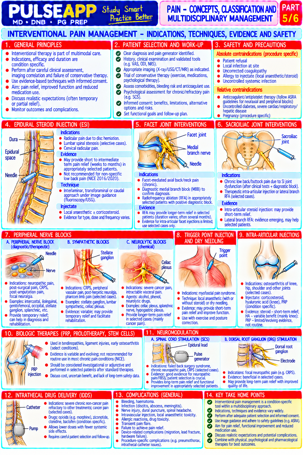
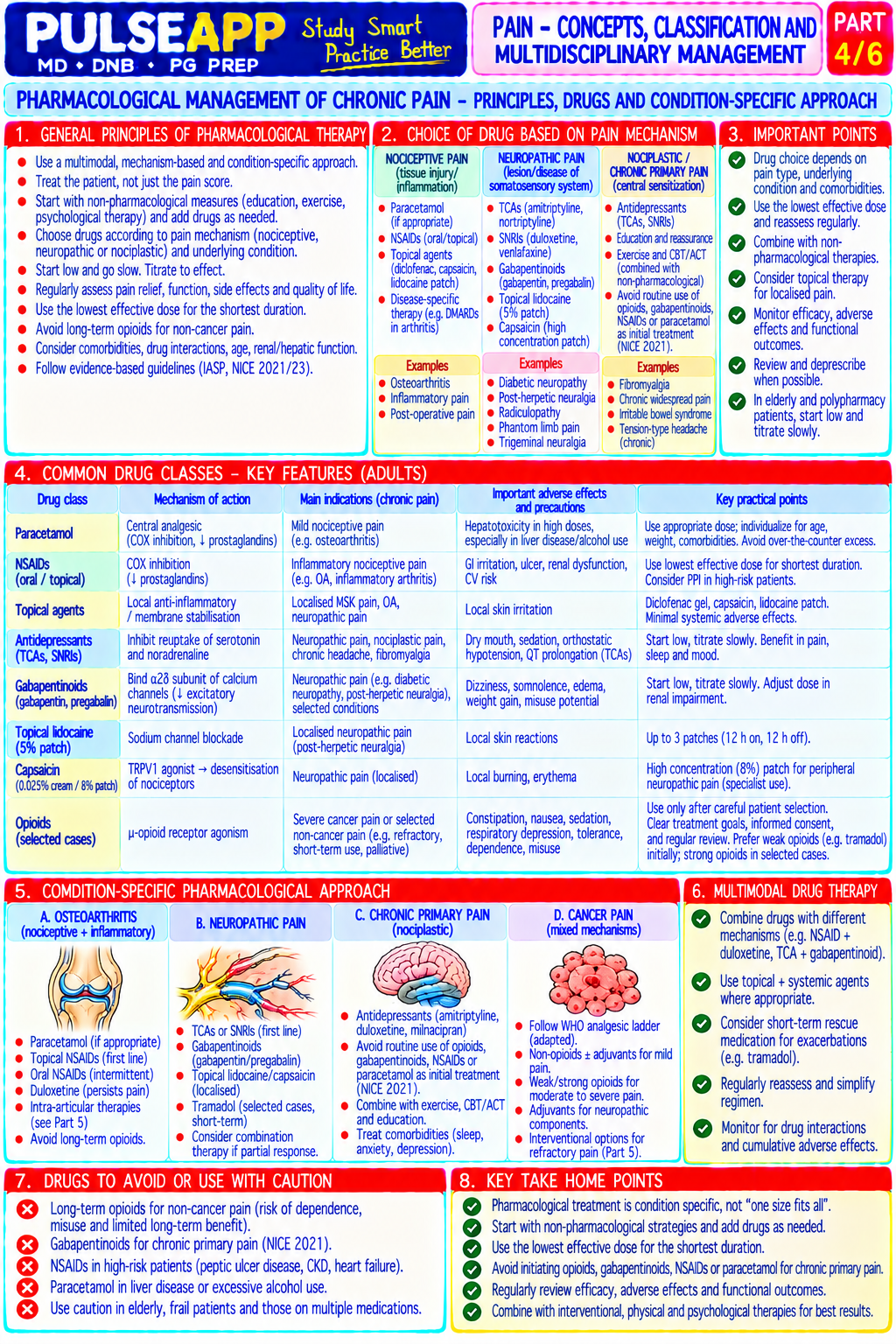
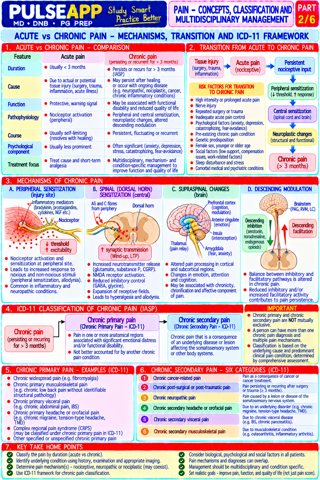
State the IASP 2020 revised definition of pain. Classify acute vs chronic pain and nociceptive vs neuropathic pain with mechanisms. Describe the multidisciplinary approach to chronic pain management including pharmacological, interventional, and psychological strategies.

collections Question Diagrams & Reference Images (1)
Question Reference Diagram
zoom_in View Image
description Clinical Response
⚙ Core Concept
The IASP revised the definition of pain in 2020 for the first time since 1979 — the update reflects 40 years of accumulated neuroscience showing that pain is not simply a signal from tissue damage but a complex conscious experience shaped by biology, psychology, and social context. Chronic pain affects over 20% of adults globally, consumes a disproportionate share of healthcare resources, and causes profound disability — yet it remains systematically undertreated. The modern multidisciplinary pain management approach, built on the biopsychosocial model, has stronger evidence than any single pharmacological or interventional treatment alone. (IASP 2020 revised definition; Merskey H; Treede RD — Chronic pain classification NCP; Turk DC — multidisciplinary pain management; Loeser J — Bonica's Management of Pain)
A. IASP 2020 Definition of Pain1 mark
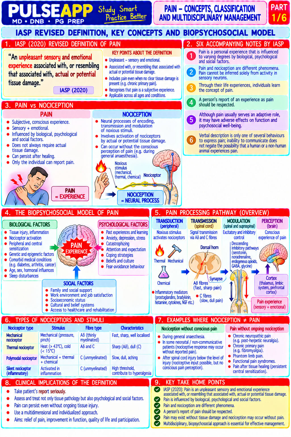
✅ IASP 2020: "An unpleasant sensory and emotional experience associated with, or resembling that associated with, actual or potential tissue
damage" The six key notes accompanying the definition: 1. Pain is always a subjective experience, influenced by biological, psychological, and social factors 2. Pain and nociception are different phenomena; pain cannot be inferred solely from activity in sensory neurons 3. Individuals learn the concept of pain through their life experiences 4. A person's report of an experience as pain should be respected — verbal description is only one of several behaviours to express pain 5. Although pain usually serves an adaptive role, it may have adverse effects on function and social and psychological well-being 6. Verbal description is only one of several behaviours to express pain; inability to communicate does not negate the possibility of pain The 2020 update explicitly includes pain without identifiable tissue damage (psychogenic pain, central sensitisation) and pain in non-communicating patients (neonates, dementia, comatose) — both excluded or poorly addressed by the 1979 definition.
B. Classification of Pain3 marks
By Duration

Feature Acute Pain Chronic Pain Duration <3 months; recent onset; expected to resolve as tissue heals Persists >3 months; beyond expected healing time; often persists after original injury has healed

Biological Adaptive — warns of tissue damage; promotes protective Maladaptive — no longer serves a protective purpose; becomes a disease in its own purpose behaviour (rest, seeking care); essential for survival right ("chronic pain syndrome") Neurobiology Nociceptive signals from tissue damage → ascending pain Central sensitisation; neuroplastic changes in the dorsal horn and brain; descending pathways → cortical pain perception; proportional to damage inhibitory pathway failure; pain independent of (or out of proportion to) peripheral stimulus Treatment Treat underlying cause; multimodal analgesia; short-term Biopsychosocial model; multidisciplinary team; minimise opioid exposure; focus on approach opioids acceptable function not pain elimination

By Mechanism

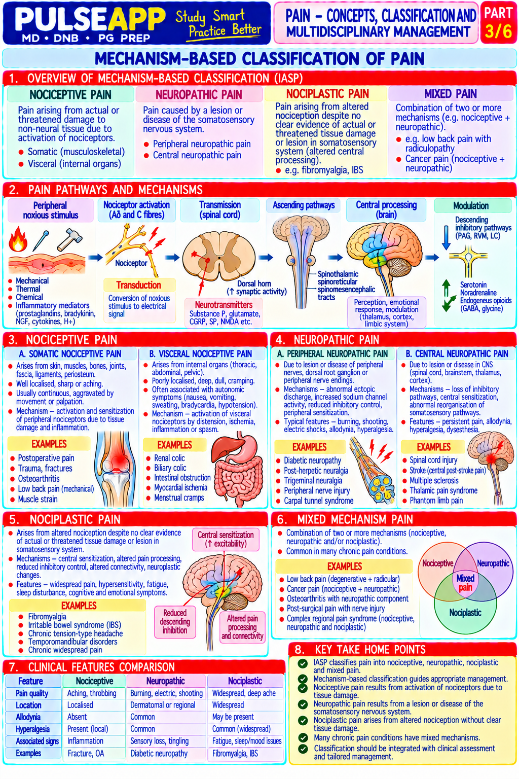
Type Mechanism Characteristics Examples Treatment Nociceptive Activation of A-delta (sharp, fast pain) and C Well-localised; proportional to stimulus; Bone pain, muscle pain, NSAIDs; — Somatic fibres (slow, burning pain) in somatic tissues improves with rest and analgesics; described incisional pain, trauma paracetamol; by noxious stimuli (mechanical, thermal, as sharp, aching, throbbing; responds to opioids; regional chemical) — signalling to the dorsal horn → NSAIDs and opioids blocks thalamus → somatosensory cortex Nociceptive Activation of visceral afferents by distension, Poorly localised; colicky or pressure-like; Bowel obstruction, biliary Opioids; N-butyl — Visceral ischaemia, or inflammation of hollow organs; associated with autonomic responses (nausea, colic, ureteric colic, scopolamine travels via sympathetic nerves; referred pain sweating, pallor); referred to somatic areas cardiac ischaemia (antispasmodic); via shared dermatomes with somatic fibres (e.g., MI → left arm pain; appendicitis → treat underlying periumbilical then RIF) cause Neuropathic Initiated or caused by a primary lesion or Burning, shooting, electric shock-like, tingling Diabetic peripheral Gabapentinoids disease in the somatosensory nervous system (dysaesthesia); allodynia (pain from normally neuropathy, post-herpetic (pregabalin, (peripheral or central); pathological changes in non-painful stimulus — light touch causes neuralgia, phantom limb, gabapentin); TCAs neural signalling — ectopic discharge from severe pain); hyperalgesia (exaggerated pain complex regional pain (amitriptyline); injured axons, central sensitisation, loss of response to painful stimulus); often associated syndrome (CRPS), SNRIs (duloxetine); descending inhibitory control, glial activation with sensory deficit in the painful area; often chemotherapy-induced, topical poorly responsive to NSAIDs/opioids post-surgical (chronic lidocaine/capsaicin; post-surgical pain) nerve blocks; spinal cord stimulation Nociplastic Altered nociception from changed functioning Widespread, diffuse pain; poor correlation with Fibromyalgia; irritable Exercise; cognitive (Central of the somatosensory nervous system without tissue findings; associated with fibromyalgia, bowel syndrome; tension- behavioural therapy Sensitisation) clear evidence of actual tissue damage or irritable bowel, tension headache; fatigue; type headache; (CBT); SNRIs nerve damage; the pain is real but arises from sleep disturbance; often has psychological widespread (duloxetine); TCAs; a sensitised central nervous system (changes comorbidities musculoskeletal pain low-dose naltrexone in dorsal horn transmission, supraspinal (experimental); processing, descending modulation) multidisciplinary rehabilitation

C. Multidisciplinary Chronic Pain Management6 marks

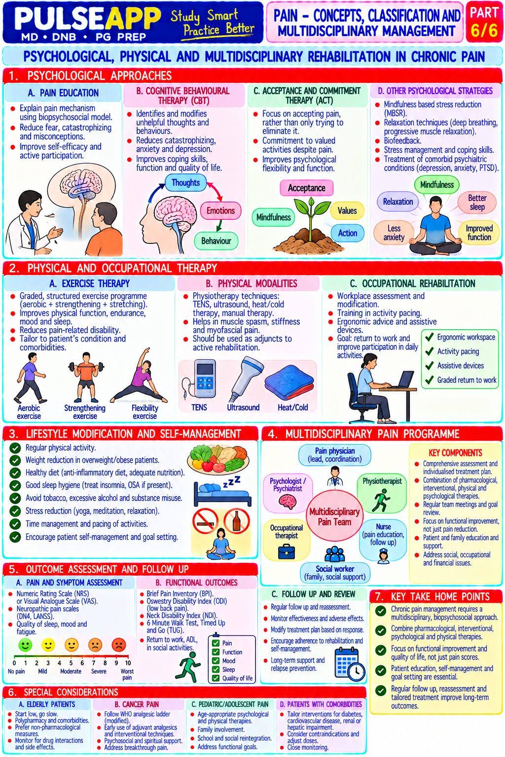
The Biopsychosocial Model — Framework for All Interventions The biopsychosocial model (Engel, 1977) recognises that chronic pain is determined by three interacting dimensions: biological (tissue pathology, neural sensitisation, genetics, pharmacology), psychological (depression, anxiety, catastrophising, fear-avoidance behaviour, sleep disturbance), and social (employment, social support, compensation claims, cultural attitudes to pain); effective chronic pain management must address all three dimensions simultaneously Component Interventions Evidence Level 1. Step 1 (mild pain): paracetamol 500–1000 mg QID; NSAIDs (ibuprofen, naproxen, celecoxib) — WHO analgesic ladder (adapted for chronic Pharmacological most evidence for acute/inflammatory pain; limit <10 days; GI/renal/cardiovascular risk monitoring pain); NICE guidelines (NG173); NNT for

Step 2 (moderate pain): weak opioids (codeine, tramadol) + step 1; tramadol (SNRI + opioid) neuropathic agents: duloxetine NNT 6.5; particularly useful for neuropathic pain; gabapentinoids (pregabalin 75–300 mg BD; gabapentin 300– pregabalin NNT 7.7; amitriptyline NNT 6.4 1200 mg TID) for neuropathic

Step 3 (severe/neuropathic): strong opioids (morphine, oxycodone, fentanyl patch) — ONLY for chronic pain with demonstrated benefit and documented risk-benefit discussion; buprenorphine patches (7-day) — less euphoric, ceiling effect, suitable for chronic pain; duloxetine 60–120 mg OD (SNRI with level 1 evidence for diabetic neuropathy, fibromyalgia, MSK pain); amitriptyline 10–75 mg nocte (low-dose TCA — analgesic, sleep aid, also antidepressant) 2. Interventional Nerve blocks: diagnostic and therapeutic; e.g., coeliac plexus block (pancreatic cancer pain), NICE (NG59) — SCS for FBSS and CRPS; / Procedural stellate ganglion block, sympathetic blocks level 1 evidence for SCS in selected

Epidural steroid injection: lumbar radiculopathy, cervical radiculopathy — reduces nerve root populations; RFA supported by multiple inflammation; short-to-medium term benefit RCTs for facet pain

Radiofrequency ablation (RFA): thermal or pulsed RFA of facet joint medial branches for chronic facet pain; cryoablation of sensory nerves

Spinal Cord Stimulation (SCS): implanted lead in the dorsal epidural space; dorsal column electrical stimulation produces paresthesia overlying the painful area; excellent evidence for CRPS, failed back surgery syndrome (FBSS), refractory angina; NNT ~2.4 for CRPS

Intrathecal drug delivery (ITDD) systems: implanted pump delivering opioids/baclofen/ziconotide directly into the CSF; for cancer pain or spasticity; reduces systemic opioid requirements

TENS (Transcutaneous Electrical Nerve Stimulation): surface electrodes; modulates pain via gate control theory (large-fibre stimulation closes the gate to C-fibre transmission); simple, noninvasive; evidence moderate but patient-driven 3. Psychological Cognitive Behavioural Therapy (CBT): the most evidence-based psychological treatment for Cochrane reviews: CBT reduces pain chronic pain; addresses catastrophising, fear-avoidance, maladaptive pain behaviours; teaches intensity (SMD −0.4) and disability (SMD coping strategies, activity pacing, relaxation; delivered individually or in group settings; best evidence −0.3) in chronic low back pain; psychological for fibromyalgia, CLBP, headache; NNT ~3–5 for significant improvement in disability interventions are NICE-recommended as

Acceptance and Commitment Therapy (ACT): focuses on accepting pain rather than eliminating it; core components of chronic pain increasing psychological flexibility; newer evidence base but growing; particularly useful in patients management where pain cannot be eliminated

Mindfulness-Based Stress Reduction (MBSR): 8-week structured programme; meditation, body scan, yoga; reduces pain catastrophising and improves quality of life; level 2 evidence for chronic pain

Pain neuroscience education (PNE): teaching patients the neuroscience of pain processing reduces catastrophising and improves outcomes; shifts the patient's conceptual model from "pain = damage" to "pain = overactive alarm system" 4. Physical / Exercise therapy: the single most evidence-based physical intervention for chronic pain; aerobic Cochrane reviews: exercise vs no treatment Rehabilitative exercise, resistance training, and specific exercise programmes (e.g., McKenzie for CLBP, graded for CLBP — moderate quality evidence for activity); mechanisms include endorphin release, central sensitisation reversal, anti-inflammatory improved pain and function; exercise is NICE effects; supervised exercise superior to home exercise first-line recommendation for CLBP

Physiotherapy: manual therapy (mobilisation, manipulation) for musculoskeletal pain; specific exercise prescription; postural correction

Hydrotherapy: warm water exercises reduce joint loading while allowing mobility; excellent for fibromyalgia, OA, rheumatoid arthritis

Occupational therapy: activity grading, adaptive devices, return-to-work planning; particularly for patients with chronic pain and disability

🎤 Viva Corner
Q. How does central sensitisation differ from peripheral sensitisation in producing chronic pain, and how does this distinction influence your choice of analgesic drug?
Peripheral sensitisation and central sensitisation are two distinct neurobiological mechanisms that together produce and maintain chronic pain, but they respond to different pharmacological interventions. Peripheral sensitisation: occurs at the level of the primary nociceptive afferent — following tissue injury, a soup of inflammatory mediators (bradykinin, prostaglandins, substance P, NGF, hydrogen ions) released from damaged cells, immune cells, and nerve terminals lowers the activation threshold and increases the firing rate of peripheral C and A-delta nociceptors; the neuron becomes more excitable — responding to lower-intensity stimuli and firing more intensely for the same stimulus. Peripheral sensitisation produces primary hyperalgesia (exaggerated pain at the injury site) and primary allodynia in the area of inflammation. The pharmacological target is the peripheral inflammatory process: NSAIDs (COX inhibition → reduced prostaglandin synthesis → reduce nociceptor sensitisation), local anaesthetics (block peripheral nerve conduction), and anti-nerve growth factor antibodies (tanezumab). Central sensitisation: occurs at the level of dorsal horn neurons in the spinal cord and supraspinal centres — repeated nociceptive input causes long-term potentiation of dorsal horn synapses (via NMDA receptor activation, removal of Mg²⁺ block by sustained depolarisation), upregulation of AMPA receptors, activation of glial cells that release proinflammatory cytokines, and failure of descending inhibitory pathways (from periaqueductal grey and locus coeruleus) to modulate incoming nociceptive input. Central sensitisation produces secondary hyperalgesia (exaggerated pain at sites remote from the injury — areas not directly innervated by injured nerves) and allodynia from non-nociceptive inputs (wind-up, temporal summation). Pharmacological targets for central sensitisation: NMDA receptor antagonists (ketamine, memantine — reduce wind-up); gabapentinoids (pregabalin, gabapentin — bind α2δ subunit of voltage-gated calcium channels on dorsal horn presynaptic terminals, reducing excitatory neurotransmitter release); SNRIs (duloxetine, venlafaxine — enhance descending noradrenergic and serotonergic inhibitory pathways); tricyclic antidepressants (amitriptyline — similar mechanism to SNRIs plus sodium channel blockade). NSAIDs and peripheral opioids are largely ineffective for the central sensitisation component — which is why patients with fibromyalgia (predominantly central sensitisation) respond poorly to anti-inflammatory drugs but better to duloxetine, pregabalin, and exercise (which modulates central processing).
Q. State the IASP 2020 definition of pain and explain the significance of the phrase "resembling that associated with actual or potential tissue damage." IASP 2020: "Pain is an unpleasant sensory and emotional experience associated with, or resembling that associated with, actual or potential tissue damage." The critical phrase "or resembling that associated with" is the key update from the 1979 definition, which stated "associated with actual or potential tissue damage." The 2020 update explicitly encompasses pain that occurs in the absence of any identifiable tissue or nerve damage — pain that is neurologically and experientially indistinguishable from pain caused by tissue injury but arises from a sensitised or dysregulated nervous system itself. This is not "imaginary" or "psychosomatic" pain in the derogatory sense — it is a real, measurable neurobiological phenomenon of central sensitisation (as seen in fibromyalgia, irritable bowel, widespread musculoskeletal pain, and chronic low back pain without structural lesion). The 2020 update ensures that these patients are not denied the diagnosis of "pain" simply because their MRI or blood tests are normal — their experience is equally valid and their condition is equally in need of treatment. It also encompasses phantom limb pain (the tissue is gone but the experience is of real pain in the amputated limb — the pain resembles tissue-damage pain but arises from cortical reorganisation and peripheral nerve stump ectopic discharge). Clinically, recognising that pain can be genuine in the absence of tissue damage changes the management approach: rather than ordering more investigations looking for a structural cause, the treatment must address the neurobiological dysfunction (central sensitisation) through psychological intervention, exercise, and centrally-acting pharmacotherapy.
★ Examiner's Pearl
State the IASP 2020 definition verbatim and explain why the 2020 update matters (explicitly includes pain without tissue damage — nociplastic/central sensitisation pain). The four pain types (nociceptive somatic/visceral, neuropathic, nociplastic) with mechanisms and specific examples for each are the classification table expected. The multidisciplinary approach structure (pharmacological/interventional/psychological/physical) with specific named drugs, specific named interventions (SCS, RFA, ITDD), and specific named psychological therapies (CBT, ACT, mindfulness) represents a comprehensive answer — incomplete answers miss the psychological and physical components which are equally weighted in modern pain medicine.
Raja SN et al. IASP revised definition of pain 2020 (Pain 2020;161:1976-1982). Treede RD et al. Chronic pain as a symptom or a disease — the IASP Classification of Chronic Pain (Pain 2019;160:19-27). NICE Guideline NG173 — Chronic pain 2021. Turk DC, Monarch ES. Biopsychosocial perspective on chronic pain (Psychol Clin North Am 1996). Bonica JJ. Loeser JD (Eds). Bonica's Management of Pain, 4th Ed.
lock Protected Clinical Document • Pulse App • Screenshots Strictly Prohibited
QUESTION 45 person Asked by .
bookmark_add

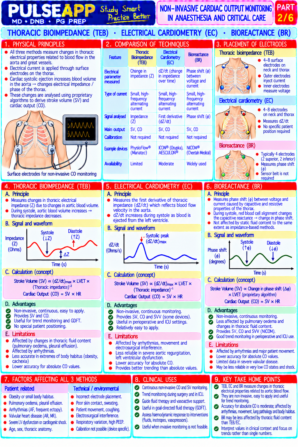
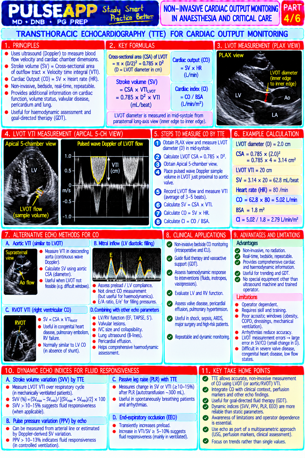
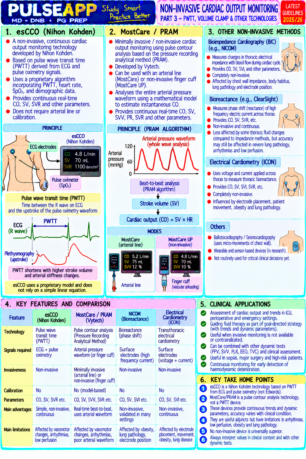
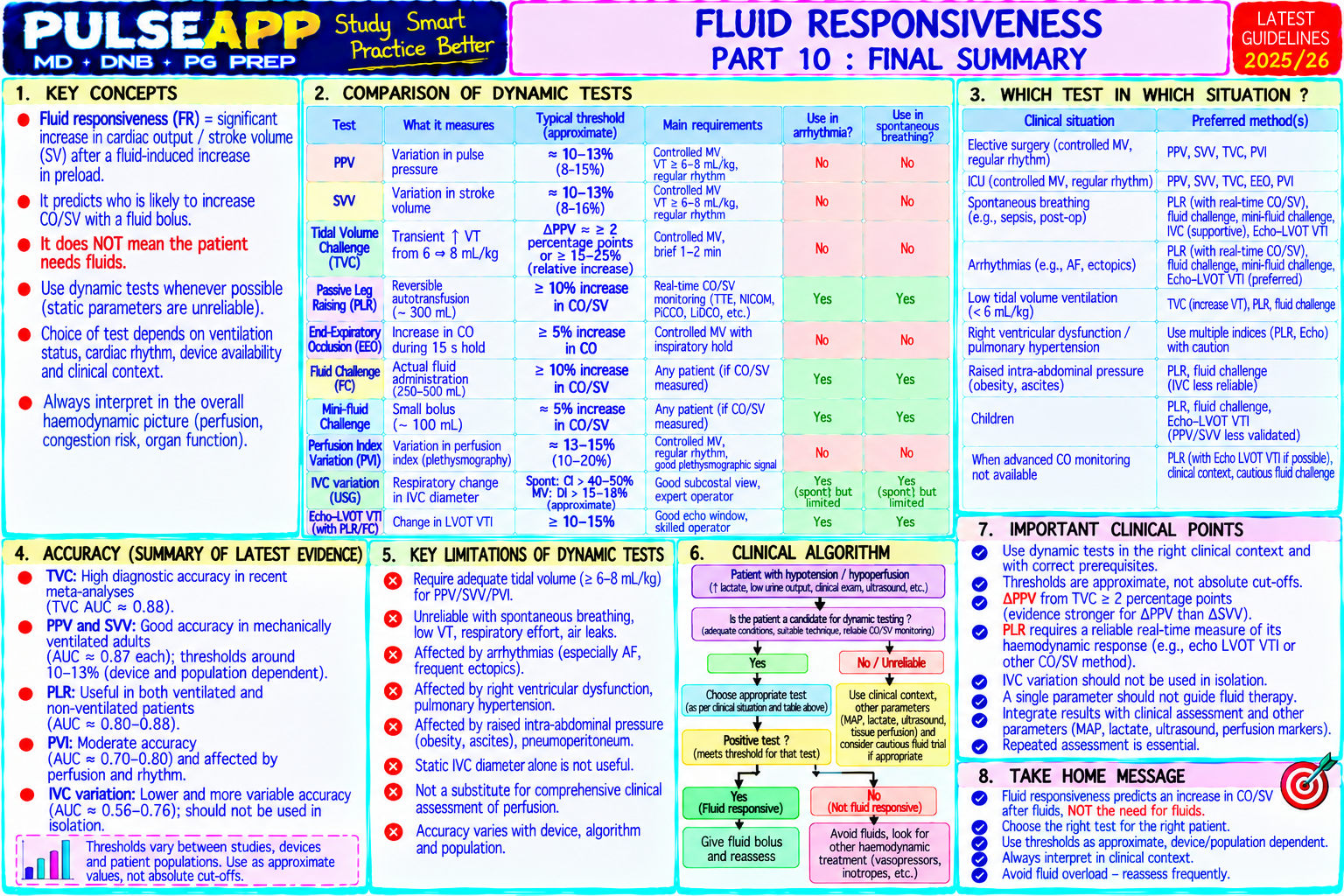
Describe the principles underlying non-invasive cardiac output monitoring modalities available in anaesthesia and critical care. Compare their accuracy, limitations, and clinical utility for goal-directed fluid therapy.
collections Question Diagrams & Reference Images (1)
Question Reference Diagram
zoom_in View Image
description Clinical Response
⚙ Core Concept
Cardiac output (CO) monitoring has historically required invasive placement of a pulmonary artery catheter — an approach associated with significant complications and not proven to improve outcomes. Non-invasive and minimally-invasive CO monitoring modalities now provide real-time haemodynamic data from surface probes, arterial waveforms, or oesophageal ultrasound, enabling goal-directed fluid therapy (GDT) and vasopressor titration without the risks of central venous or pulmonary artery catheterisation. GDT guided by non-invasive CO monitoring reduces postoperative complications in major surgery. (Miller's Anaesthesia 9th Ed; Pearse R et al. — OPTIMISE trial; Cecconi M; Gan TJ — goal-directed therapy; Marik P — non-invasive CO)
A. Principles of Cardiac Output and What We Measure1 mark

Cardiac Output (CO): the volume of blood ejected by the left ventricle per minute; CO = Stroke Volume (SV) × Heart Rate (HR); normal = 4–8 L/min; Cardiac Index (CI) = CO/BSA; normal CI = 2.5–4.0 L/min/m²

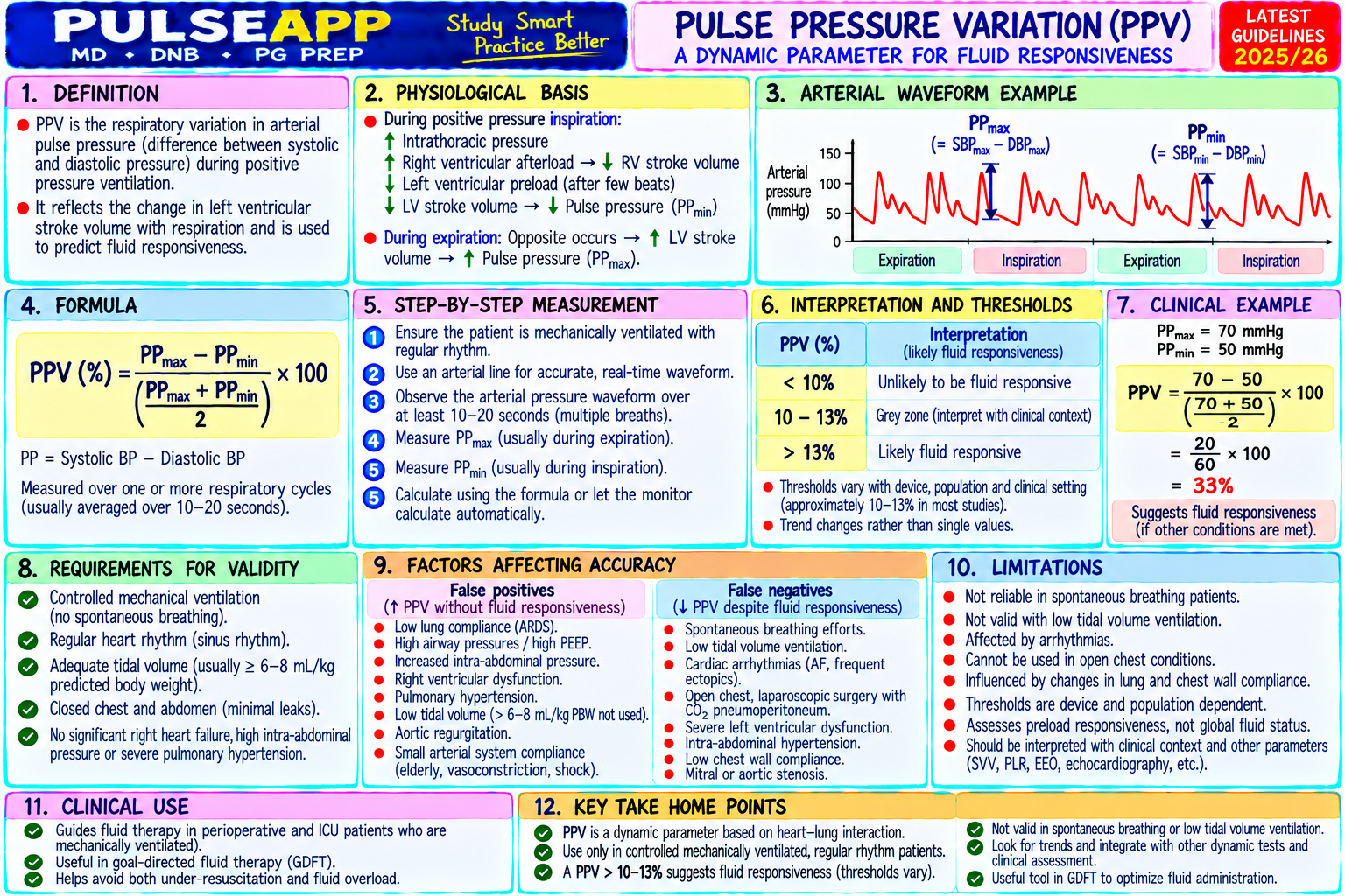
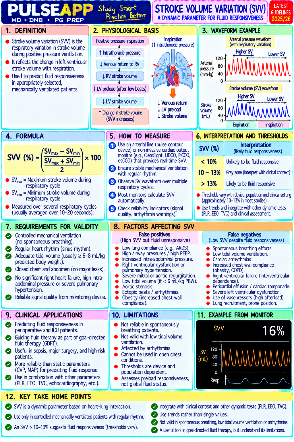
Stroke Volume (SV): the volume ejected per beat (normal 60–100 mL); determined by preload, afterload, and contractility; SV variation (SVV) and pulse pressure variation (PPV) are dynamic indices of preload responsiveness in mechanically-ventilated patients — if SVV >13% or PPV >13%, the patient is likely preload-responsive (will increase SV with fluid challenge) Most non-invasive monitors estimate CO or SV indirectly through physical principles (thermodilution, Fick, bioreactance, Doppler) — each with its own assumptions, limitations, and sources of error

B. Non-Invasive and Minimally-Invasive CO Monitoring Methods7 marks

Accuracy vs Method / Device Principle Invasiveness Key Advantages Key Limitations PAC Oesophageal A flexible probe (6 mm) placed in the Minimally Good Real-time continuous; the Requires intubation and Doppler Monitor oesophagus at the level of the invasive — concordance most validated non-PAC CO sedation; operator skill for (ODM) — CardioQ descending thoracic aorta; emits requires with PAC (bias monitor; FTc provides probe positioning; not continuous Doppler ultrasound at 4 MHz; intubated/sedated within ±20%); unique stroke volume index reliable in AF (irregular measures red blood cell velocity in the patient; placed validated in and preload assessment; stroke volumes); descending aorta → waveform analysis like NGT; not multiple large OPTIMISE trial showed overestimates CO if probe provides stroke volume, corrected flow possible in awake trials ODM-GDT reduces not correctly aligned with time (FTc), and cardiac output estimate; patients or with complications in major aortic blood flow; only assumes a fixed proportion of CO passes oesophageal surgery; NICE-approved measures CO indirectly through the descending aorta (roughly pathology (MTG3) for use in major (assumes fixed descending 70%) surgery aorta fraction) Pulse Contour Analyses the arterial pressure waveform Minimally Moderate Continuous real-time CO; Accuracy degrades

Analysis — to calculate stroke volume from the area invasive — concordance also provides SVV and PPV significantly with arterial PiCCO, LiDCO, under the systolic portion of the pressure requires arterial with PAC; (fluid responsiveness waveform distortion FloTrac/Vigileo wave; requires an arterial line (radial or line (standard accuracy indices); PiCCO additionally (vasopressors, arrhythmia, femoral); some systems require anaesthetic decreases in: provides extravascular lung vasoconstriction); intermittent external calibration (PiCCO monitoring); no high-dose water and intrathoracic calibrated systems need uses transpulmonary thermodilution; additional central vasopressors, blood volume; suitable for periodic recalibration LiDCO uses lithium dilution); venous access irregular rhythms major surgery and ICU; (position change, FloTrac/Vigileo uses a proprietary needed for (AF), severe FloTrac requires only a haemodynamic instability); algorithm based on patient demographics FloTrac; PiCCO peripheral standard arterial line FloTrac less accurate than without external calibration and LiDCO vasoconstriction, calibrated systems in require calibration cardiac haemodynamically bolus arrhythmias, unstable patients aortic regurgitation Thoracic Alternating electrical current is passed Non-invasive — Moderate Completely non-invasive — Lower accuracy than ODM Electrical through the thorax via surface electrodes surface concordance no needles; suitable for or calibrated pulse contour Bioimpedance / on the neck and chest; changes in electrodes; no with PAC in non-intubated patients; can in high acuity; affected by Bioreactance transthoracic impedance (related to the arterial line stable patients; monitor awake patients in patient movement (NICOM, Cheetah) aortic blood volume change with each required; fully poor accuracy in: ICU or stepdown; useful for (artefact); unreliable in heartbeat) are used to calculate SV; non-invasive obesity, trend monitoring pulmonary oedema, severe bioreactance (NICOM) measures the cardiac pulmonary obesity, thoracic surgery phase shift of the AC signal rather than monitoring oedema (fluid patients amplitude — more specific to aortic flow, alters less affected by pleural fluid or body impedance), composition arrhythmias, after cardiac surgery (altered thoracic anatomy and fluid) Transoesophageal A phased-array ultrasound transducer on Minimally Excellent Provides qualitative AND Requires intubated patient; Echocardiography an oesophageal probe; provides direct invasive — accuracy when quantitative data; directly needs formal training (TOE/TEE) imaging of cardiac chambers, valves, and requires performed by a visualises the cause of (BSE/EACVI TOE great vessels; SV calculated from the intubation and competent haemodynamic instability accreditation); not portable velocity-time integral (VTI) of the LVOT sedation; operator; (tamponade, LV/RV or continuously available in Doppler multiplied by the LVOT cross- operator considered a dysfunction, hypovolaemia, all settings; time-intensive sectional area (2D echo measurement); expertise reference PE, valve per measurement; relative provides the gold standard for direct required; requires standard for regurgitation/stenosis); contraindications visualisation of cardiac function, wall formal TOE perioperative cannot be replicated by any (oesophageal disease, motion, pericardial effusion, and competency haemodynamics; other non-invasive monitor; variceal bleeding risk) haemodynamic instability training limited not by provides wall motion (EACVI/ASE accuracy but by assessment for ischaemia accreditation) operator skill and detection access Non-Invasive Continuous non-invasive blood pressure Non-invasive — Moderate Completely non-invasive; Accuracy reduced by Haemodynamic measurement from a finger cuff using the finger cuff; accuracy for suitable for monitoring in peripheral vasoconstriction; Monitor (CNAP — volume clamp method (Finapres suitable for trending; may awake patients, procedural requires regular calibration continuous non- technology); uses the continuous BP awake patients underestimate in sedation, obstetrics (non- cycles; less accurate than invasive arterial waveform to compute CO via pulse peripheral invasive BP + CO), arterial line-based systems pressure + Nexfin) contour analysis; completely non-invasive vasoconstriction monitored care settings in haemodynamically — finger cuff only and overestimate unstable patients in warm vasodilated patients

C. Goal-Directed Therapy (GDT) — Clinical Application2 marks

Concept: GDT uses real-time CO/SV monitoring to guide fluid and vasopressor therapy toward specific haemodynamic targets — aiming to optimise oxygen delivery (DO₂) to tissues rather than simply normalising conventional vital signs (MAP, HR, urine output); key targets: CO/CI, SV, SVV/PPV (fluid responsiveness), and derived DO₂ OPTIMISE Trial (Pearse et al., BMJ 2014; n=734): high-risk major gastrointestinal surgery patients randomised to ODM-guided GDT vs standard care; GDT group received targeted colloid boluses to maximise SV and maintain FTc 35–40 ms; GDT significantly reduced postoperative complications (44.3% vs 51.0% — adjusted risk ratio 0.84) — the largest pragmatic RCT of CO-guided therapy in major surgery

Fluid responsiveness assessment: SVV >13% (mechanically-ventilated patients in sinus rhythm) or a passive leg raise (PLR) test predicts fluid responsiveness; if fluid responsive → 250 mL crystalloid/colloid bolus → reassess SV; if SV increases >10% → repeat; if SV increase <10% → patient is on the flat part of the Frank-Starling curve → additional fluid will not increase SV → use vasopressors instead

🎤 Viva Corner
Q. A patient is mechanically ventilated, sinus rhythm, after major colonic resection. SVV is 18%. What does this tell you, and what do you do?
SVV (Stroke Volume Variation) of 18% in a mechanically-ventilated patient in sinus rhythm indicates that the patient is preload-responsive — they are on the ascending portion of their Frank-Starling curve, meaning that ventricular stroke volume is significantly varying with the respiratory cycle (rising with inspiration as intrathoracic pressure increases and falling with expiration), reflecting that the right and left ventricles are operating in a preload-dependent range. The normal threshold is SVV >13% indicating likely preload responsiveness. This means that administering a fluid challenge is likely to increase stroke volume by >10% and therefore increase cardiac output meaningfully. Management: administer a 250–500 mL crystalloid (or colloid) fluid challenge over 10–15 minutes; reassess SVV and stroke volume after the challenge. If SV has increased by >10% (or SVV has fallen below 13%): the patient was indeed preload-responsive, the fluid improved haemodynamics, and further fluid boluses may be given as long as the patient remains responsive. If SV did not increase by >10% (and SVV has not fallen below 13%): the patient is non-responsive despite an elevated SVV — this is unusual and may indicate the SVV was elevated from a cause other than hypovolaemia (arrhythmia? Low tidal volume? Open chest?). In this case, consider vasopressors (noradrenaline) if MAP is inadequate, and investigate other causes of low CO. Important caveats for SVV interpretation: SVV is only valid in mechanically-ventilated patients in sinus rhythm, with TV ≥8 mL/kg IBW, and without spontaneous breathing effort or arrhythmias — any of these conditions invalidate the SVV as a fluid responsiveness index.
Q. Compare the oesophageal Doppler monitor (ODM) with FloTrac/Vigileo pulse contour analysis for intraoperative CO monitoring. Which would you choose for a high-risk major abdominal surgery patient and why?
Both the ODM (CardioQ) and FloTrac/Vigileo provide minimally-invasive continuous CO monitoring using different physical principles, and both have been used for GDT in major surgery. For a high-risk major abdominal surgery patient, I would choose the Oesophageal Doppler Monitor (ODM). The primary reason is the evidence base: the OPTIMISE trial (Pearse et al., BMJ 2014) demonstrated that ODM-guided GDT reduces postoperative complications in major gastrointestinal surgery — this is the largest pragmatic RCT validating this specific monitoring approach in this specific patient population, and is the basis for NICE's MedTech guidance (MTG3) recommending the ODM for major surgery. The FloTrac/Vigileo does not have equivalent-quality trial data demonstrating similar outcome benefits in major surgery, and its accuracy is a specific concern for this patient: FloTrac uses an uncalibrated algorithm that relies on assumptions about the arterial waveform that are violated in the presence of high-dose vasopressors, arrhythmias, and significant peripheral vasoconstriction — all of which may occur in a highrisk intraoperative patient. The ODM, by contrast, measures aortic blood velocity directly via Doppler (a physical measurement, not a model-based estimate), making it more reliable when haemodynamics are changing rapidly. Additionally, the ODM provides the corrected flow time (FTc) — a unique parameter that estimates preload and vascular tone that FloTrac does not provide. The practical disadvantage of the ODM (requires an intubated, sedated patient with the probe in situ throughout the case) is acceptable in a patient undergoing major abdominal surgery under general anaesthesia.
★ Examiner's Pearl
Name all five modalities with their underlying physical principle — examiners specifically check whether candidates know the principle (Doppler for ODM; impedance/bioreactance for NICOM; pulse contour analysis for FloTrac; direct echo measurement for TOE). The OPTIMISE trial (Pearse, BMJ 2014) with its result (reduced postoperative complications in major GI surgery) and the NICE guidance (MTG3) for ODM are the specific evidence landmarks that distinguish comprehensive answers. SVV >13% as the fluid responsiveness threshold with its specific limitations (sinus rhythm, mechanical ventilation, TV ≥8 mL/kg) must be stated precisely.
Pearse RM et al. Effect of a perioperative cardiac output-guided haemodynamic therapy algorithm on outcomes — OPTIMISE trial (BMJ 2014;348:g2082). Cecconi M et al. Consensus on perioperative haemodynamic monitoring (Intensive Care Med 2014;40:1795-1815). NICE MTG3 — CardioQ-ODM oesophageal Doppler monitor for guidance of intravenous fluid administration 2011. Marik PE. Noninvasive cardiac output monitors (J Intensive Care Med 2013;28:121-134). Miller RD et al. Miller's Anaesthesia, 9th Ed, Chapter 45.
lock Protected Clinical Document • Pulse App • Screenshots Strictly Prohibited
QUESTION 46 person Asked by .
bookmark_add

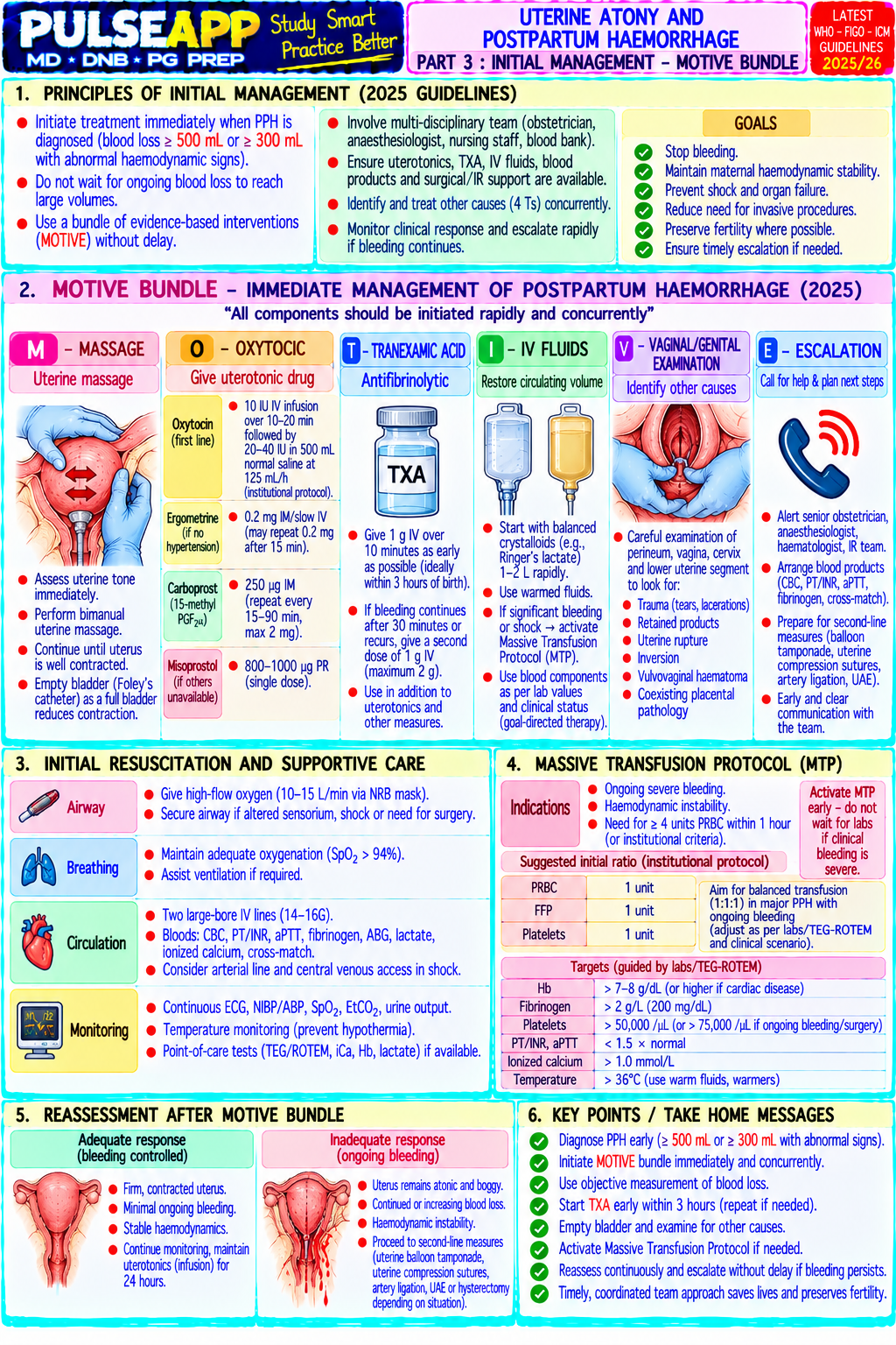
Define uterine atony and describe its pathophysiology. List the risk factors (the 4 Ts). Outline the stepwise management including uterotonic drugs (mechanisms, doses, side effects), balloon tamponade, surgical procedures, and interventional radiology options.

description Clinical Response
⚙ Core Concept
Postpartum haemorrhage (PPH) — defined as blood loss >500 mL after vaginal delivery or >1000 mL after caesarean section — is the leading cause of maternal mortality globally, accounting for 27% of all maternal deaths. Uterine atony (failure of the uterus to contract after delivery of the placenta) is responsible for approximately 80% of all PPH cases. The normal uterus after delivery contracts to compress the spiral arteries of the placental bed — in atony, this physiological tourniquet fails, and the open spiral arteries bleed profusely. Early recognition, systematic escalation of uterotonic therapy, and anaesthetic preparedness for massive obstetric haemorrhage are non-negotiable competencies. (Miller's Anaesthesia 9th Ed; RCOG Green-top Guideline 52; WHO PPH Guidelines; Evensen A; Mousa HA)
A. Pathophysiology of Uterine Atony2 marks

During third stage of labour, the placenta separates and the uterus must contract rapidly to compress the spiral arteries of the placental bed — these arteries supply the placenta at a rate of approximately 500 mL/min at term; without uterine contraction, these open vessel sinuses bleed at this rate, producing potentially fatal haemorrhage within minutes Normal uterine contraction is mediated by oxytocin (from the posterior pituitary) acting on uterine myocyte oxytocin receptors → intracellular Ca²⁺ → myosin light chain kinase activation → smooth muscle contraction; prostaglandins (PGE₂, PGF₂α) and ergometrine (α-adrenergic stimulation + 5-HT-mediated) independently support uterine tone

Atony occurs when: oxytocin receptor downregulation (from prolonged IV oxytocin during labour); myometrial fatigue (prolonged labour, grand multiparity); mechanical failure to contract (fibroids, distension from polyhydramnios, multiple gestation, macrosomia); retained placental tissue preventing uniform contraction; drug-induced uterine relaxation (volatile anaesthetic agents, magnesium sulphate, tocolytics, nifedipine)

B. The 4 Ts — Risk Factors for Uterine Atony2 marks

T Category Specific Risk Factors

T1 — Uterine atony Grand multiparity (≥5 deliveries — exhausted myometrium); overdistended uterus (multiple pregnancy, polyhydramnios, macrosomia —

TONE — failure of LGA baby >4 kg); prolonged labour (myometrial fatigue); precipitate labour; induction/augmentation with oxytocin (receptor uterine downregulation); uterine fibroids (prevent uniform contraction); general anaesthesia with volatile agents (dose-dependent uterine muscle to relaxation); magnesium sulphate infusion (tocolytic); chorioamnionitis contract
T2 — Retained Retained placenta or placental fragments (prevents normal uterine retraction and contraction); succenturiate lobe (accessory lobe — TISSUE products of frequently retained); placenta accreta spectrum (accreta/increta/percreta — placenta abnormally adherent, cannot separate normally; conception major risk for catastrophic PPH); placenta praevia (abnormally located, prevents normal third stage)

T3 — Genital tract Cervical tears (especially after rapid delivery); vaginal lacerations; perineal tears (1st–4th degree); uterine rupture (rare but catastrophic); TRAUMA lacerations uterine inversion (fundus turns inside out — rare; causes profound haemorrhage and shock); episiotomy; forceps/ventouse delivery; and injuries caesarean incision extension

T4 — Coagulopathy Pre-existing coagulopathy (ITP, vWD, haemophilia carrier); obstetric causes of coagulopathy: abruptio placentae (DIC from thromboplastin THROMBIN release), amniotic fluid embolism (DIC), HELLP syndrome, pre-eclampsia; dilutional coagulopathy from massive transfusion; anticoagulant therapy (LMWH, warfarin)

C. Stepwise Management of Uterine Atony6 marks
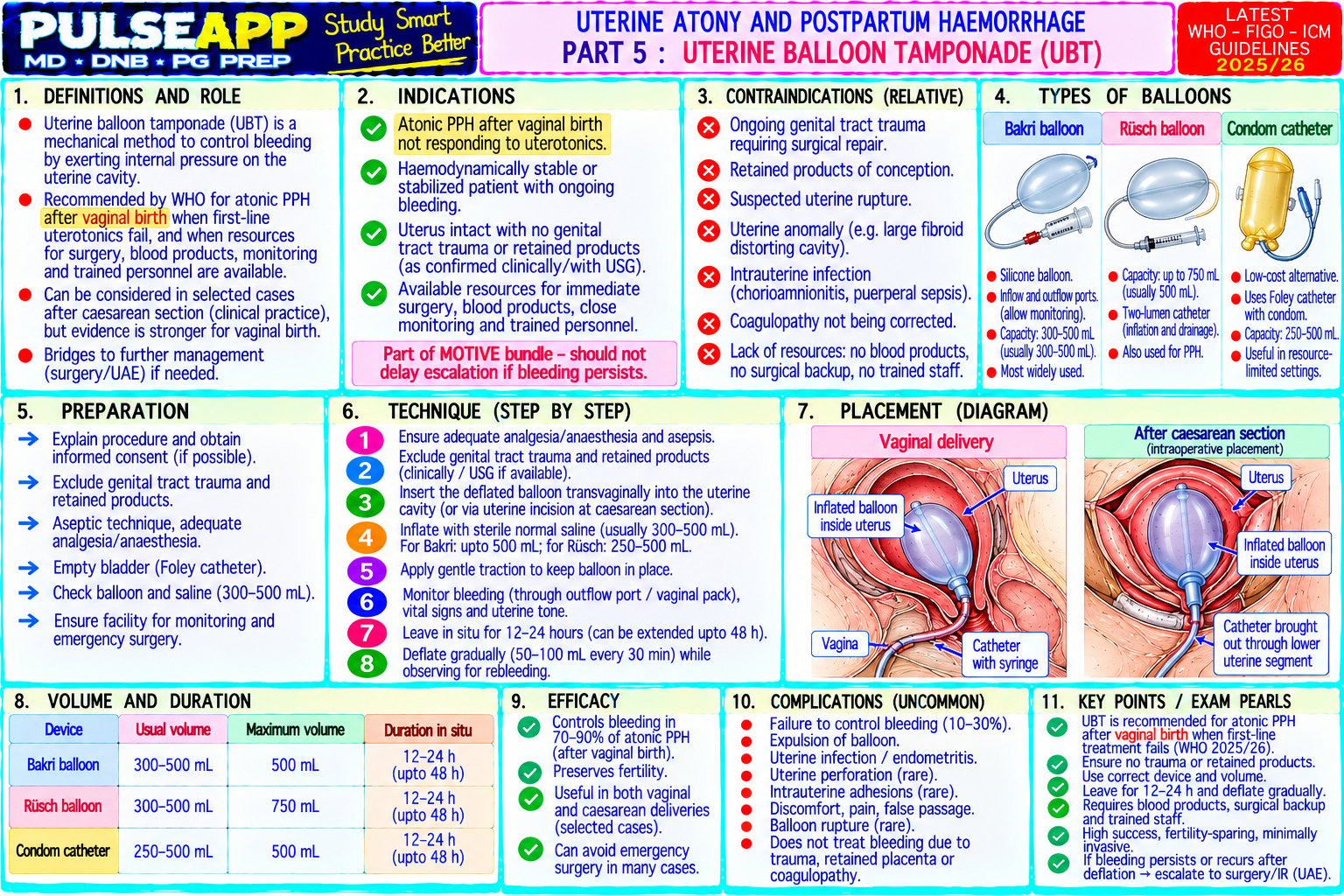
⚠ Call for help IMMEDIATELY — obstetric emergency team; anaesthesia; blood bank; theatre
Step 1 — Initial Assessment & Resuscitation Assess blood loss (weigh swabs, measure suction), vital signs, uterine tone (bimanual examination — boggy, enlarged, hypotonic uterus = atony); call for help; establish two large-bore IV cannulae (16G or 14G); send urgent bloods: FBC, clotting (PT, APTT, fibrinogen, TEG), G&S/crossmatch; IV crystalloid/colloid resuscitation; warm fluids; cell salvage if available; activate massive transfusion protocol if blood loss >1000 mL ongoing Uterine massage: bimanual uterine compression (hand in vagina anteriorly, abdominal hand posteriorly) — provides direct mechanical stimulation while uterotonics take effect Step 2 — Uterotonic Drug Therapy Drug Mechanism Dose Contraindications & Side Effects Oxytocin Synthetic posterior pituitary hormone; binds 5 IU slow IV push (over 1–2 minutes) at CS IV bolus causes systemic vasodilation → hypotension (Syntocinon) oxytocin receptors on uterine smooth muscle or PPH; THEN 40 IU in 500 mL NS at 125 + tachycardia (give slowly over 1–2 min, NEVER as → ↑ intracellular Ca²⁺ → uterine contraction; mL/hr (infusion); or IM 10 IU; prophylactically rapid IV bolus in cardiac disease); nausea; fluid also stimulates milk ejection in third stage retention; prolonged infusion → receptor downregulation (reduces efficacy) Ergometrine Ergot alkaloid; stimulates α-adrenergic and 5- 0.2–0.5 mg IM or slow IV; also available CONTRAINDICATED in hypertension, pre(Ergonovine) HT receptors on uterine smooth muscle → combined with oxytocin as Syntometrine (0.5 eclampsia, cardiovascular disease (causes sustained tonic contraction (different from mg ergometrine + 5 IU oxytocin IM) peripheral vasoconstriction → severe hypertension, oxytocin's rhythmic contractions); stronger and angina, MI, stroke); nausea and vomiting common; more prolonged than oxytocin alone avoid in Raynaud's Carboprost Synthetic prostaglandin F₂α analogue; 250 mcg IM every 15–90 minutes up to CONTRAINDICATED in asthma (bronchoconstriction (15-methyl stimulates FP receptors on uterine smooth maximum 8 doses (2 mg) — can cause fatal bronchospasm); severe diarrhoea PGF₂α — muscle → powerful sustained contraction; and vomiting; flushing; fever; relative contraindication Hemabate) bronchoconstrictive in hypertension; avoid IV administration (bronchospasm risk) Misoprostol Synthetic prostaglandin E₁; stimulates EP2 600–800 mcg sublingually or rectally Shivering (very common — up to 50%), fever, (PGE₁ and EP3 receptors on uterine smooth muscle (rectal has slower but more sustained diarrhoea, nausea; less potent than oxytocin for PPH analogue) → uterotonic effect; thermostable (can be effect); for PPH when IV access unavailable treatment but essential as second-line or when IV stored without refrigeration) or in resource-limited settings; also used unavailable; WHO recommended for resource-limited prophylactically 600 mcg oral at third stage settings Tranexamic Antifibrinolytic — competitive inhibitor of 1 g IV over 10 minutes; repeat 1 g IV if Well tolerated; nausea, vomiting; theoretical acid (TXA) plasminogen activation → prevents fibrinolysis bleeding continues after 30 minutes or thrombosis risk (used within the first 3 hours gives → stabilises formed clots; does not cause restarts within 24 hours (WOMAN trial maximal benefit — WOMAN trial); no significant uterine contraction but reduces fibrinolytic protocol) adverse effects in obstetric doses; NOW coagulopathy contributing to PPH RECOMMENDED in all cases of PPH by WHO 2022 and RCOG Step 3 — Balloon Tamponade If uterotonics fail: intrauterine balloon tamponade (Bakri balloon — placed into the uterine cavity and inflated with saline 250–500 mL) applies hydrostatic pressure to the placental bed, mechanically compressing the open spiral arteries; simple, rapid to insert, can be applied vaginally or at CS "Sandwich technique" (B-Lynch or compression suture + Bakri balloon) provides both mechanical compression and internal tamponade The "tamponade test" — if bleeding stops with balloon inflated → balloon tamponade is effective → continue for 12–24 hours with gradual deflation; if bleeding continues despite balloon → proceed to surgical/radiological intervention Step 4 — Surgical Management B-Lynch compression suture: a brace suture around the uterus compressing it like two hands squeezing — highly effective, preserves fertility; success rate ~80% Bilateral uterine artery ligation: reduces blood flow to uterus by 90% when both uterine arteries are ligated; preserves fertility; success ~90% in skilled hands Internal iliac (hypogastric) artery ligation: complex procedure; reduces pulse pressure to the uterine vasculature; less commonly performed now due to availability of uterine artery embolisation Peripartum hysterectomy: definitive control of PPH; life-saving but results in permanent infertility; subtotal hysterectomy faster than total; decision should be made early (not as a last resort when the patient is in DIC) Step 5 — Interventional Radiology Uterine artery embolisation (UAE): radiological catheter-based occlusion of the uterine arteries using gelfoam or polyvinyl alcohol particles; preserves fertility; requires haemodynamically stable patient and IR team availability; not suitable for acute life-threatening haemorrhage Prophylactic iliac artery balloon occlusion: balloons placed in iliac arteries before CS for known placenta accreta — inflated at time of delivery to reduce haemorrhage during hysterectomy
🎤 Viva Corner
Q. A patient receives oxytocin 5 IU rapid IV bolus after caesarean section and immediately develops severe hypotension (BP 70/40) and tachycardia (HR 140). Explain the mechanism and how you manage it. The rapid IV oxytocin bolus has caused the well-documented but often under-appreciated cardiovascular side effects of IV oxytocin — specifically systemic vasodilation and reduced cardiac output. Mechanism: oxytocin receptors are present not only on the uterine myometrium but also on vascular smooth muscle endothelium and the myocardium; systemic IV oxytocin (especially as a rapid bolus) stimulates these vascular receptors → nitric oxide release from endothelium → profound vasodilation → drop in systemic vascular resistance → hypotension; simultaneously, the heart attempts to compensate with tachycardia; the rapid bolus also produces a direct negative inotropic effect on the myocardium at high plasma concentrations (reduced cardiac contractility). These effects are amplified by the pre-existing vasodilation of pregnancy and spinal anaesthesia (if used). The cardiovascular effects of oxytocin are dose-rate-dependent — a slow infusion of the same 5 IU dose produces dramatically less haemodynamic disturbance than a rapid bolus. Management: the hypotension should be treated with IV vasopressor — phenylephrine 100 mcg IV bolus or ephedrine 6–12 mg IV (depending on heart rate — phenylephrine is better if tachycardic as it does not worsen tachycardia; ephedrine is better if bradycardic) and IV fluid bolus 250 mL crystalloid rapidly. For future obstetric practice: oxytocin must NEVER be administered as a rapid IV bolus in any patient with cardiovascular disease or instability; the recommended administration is 5 IU slow IV over 1–2 minutes (or IM); followed by an infusion of 40 IU in 500 mL at 125 mL/hr; for high-risk patients with cardiac disease, carbetocin 100 mcg IV (the synthetic long-acting oxytocin analogue with fewer cardiovascular effects) or misoprostol should be considered as the primary uterotonic. Q. A patient with PPH from uterine atony has received oxytocin, ergometrine, and carboprost but continues to bleed. Blood loss is estimated at 2000 mL. Describe your management of the haematological/coagulation aspect simultaneously with the surgical management. At 2000 mL blood loss with ongoing haemorrhage despite three uterotonics, this patient meets the criteria for massive PPH and I need to activate the massive transfusion protocol and manage haematological failure simultaneously with the surgical escalation. Immediately: activate the MTP (massive transfusion protocol — the "code obstetric major haemorrhage" call); simultaneously inform the obstetrician that medical management has failed and surgical escalation (balloon tamponade, compression sutures, possible hysterectomy) must begin without further delay. Haematological management: order urgent TEG/ROTEM or clotting screen including fibrinogen (critically important in obstetric haemorrhage — fibrinogen is consumed early and significantly; a fibrinogen <2 g/L in obstetric haemorrhage is a strong predictor of progression to severe PPH); give packed red blood cells (pRBC) and fresh frozen plasma (FFP) in a 1:1 ratio (same as military/trauma massive transfusion experience); tranexamic acid 1 g IV immediately and repeat 1 g in 30 minutes if bleeding continues (WOMAN trial protocol — reduces mortality from bleeding when given within 3 hours of delivery); if fibrinogen is low (<2 g/L): cryoprecipitate 10 units IV (provides concentrated fibrinogen ~400 mg per unit × 10 = 4 g fibrinogen) OR Fibrinogen concentrate 2–4 g IV (faster, pathogen-inactivated); if platelets <75,000 → platelet transfusion. Simultaneously: the obstetrician should be attempting Bakri balloon insertion; if no response (tamponade test negative) → B-Lynch suture or compression sutures; if still failing → uterine artery ligation; if all measures fail → timely peripartum hysterectomy — it should be performed before the patient enters DIC, NOT as a last resort after DIC is established. Maintain temperature ≥36°C (hypothermia worsens coagulopathy); warm all blood products; active warming measures throughout; check arterial blood gas and electrolytes (hypocalcaemia from citrate in FFP → treat with calcium gluconate 10 mL 10% IV).
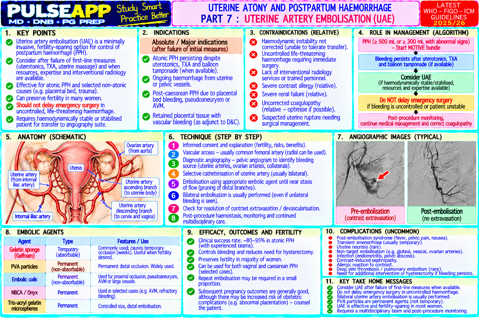
★ Examiner's Pearl
The 4 Ts framework (Tone, Tissue, Trauma, Thrombin) is a mandatory mnemonic to reproduce — with examples for each T. The uterotonic drug table must include ergometrine contraindications (hypertension/pre-eclampsia = absolute contraindication) and carboprost contraindication (asthma = absolute contraindication) — these are specifically tested as safety questions. Tranexamic acid dose (1 g IV, repeat 1 g) with the WOMAN trial citation is now a mandatory component of PPH management answers. The stepwise approach (1.uterotonics → 2.balloon → 3.sutures → 4.hysterectomy) must be presented in the correct escalating sequence.
RCOG Green-top Guideline No. 52 — Postpartum Haemorrhage (2016). WHO Recommendations for the Prevention and Treatment of PPH (2012). WOMAN Trial Collaborators. Effect of tranexamic acid on death, disability, vascular occlusive events and other morbidities in women with post-partum haemorrhage (WOMAN trial) (Lancet 2017;389:2105-2116). Miller RD et al. Miller's Anaesthesia, 9th Ed, Chapter 77 (Obstetric Anaesthesia). Evensen A et al. Postpartum haemorrhage (Am Fam Physician 2017;95:442-449).
lock Protected Clinical Document • Pulse App • Screenshots Strictly Prohibited
QUESTION 47 person Asked by .
bookmark_add

Describe the mechanism of organophosphorus poisoning. Classify the clinical features into muscarinic, nicotinic, and CNS manifestations using the SLUDGE or DUMBELS mnemonic. Outline the grading of severity and the emergency management including antidotes, airway management, and pralidoxime timing.

collections Question Diagrams & Reference Images (5)
Question Reference Diagram
zoom_in View Image
Question Reference Diagram
zoom_in View Image
Question Reference Diagram
zoom_in View Image
Question Reference Diagram
zoom_in View Image
Question Reference Diagram
zoom_in View Image
description Clinical Response
⚙ Core Concept
Organophosphorus (OP) compounds — used as pesticides (parathion, malathion, chlorpyrifos), nerve agents (sarin, VX, novichok), and historically as chemical warfare agents — are the most common cause of pesticide-related death globally, with India having one of the highest incidence rates of OP poisoning in the world. The mechanism is elegant and deadly: irreversible inhibition of acetylcholinesterase (AChE) → accumulation of ACh at all cholinergic synapses → cholinergic hyperstimulation — a crisis of too much neurotransmitter. The anaesthesiologist's specific roles in OP poisoning are: airway management (copious secretions + bronchospasm + respiratory muscle failure = one of the most challenging airway scenarios); avoiding succinylcholine (pseudocholinesterase inhibited → fatal prolonged paralysis); and judicious atropine titration. (Tintinalli's Emergency Medicine 9th Ed; Eddleston M — OP poisoning; Worek F — pralidoxime efficacy; Vale JA)
A. Mechanism of Toxicity2 marks

OP compound (lipid-soluble; absorbed via skin, inhalation, ingestion, conjunctiva) → covalently phosphorylates the serine hydroxyl group at the active site of acetylcholinesterase (AChE) → AChE is IRREVERSIBLY INHIBITED → cannot hydrolyse acetylcholine (ACh) at cholinergic synapses → ACh accumulates at: (1) all parasympathetic postganglionic nerve-effector junctions (muscarinic receptors); (2) nicotinic receptors at the NMJ (skeletal muscle); (3) autonomic ganglia — sympathetic and parasympathetic (nicotinic ganglionic receptors); (4) CNS synapses (muscarinic and nicotinic receptors in brain and spinal cord) → prolonged, excessive stimulation of ALL cholinergic receptors simultaneously → the CHOLINERGIC CRISIS.

⚠ "Ageing" — The Irreversibility Clock
The OP-AChE bond undergoes a process called "ageing" — a further chemical rearrangement of the covalent bond that makes it permanently irreversible, even to pralidoxime (the oxime antidote). Ageing rate varies by compound: soman (nerve agent) ages within minutes; parathion ages in hours; some OP pesticides take days. Once ageing is complete, pralidoxime cannot regenerate AChE — recovery depends entirely on synthesis of new AChE enzyme (which takes 2–3 weeks). This is why early pralidoxime administration (before ageing) is the critical time-dependent intervention.
B. Clinical Features — Three Receptor Systems3 marks

SLUDGE (Muscarinic) = Salivation, Lacrimation, Urination, Defecation, GI cramps/Emesis DUMBELS = Defecation/Diarrhoea, Urination, Miosis, Bradycardia/Bronchospasm/Bronchorrhoea, Emesis, Lacrimation, Salivation System Receptor Features Mnemonic Aid MUSCARINIC M1, M2, Salivation (excessive drooling), Lacrimation (tearing), Urination (incontinence), Defecation (diarrhoea), GI SLUDGE + DUMBELS; (parasympathetic M3 cramps, Emesis; ADDITIONALLY: MIOSIS (pinpoint pupils — pathognomonic), bradycardia, "wet and small" — nerve-effector receptors bronchospasm, BRONCHORRHOEA (copious secretions in airways — the most life-threatening feature), excessive secretions + junctions) hypotension, diaphoresis, urinary incontinence miosis NICOTINIC (NMJ N1 NMJ stimulation → initial FASCICULATIONS (visible muscle twitching — brief initial stimulation phase) → "Paralysis follows + autonomic (ganglionic) progressing to PARALYSIS (continuous receptor depolarisation → end-plate depolarisation block → fasciculations" — never ganglia) and N2 flaccid paralysis); diaphragm paralysis → respiratory failure (the primary cause of death); GANGLIONIC use succinylcholine (NMJ) stimulation → tachycardia (may coexist with or override bradycardia), hypertension (early), pallor, (pseudocholinesterase receptors mydriasis (may counteract or compete with muscarinic miosis) inhibited → prolonged paralysis) CNS Central Anxiety, restlessness, delirium, seizures (classically intractable — resistant to standard anticonvulsants "Seizures + coma" — muscarinic alone; require benzodiazepines + atropine for adequate control); coma; central respiratory depression CNS cholinergic storm and (compounding NMJ paralysis); in severe poisoning, all three mechanisms combine to produce apnoea nicotinic receptors

C. Severity Grading (Peradeniya Scale)1 mark

Grade Clinical Features Management Mild Alert; salivation, lacrimation, miosis, diarrhoea; RR normal; no Observation; atropine titration; supportive respiratory compromise Moderate Confusion; significant bronchorrhea, bronchospasm; mild respiratory ICU admission; IV atropine; pralidoxime; monitor RR closely — may deteriorate distress; RR increased; fasciculations prominent rapidly Severe Coma; seizures; severe bronchorrhoea and bronchospasm; respiratory Immediate intubation + mechanical ventilation; IV atropine titration; pralidoxime; failure (apnoea); bradycardia + hypotension; paralysis benzodiazepines for seizures; ICU; prolonged ventilatory support

D. Emergency Management4 marks
Immediate Measures

PERSONAL PROTECTION FIRST: OP compounds are highly lipid-soluble and absorbed through skin; all healthcare staff must wear gloves, gown, and eye protection before approaching the patient; remove and bag the patient's contaminated clothing before they enter the department

Decontamination: thorough washing of all exposed skin surfaces with soap and water (for 10–15 minutes) if dermal exposure; flush eyes with saline for conjunctival exposure; in ingestion cases: gastric lavage via NGT (within 1 hour of ingestion) + activated charcoal 50 g via NGT (if airway protected); emesis is CONTRAINDICATED (risk of aspiration, and rapid loss of consciousness)

Airway Management — The Critical Challenge
⚠ NEVER USE SUCCINYLCHOLINE IN OP POISONING
OP compounds inhibit plasma pseudocholinesterase (butyrylcholinesterase) in addition to AChE — pseudocholinesterase normally hydrolyses succinylcholine; with pseudocholinesterase inhibited, succinylcholine (whose action normally lasts 5–10 minutes) may produce paralysis lasting HOURS, potentially contributing to respiratory failure. Use rocuronium (with sugammadex reversal available) for RSI in OP poisoning. Indication for intubation: GCS ≤8, respiratory failure (RR >30 or <8, SpO₂ <90%), inability to handle secretions, impending respiratory muscle paralysis RSI with rocuronium 1.2 mg/kg (sugammadex 16 mg/kg available); atropine 0.6 mg IV before laryngoscopy (reduces excessive secretions and bronchospasm during intubation); manual inline suction of secretions before intubating High suction demand in ventilated OP patients — copious secretions require frequent ETT suctioning; consider HME filter over tracheal tube (absorbs some secretions)
Atropine — The Primary Antidote
✅ Atropine — Titrate to DRY Secretions, NOT to Heart Rate or Pupils
Atropine is a competitive muscarinic antagonist — it blocks the muscarinic effects of accumulated ACh (drying secretions, reversing bronchospasm, increasing heart rate, dilating pupils). The TITRATION ENDPOINT is drying of bronchial secretions (the patient can be adequately ventilated without constant suctioning) — NOT heart rate normalisation or pupil dilation. Dose: 2–4 mg IV bolus initially; then 2 mg IV every 5–10 minutes until secretions dry; total dose may be massive (10–50 mg or more over 24 hours in severe poisoning — doses of 100–200 mg over 24 hours have been reported in severe cases); then atropine infusion to maintain drying of secretions; titrate down over subsequent days as OP effects wear off (new AChE synthesised over 2–3 weeks)
Pralidoxime — The Enzyme Reactivator (Oxime)

Mechanism: pralidoxime (PAM; 2-PAM chloride) is an oxime compound that attacks the OP-AChE bond BEFORE ageing occurs → reactivates AChE by removing the OP group from the serine active site → restored AChE can resume hydrolysing ACh; this specifically reverses the NMJ (nicotinic) effects of OP poisoning (reverses fasciculations and paralysis) in addition to reducing the ACh burden at muscarinic synapses

Critical timing: pralidoxime MUST be given BEFORE AGEING; ageing rate depends on the specific OP compound — for most pesticide OPs, ageing occurs over hours; give pralidoxime as soon as diagnosis is made (ideally within 6–12 hours for most pesticides)

Dose: 1–2 g IV over 15–30 minutes; then 200–500 mg/hr infusion for 24–48 hours; continue until patient is asymptomatic and off atropine

Controversy: a large RCT from Sri Lanka (Eddleston et al., Lancet 2009) showed no mortality benefit of pralidoxime in agricultural OP poisoning — ongoing debate about its clinical utility in pesticide poisoning; still recommended in WHO and Indian guidelines; most useful early, before ageing

Other Measures

Seizures: benzodiazepines (diazepam 10 mg IV or midazolam 5–10 mg IV) — first-line; phenytoin is relatively ineffective for OP seizures (which are cholinergic in origin); continue until seizures stop

Monitor: ECG (QT prolongation from OP toxicity + atropine; AF, VT); blood glucose (OP poisoning causes hyperglycaemia from glycogen mobilisation); RBC cholinesterase activity (reflects AChE inhibition — correlates with severity); plasma cholinesterase (pseudocholinesterase — also inhibited but recovers faster)

🎤 Viva Corner
Q. A farmer is brought to the ED unconscious with pinpoint pupils, excessive salivation, and copious frothy secretions from his mouth. You prepare for intubation. What specific anaesthetic agents must you AVOID and why, and what will you use instead?
The confirmed diagnosis is organophosphorus poisoning from the triad of miosis (pinpoint pupils — M3 muscarinic iris sphincter stimulation), salivation, and bronchorrhoea in an agricultural worker. For intubation, the specific contraindication is succinylcholine — I will not use it under any circumstances in this patient. The reason: OP compounds inhibit not only acetylcholinesterase (AChE) at neural synapses but also plasma pseudocholinesterase (butyrylcholinesterase, PChE) — the enzyme that normally hydrolyses and terminates the action of succinylcholine within 5–10 minutes of IV administration. With PChE significantly inhibited by the OP compound, succinylcholine will not be degraded at the normal rate; its neuromuscular blocking action will be dramatically prolonged — potentially lasting hours rather than minutes. In a patient who already has respiratory compromise from bronchospasm and bronchorrhoea, adding hours of succinylcholine-induced apnoeic paralysis on top of the OP-induced NMJ block would be immediately fatal if intubation is unsuccessful. What I will use instead: rocuronium 1.2 mg/kg IV for RSI — rocuronium is metabolised by the liver (not by pseudocholinesterase), so its pharmacokinetics are unaffected by OP poisoning; its duration is predictable; and crucially, sugammadex 16 mg/kg can completely and rapidly reverse even profound rocuronium block within 2–3 minutes if intubation fails. Before laryngoscopy: atropine 0.6–1.2 mg IV to reduce the copious secretions that will impede laryngoscopy; suction aggressively before and during intubation; choose a cuffed ETT and inflate the cuff immediately after intubation to prevent aspiration of the abundant secretions pooling in the pharynx.
Q. Why is the titration endpoint for atropine in OP poisoning specifically "dry secretions" rather than normal heart rate or pupil dilation?
Atropine is a competitive muscarinic antagonist — it competes with accumulated ACh at muscarinic receptors. The goal of atropine therapy is not to reverse all muscarinic features (which would require enormous doses and produce atropine toxicity — tachyarrhythmias, hyperthermia, urinary retention, delirium) but to specifically protect the LIFE-THREATENING muscarinic effects — the bronchospasm and bronchorrhea that cause respiratory failure and death. The specific endpoint of "dry secretions" (bronchial secretions manageable, patient can be ventilated without constant suctioning, SpO₂ maintained) reflects this targeted approach: when secretions are dry, the airways are clear and ventilation is possible, which is the immediate life-saving goal of atropine. Using heart rate as the endpoint would result in gross under-dosing: the heart rate in OP poisoning may already be high from nicotinic ganglionic stimulation (competing with muscarinic bradycardia), and "normalising" it with atropine would be ambiguous and unreliable as a marker of adequate muscarinic blockade in the airways. Using pupil dilation (mydriasis from atropine competing with OP-induced miosis) would require even higher atropine doses and would over-titrate the drug with attendant toxicity risk. The pupils are NOT a reliable endpoint because the CNS muscarinic tone and the peripheral muscarinic tone at the iris do not necessarily parallel the bronchial muscarinic state. The practical guidance: give 2–4 mg IV initially and repeat every 5–10 minutes while actively suctioning and listening to breath sounds; once the secretions are manageable (chest clear, SpO₂ stable, ETT suction less frequent and less productive), maintain that level with a continuous infusion adjusted to keep the airways clear without producing atropine toxicity.
★ Examiner's Pearl
SLUDGE mnemonic must be fully expanded (Salivation/Lacrimation/Urination/Defecation/GI cramps/Emesis) with the addition of the most dangerous feature: BRONCHORRHOEA (not part of SLUDGE but the most life-threatening muscarinic effect). The succinylcholine contraindication reason (pseudocholinesterase inhibited by OP → prolonged paralysis) is the most commonly tested anaesthesia-specific OP fact. Atropine endpoint = DRY SECRETIONS (not heart rate, not pupils) with the specific mechanistic explanation — this is the most discriminating viva question in OP poisoning management.
Tintinalli JE et al. Tintinalli's Emergency Medicine, 9th Ed, Chapter 179 (Insecticides and Herbicides). Eddleston M et al. Pralidoxime in organophosphorus insecticide self- poisoning (Lancet 2009;374:592-600). Worek F et al. Reactivation of organophosphate-inhibited human acetylcholinesterase by oximes (Toxicology 2012;294:91-99). Vale JA, Lotti M. Organophosphorus compound and nerve agent poisoning (Handb Clin Neurol 2015;131:149-168). WHO. Organophosphorus pesticide poisoning — management guidelines 2016.
lock Protected Clinical Document • Pulse App • Screenshots Strictly Prohibited
QUESTION 48 person Asked by .
bookmark_add

Describe the key pharmacokinetic parameters of IV anaesthetic agents including volume of distribution, clearance, half- life, and context-sensitive half-time (CSHT). Explain how these parameters determine the clinical behaviour of propofol, thiopentone, ketamine, and etomidate.

collections Question Diagrams & Reference Images (1)
Question Reference Diagram
zoom_in View Image
description Clinical Response
⚙ Core Concept
Pharmacokinetics — "what the body does to the drug" — determines how quickly a drug reaches the brain, how long it stays there, and how predictably it leaves. The CSHT, not the elimination half-life, is the clinically relevant parameter for infusions: it describes how long it takes for the plasma concentration to halve after stopping an infusion of a specific duration, accounting for the redistribution dynamics that change with infusion length. (Miller's Anaesthesia 9th Ed; Shafer SL, Varvel JR; Stanski DR; Hughes MA — CSHT)
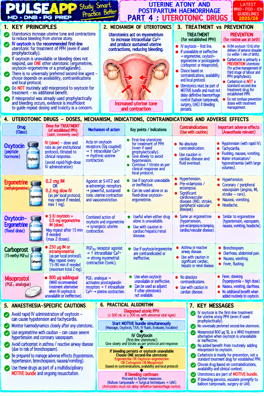
A. Key Pharmacokinetic Parameters3 marks

Parameter Definition Clinical Meaning Volume of The apparent volume into which a drug distributes to produce the observed Large Vd → drug distributes extensively into peripheral tissues →

Distribution plasma concentration: Vd = Dose / Cp; expressed in L or L/kg smaller plasma concentration for a given dose → longer elimination half(Vd) life; e.g., propofol Vd ~200–500 L (large — distributes into fat widely) Clearance Volume of plasma cleared of drug per unit time (mL/min or L/hr); CL = Dose / High clearance → drug eliminated rapidly from plasma → short duration; (CL) AUC; depends on hepatic blood flow (for flow-limited drugs like propofol, propofol CL ~1.5–2 L/min (very high — entire hepatic blood flow) fentanyl) or hepatic enzyme capacity (capacity-limited) Half-life t½ = 0.693 × Vd / CL; the time for plasma concentration to halve; for drugs Terminal t½ is often misleading for clinical practice — a drug with large (t½) with large Vd or low CL → long t½ Vd has long t½ even with high CL because drug continues to return from peripheral compartments; CSHT is more clinically relevant Context- The time for plasma concentration to fall 50% after stopping an infusion of a The most clinically relevant parameter for infusions: remifentanil CSHT = Sensitive specific duration ("context" = infusion duration); accounts for redistribution from 3 min (flat — no accumulation, organ-independent ester hydrolysis); Half-Time peripheral compartments back into plasma after stopping; CSHT INCREASES propofol CSHT rises slowly (short cases ~5 min; 8 hr case ~40 min — (CSHT) with longer infusion duration as peripheral compartments become still manageable); thiopentone CSHT rises steeply (very long — not progressively saturated suitable for infusion)

B. Comparison of IV Anaesthetic Agents5 marks

Drug Vd CL t½β CSHT Protein Binding Key Clinical Feature

Propofol Large: 200– Very high: Biphasic: α- Short and 98% protein Rapid induction AND relatively predictable recovery from 500 L (3- 1.5–2 L/min phase 2–4 only bound (albumin); infusions; suitable for TIVA; cardiorespiratory depression compartment) (exceeds min moderately reduced in dose-dependent; pain on injection (use large vein, lidocaine hepatic blood (redistribution, increasing: 5 hypoalbuminaemia pre-treatment) flow — responsible min after 1- → enhanced extrahepatic for clinical hr infusion; effect metabolism: recovery); β- ~40 min lung, kidney) phase 1–3 after 8-hr hours infusion

Thiopentone Large: ~400 Low: 0.15– Very long: 8– Rises very 80% protein Rapid induction from redistribution; single induction dose = L 0.25 L/min 12 hours (but steeply — bound brief action because drug redistributes to fat/muscle; repeat clinical offset becomes doses = accumulation in peripheral compartments → is from clinical prolonged recovery; still used for RSI and ICP reduction redistribution hangover (burst-suppression) not with elimination) repeated doses or infusion; NOT suitable for maintenance

Ketamine Large: 3 L/kg Moderate: 1 2–3 hours Moderate — 27% protein Dissociative anaesthesia; NMDA antagonist; preserves (highly lipid L/min suitable for bound airway reflexes and spontaneous ventilation; soluble) (hepatic N- bolus or sympathomimetic (raises BP/HR — useful in demethylation short haemodynamically compromised patients); bronchodilator; to infusion; increases ICP and IOP (controversial — may be used with norketamine norketamine sedation in ventilated patients); emergence delirium — active (active (benzodiazepine premedication reduces incidence) metabolite) metabolite) prolongs clinical effects

Etomidate Moderate: 4 High: 1 L/min 2–5 hours Moderate — 77% protein Most cardiovascularly stable induction agent — minimal L/kg (hepatic ester suitable for bound effect on MAP, HR, cardiac output; drug of choice for hydrolysis to induction; haemodynamically compromised patients (septic shock, inactive acid) not used for cardiogenic shock, cardiac tamponade) and for cardiac maintenance surgery induction; adrenal suppression (inhibits 11βhydroxylase → reduces cortisol and aldosterone synthesis for up to 12 hours after single dose — controversial, avoid in septic shock where adrenal function already stressed); myoclonus and pain on injection; increases seizure threshold (used in ECT)

C. Three-Compartment Model & Clinical Implications2 marks

Central compartment (V1 — blood + highly perfused organs) ↔ Peripheral compartment 1 (V2 — muscle — rapid equilibration) ↔ Peripheral compartment 2 (V3 — fat — slow equilibration)

After IV induction: drug rapidly enters the brain (part of V1 — high blood flow) → onset of effect; drug simultaneously redistributes to V2 (muscle) → plasma concentration falls → drug leaves the brain along the new gradient → patient wakes up; this redistribution-mediated recovery is why even thiopentone (long t½) produces short induction duration from a single bolus

With prolonged infusion: V2 and V3 progressively fill with drug; when the infusion stops, drug continues to return from V2 and V3 back to plasma → slows the fall in plasma concentration → CSHT rises with infusion duration; the extent of this depends on the lipid solubility and Vd of V3 (fat compartment) — drugs with very high fat solubility (thiopentone) have steeply rising CSHT; drugs with moderate fat solubility (propofol) have slowly rising CSHT

🎤 Viva Corner
Q. Why does a patient wake up quickly after a single induction dose of thiopentone but show prolonged sedation after a thiopentone infusion, even though the drug's elimination half-life is the same in both cases?
This is the classic illustration of redistribution vs elimination in IV anaesthetic pharmacokinetics. After a single induction bolus of thiopentone: drug enters the central compartment rapidly and reaches peak brain concentration within 30–60 seconds (because the brain is part of the highly perfused central compartment); the patient loses consciousness. Simultaneously, thiopentone rapidly redistributes to the large peripheral compartments — muscle (V2) and fat (V3) — which have much larger capacities; plasma concentration falls steeply as drug moves out of V1 into V2 and V3; because plasma concentration falls faster than brain concentration can fall, the brain-plasma gradient reverses, drug leaves the brain following the new gradient → the patient wakes up typically within 5–8 minutes despite thiopentone's 8–12 hour elimination half-life. This recovery is entirely from redistribution, not elimination. With a thiopentone infusion: initially the same redistribution occurs, providing "recovery" between boluses; however, with prolonged infusion, V2 (muscle) reaches equilibrium and can no longer act as a reservoir (it is full); then V3 (fat, with its massive capacity) begins filling but fat equilibration takes hours; as the infusion continues, peripheral compartments progressively saturate; when the infusion is stopped, drug continues to return from these saturated compartments back to plasma at the same rate it returns from fat — very slowly; plasma concentration does not fall as fast because the V2 and V3 "sponge" that previously soaked up the drug is now full and returning it; the CSHT becomes very long. This is why thiopentone, despite being a rapid-acting induction agent for single boluses, is completely unsuitable for infusion maintenance — its steeply rising CSHT from fat accumulation makes recovery unpredictably prolonged.
★ Examiner's Pearl
CSHT definition (time for 50% plasma concentration fall after stopping an infusion of specific duration) with the word "context" specifically referring to infusion duration is the most tested PK definition in this topic. The remifentanil flat CSHT (3 min regardless of infusion duration — organ-independent ester hydrolysis) vs thiopentone steeply rising CSHT is the specific comparison examiners use to illustrate clinical pharmacokinetic principles. Etomidate adrenal suppression (11β-hydroxylase inhibition → impairs cortisol synthesis for 12 hours after single dose) is the safety fact most tested about etomidate.
Hughes MA et al. Context-sensitive half-time in multicompartment pharmacokinetic models for IV drugs (Anesthesiology 1992;76:334-341). Shafer SL, Varvel JR. Pharmacokinetics, pharmacodynamics and rational opioid selection (Anesthesiology 1991;74:53-63). Miller RD et al. Miller's Anaesthesia, 9th Ed, Chapter 24 (Pharmacokinetics and Pharmacodynamics of IV Agents).
lock Protected Clinical Document • Pulse App • Screenshots Strictly Prohibited
QUESTION 49 person Asked by .
bookmark_add

Describe the principles of neuromuscular monitoring. Compare qualitative (visual/tactile) vs quantitative (acceleromyography) methods. Define TOF, DBS, tetanus, and post-tetanic count. Explain their clinical applications and why quantitative monitoring is essential.

description Clinical Response
⚙ Core Concept
Neuromuscular monitoring is the objective measurement of the degree of block at the neuromuscular junction — essential for safe NMB use because residual block (TOFR <0.9) cannot be detected by clinical assessment alone and causes dangerous post-operative airway compromise. Despite being a patient safety imperative, quantitative NMJ monitoring is used in fewer than 20% of cases where NMBs are administered in most audit data — a persistent safety gap. (Naguib M et al. — Consensus Statement Anesthesiology 2018; Murphy GS; Miller's Anaesthesia 9th Ed)
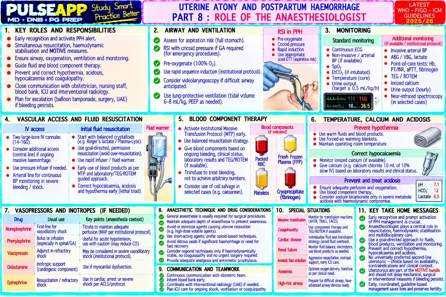
A. Principles of NMJ Stimulation2 marks

Nerve stimulation: a peripheral nerve (ulnar nerve at the wrist — most common; facial, posterior tibial as alternatives) is stimulated with surface or needle electrodes delivering supramaximal current pulses (20–60 mA for 0.1–0.2 ms; calibrated to exceed the maximum that produces maximum twitch — ensures all motor units are activated)

Response measurement: the mechanical or electrical response of the innervated muscle is measured; at the adductor pollicis (ulnar nerve → thumb adduction); quantitative devices measure the force or acceleration of thumb movement with each stimulus

Principle: residual NMB reduces the amplitude or number of twitches in proportion to the degree of receptor occupancy; the specific pattern of response varies by stimulation pattern and type of block (depolarising vs non-depolarising)

B. Stimulation Patterns3 marks

Pattern Description What It Detects Clinical Application Single Single supramaximal stimulus at 0.1 Hz (every 10 sec); Overall degree of block vs baseline; T1 = 0% Basic monitoring; requires a baseline preTwitch compares height to baseline (pre-NMB) twitch height; T1 = complete block; T1 = 25% → adequate NMB measurement; less used than TOF in expressed as % of baseline intubating conditions with most NMBs practice Train-of- Four supramaximal stimuli at 2 Hz (0.5 sec intervals) TOF count: 0 = profound block; 1 = deep The primary monitoring tool; no baseline Four (TOF) every 10–15 seconds; counts responses (TOF count = 0, block; 2–3 = moderate block; 4 = recovering; needed (counts 0–4 twitches); TOFR is the 1, 2, 3, or 4); measures ratio T4/T1 = TOFR TOFR: ratio of 4th to 1st twitch; TOFR <0.9 = criterion for safe extubation; fade pattern clinically significant residual block; TOFR (T4<T1) is pathognomonic for NON≥0.9 = adequate recovery depolarising block; no fade in depolarising (Phase I) block Double Two short bursts of 3 stimuli each at 50 Hz, separated by Detects residual block at TOFR 0.6–0.9 Useful for tactile/visual assessment when Burst 750 ms; easier to detect fade visually than TOF because better than TOF tactile assessment; fade in quantitative monitors unavailable; better Stimulation two larger responses are compared rather than four DBS (second response weaker than first) sensitivity than TOF for detecting residual (DBS) smaller ones indicates residual block block near extubation Tetanic Sustained high-frequency stimulation (usually 50 Hz for 5 Fade to tetanus indicates non-depolarising Confirms non-depolarising block character; Stimulation seconds); produces sustained muscle contraction block; sustained tetanus indicates either no used before PTC assessment (tetanus (50 or 100 (tetanus); in the presence of NON-depolarising block: block or Phase I depolarising block; NOT mobilises ACh stores for post-tetanic Hz) tetanic stimulation is not sustained → FADE of the tetanic routinely used (painful in awake patients); potentiation) contraction; in DEPOLARISING block (Phase I): no fade used in research Post- A 50 Hz tetanic stimulus for 5 seconds → 3-second Used when TOF count = 0 (no response to Guides decision between routine reversal Tetanic pause → single twitches at 1 Hz counted; the tetanic TOF); PTC 1–2 = deep block (→ (TOF≥2 → sugammadex 2 mg/kg) and deep Count stimulation temporarily mobilises ACh → post-tetanic sugammadex 4 mg/kg for reversal; reversal (PTC 1–2 → sugammadex 4 (PTC) potentiation → some twitches visible that were not neostigmine inadequate); PTC 0 = profound mg/kg) present before tetany; count the number of single block; PTC >10 = recovery imminent (TOF twitches detectable count about to reappear)

C. Qualitative vs Quantitative Monitoring3 marks

Feature Qualitative (Tactile/Visual) Quantitative (Acceleromyography — TOF-Watch) Method Anaesthesiologist feels or sees the response to TOF stimulation; Accelerometer attached to the thumb; piezoelectric transducer measures estimates fade by comparing T4 to T1 feel acceleration of thumb adduction; outputs TOFR as a number (0–1.0) TOFR threshold Fade detected only when TOFR <0.4 (60% of receptor occupancy); Provides exact TOFR; can detect TOFR 0.7, 0.8, 0.9 — the clinically for detection at TOFR 0.4–0.9 — "feels normal" to the clinician despite clinically dangerous range that qualitative monitoring misses completely dangerous residual block Clinical Patients extubated with TOFR 0.4–0.9 → postoperative residual Confirms TOFR ≥0.9 before extubation → eliminates PORC; Grosseconsequence of curarisation (PORC) → impaired airway protection, aspiration, Sundrup Lancet 2012: sugammadex + quantitative monitoring → significantly limitation hypoventilation, PTSD reduced pulmonary complications Evidence-based Insufficient for routine practice ASRA/AHA consensus 2018: quantitative monitoring should be the standard recommendation of care whenever NMBs are used; endorsed by major anaesthesia societies globally
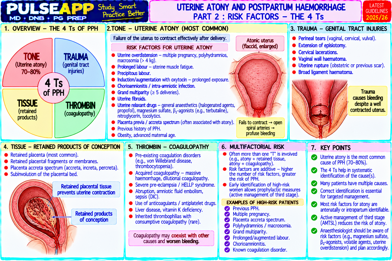
D. Specific Monitoring Issues2 marks

Temperature effect: cool fingers → reduced acceleromyography signal → falsely low apparent block may be underestimated; ensure hand and monitoring site are warm; consider TOF at the corrugator supercilii (facial nerve) — less temperature-sensitive and more relevant for the diaphragm/airway muscles than adductor pollicis (which is more resistant to NMB than facial muscles)

Neuromuscular junction variability: different muscles have different sensitivities; adductor pollicis is MORE RESISTANT to NMB than the diaphragm and genioglossus; paradox: if TOF is measured at the thumb and TOFR = 0.7, the critical airway muscles (genioglossus, pharynx) may have TOFR = 0.4–0.5 → inadequate for safe extubation; this is why the TOFR threshold for extubation must be ≥0.9 at the adductor pollicis to ensure airway muscles are adequately recovered

🎤 Viva Corner
Q. At end of LSCS, the TOF count is 3. Should you give neostigmine or sugammadex?
TOF count of 3 means three out of four twitches are detectable, indicating moderate residual non-depolarising block. Both neostigmine and sugammadex can be used at TOF count 3, but they differ significantly in reliability and speed. Neostigmine at TOF count 2–3: administer 0.04–0.07 mg/kg IV with glycopyrrolate 0.2 mg per 1 mg neostigmine; at TOF count 3, neostigmine may achieve adequate reversal (TOFR ≥0.9) in approximately 10–15 minutes in most patients, but residual block (TOFR <0.9) persists in a significant minority even with full neostigmine doses — particularly relevant in a post-CS patient who needs a safe airway for potential aspiration risk. Sugammadex 2 mg/kg (for TOF ≥2): achieves TOFR ≥0.9 reliably in >98% of patients within 3 minutes regardless of depth of block; faster, more reliable, no need for anticholinergic co-administration. For this LSCS patient: I would use sugammadex 2 mg/kg IV — the combination of a full-stomach aspiration risk (post-CS patients are not reliably fasted, have raised gastric pressure from postpartum uterine contractile activity) and the need for rapid reliable reversal to TOFR ≥0.9 before extubation makes sugammadex the superior choice. I must also counsel the patient on the OCP interaction (use additional contraception for 7 days). Confirm TOFR ≥0.9 with quantitative acceleromyography before extubation.
★ Examiner's Pearl
The five stimulation patterns (single twitch/TOF/DBS/tetanus/PTC) must all be described with their specific use — PTC is specifically for TOF count = 0 (deep block) and guides the 4 mg/kg sugammadex dose decision. The qualitative vs quantitative comparison with the specific threshold (qualitative detects fade only at TOFR <0.4; clinically dangerous TOFR 0.4–0.9 is invisible to tactile assessment) is the central safety argument for quantitative monitoring. TOF fade pattern in NDMR vs no fade in depolarising block is a mechanistic pharmacology distinction tested in written papers.
Naguib M et al. Consensus Statement on Perioperative Use of NMJ Monitoring (Anesthesiology 2018;128:1021-1049). Murphy GS et al. Residual NMB is associated with impaired clinical recovery (Anesth Analg 2010). Grosse-Sundrup M et al. (Lancet 2012;380:1273-1281). Miller RD et al. Miller's Anaesthesia, 9th Ed, Chapter 35.
lock Protected Clinical Document • Pulse App • Screenshots Strictly Prohibited
QUESTION 50 person Asked by .
bookmark_add

Briefly describe sugammadex mechanism, depth-specific doses (2/4/16 mg/kg), role in CICO emergency, and contraindications or interactions including renal failure and oral contraceptives.

collections Question Diagrams & Reference Images (1)
Question Reference Diagram
zoom_in View Image
description Clinical Response
⚙ Core Concept
Sugammadex is a modified gamma-cyclodextrin that physically encapsulates aminosteroid NMBs (rocuronium/vecuronium) into an inert inclusion complex, removing them from the NMJ — providing complete, reliable reversal at ANY depth of block in 2–3 minutes. This transforms the rocuronium RSI into a "fast-on, fast-off" technique comparable to succinylcholine but without its dangerous contraindications. (Bridion PI; DAS 2015; Naguib M — Anesthesiology 2007)
A. Mechanism1 mark

8 glucose units in a ring (gamma-cyclodextrin); hydrophobic core encapsulates the steroidal nucleus of rocuronium or vecuronium → 1:1 high-affinity inclusion complex (Kd ~10⁻²⁵ mol/L); free plasma rocuronium concentration falls → rocuronium dissociates from NMJ receptors → NMJ function restored; no receptor activity — purely pharmacokinetic mechanism; inactive complex excreted renally

B. Depth-Specific Dosing2 marks

Clinical Situation NMJ Monitor Dose Expected Recovery Routine reversal (moderate block) TOF count ≥2 2 mg/kg actual body weight TOFR ≥0.9 within ~3 min Deep block reversal PTC 1–2 (TOF count = 0) 4 mg/kg actual body weight TOFR ≥0.9 within ~3 min

CICO emergency — immediate Within 3 min of rocuronium 1.2 mg/kg RSI 16 mg/kg actual body weight (push Spontaneous ventilation restored in ~2– reversal dose dose) 3 min

C. Special Considerations2 marks

Issue Management Renal failure (eGFR <30) Complex excreted renally; accumulation with renal failure may cause delayed re-paralysis; use with caution; extended monitoring ≥24 hours; dialysis removes complex Oral contraceptives Progesterone may bind cyclodextrin cavity → reduced contraceptive efficacy equivalent to missing one OCP dose; advise additional barrier (OCP) contraception for 7 days post-administration Toremifene (breast High cyclodextrin affinity → displaces rocuronium → delayed/incomplete reversal; avoid sugammadex; use neostigmine or alternative NMB cancer treatment) Re-paralysis after 16 Sugammadex still present for hours; subsequent rocuronium/vecuronium doses will be unpredictably bound; if NMB needed within 24 hours mg/kg dose → use cisatracurium (not encapsulated by sugammadex)

🎤 Viva Corner
Q. Why is 16 mg/kg recommended for the CICO scenario specifically, and not 4 mg/kg which reverses deep block?
The CICO dose (16 mg/kg) is 4× the deep block dose (4 mg/kg) because the pharmacokinetic situation immediately after an RSI dose is fundamentally different from a deep block during ongoing anaesthesia. At 3 minutes after rocuronium 1.2 mg/kg, the plasma rocuronium concentration is at its absolute peak — the drug has been administered as a bolus and has distributed only to the rapidly-equilibrating central compartment and muscle; it has not yet distributed to fat. This peak plasma concentration is far higher than the plasma concentration present when a deep block (PTC 1–2) is confirmed after ongoing redistribution of a maintenance dose. The sugammadex must provide a sufficiently large molar excess to immediately encapsulate all the rocuronium present at this high peak plasma concentration AND leave enough free sugammadex to maintain a binding equilibrium that drives any remaining NMJ-bound rocuronium off the receptor. A molar ratio of approximately 2:1 (sugammadex:rocuronium) is needed for clinical reversal; 16 mg/kg provides approximately a 15–20:1 molar excess over the rocuronium from a 1.2 mg/kg RSI dose, ensuring immediate and complete encapsulation. At 4 mg/kg, reversal of a post-RSI dose would be incomplete — residual high plasma concentrations of unencapsulated rocuronium would continue to block the NMJ despite the sugammadex dose, potentially delaying recovery of spontaneous ventilation in the critical CICO scenario.
★ Examiner's Pearl
All three doses (2/4/16 mg/kg) with their exact NMJ monitoring triggers and expected recovery times must be reproduced as a table. The OCP interaction (equivalent to missing one dose → 7 days additional contraception) is specifically testable as a patient counselling safety point. "Actual body weight" not IBW for dosing — specifically tested for obese patients.
Bridion prescribing information 2021. Naguib M. Sugammadex (Anesth Analg 2007;104:575). DAS Guidelines 2015 (BJA 2015;115:827). Miller RD et al. Miller's Anaesthesia, 9th Ed, Chapter 34.

Showing 4150 of 337 questions

account_tree

Subcategory Tree

Explore Anesthesia subcategories
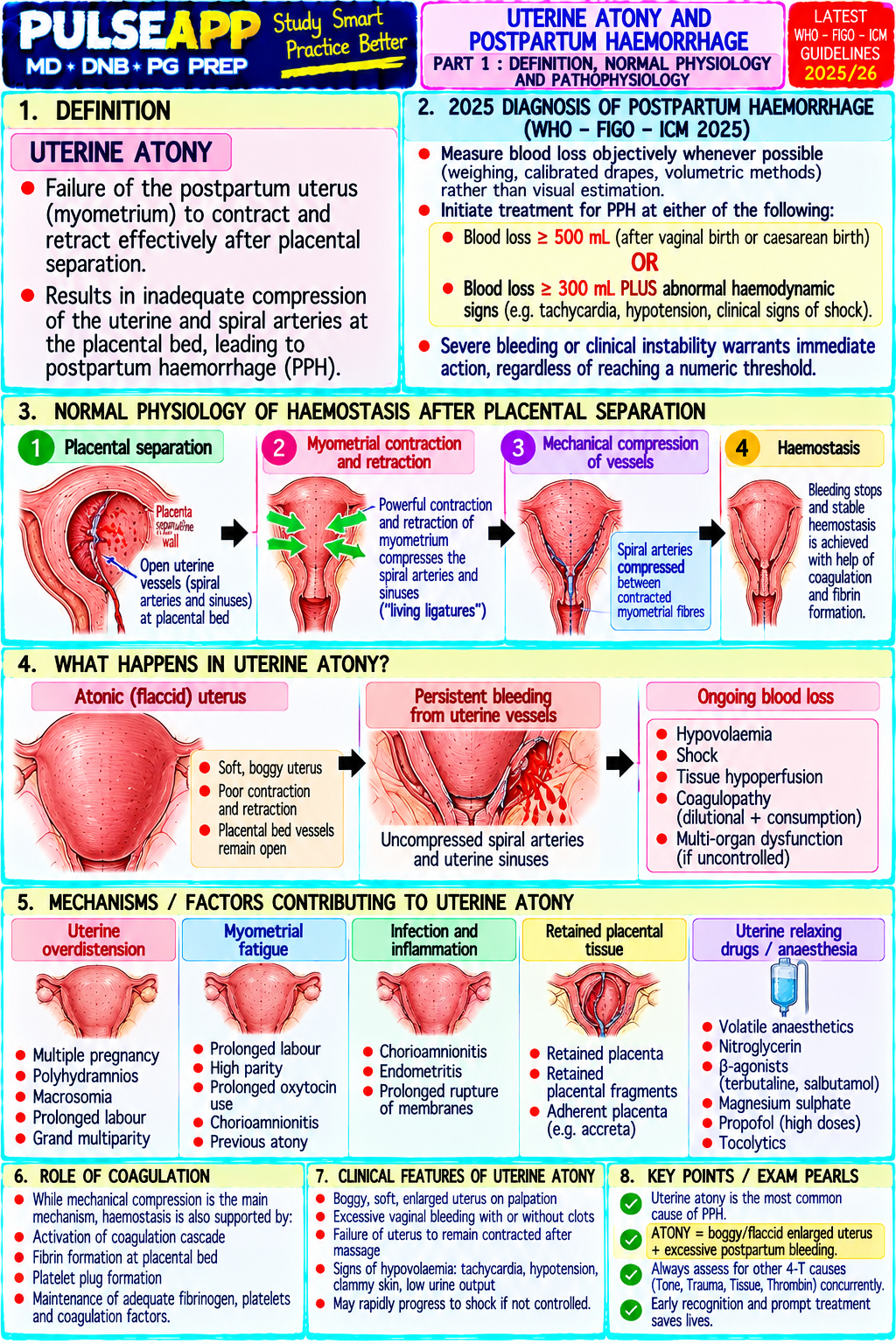
folder_special Anesthesia
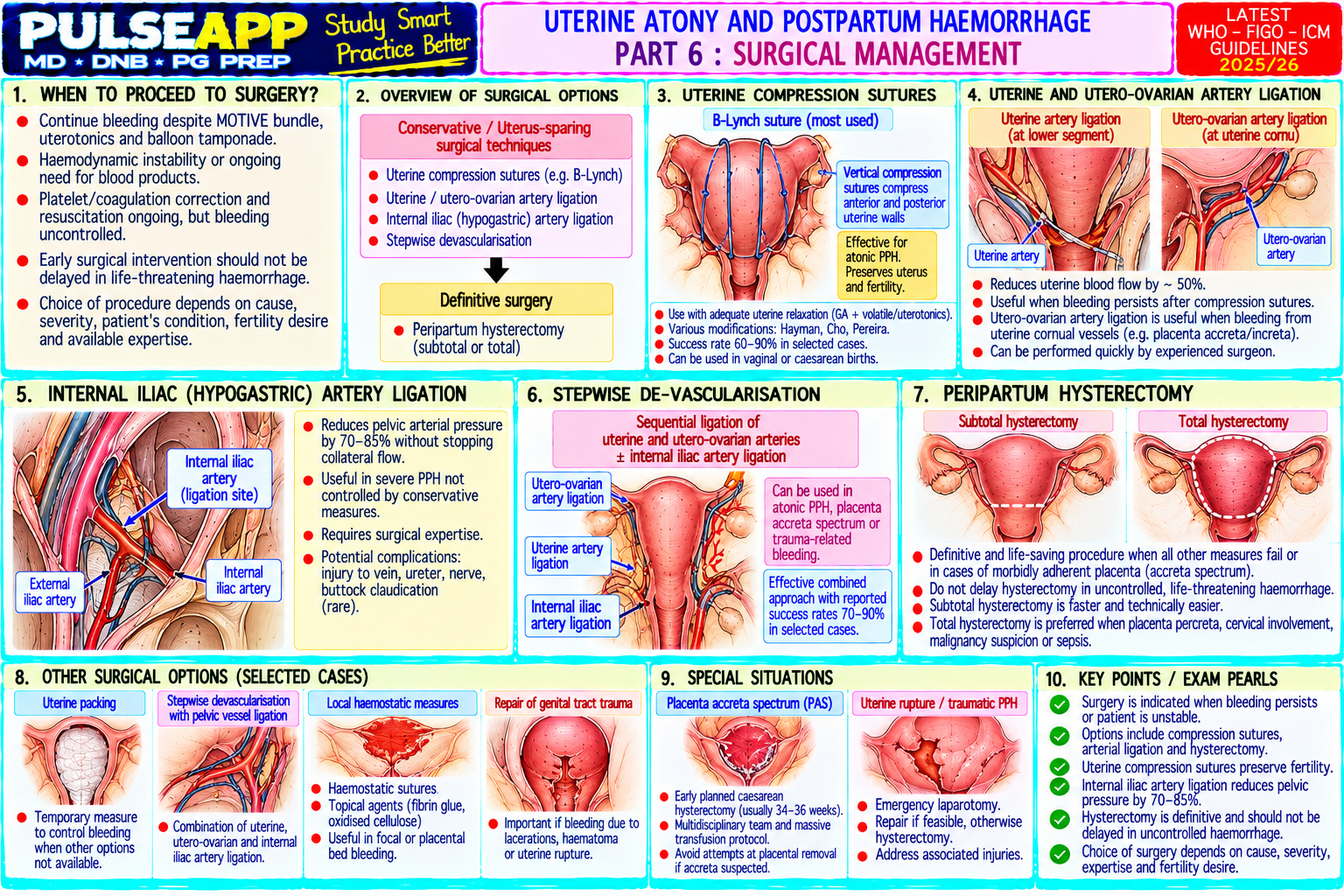
Main
lock

Category Subscription

Subscribe to Anesthesia to unlock this module and all nested subcategories.

  • check_circle Access Anesthesia & all subcategories
  • check_circle Detailed, Peer-Reviewed Answers
  • check_circle High-yield visual aids & imaging
Get Category Subscription arrow_forward
Secure 256-bit SSL Connection

Anatomical Models

Explore high-fidelity 3D visualizations included in premium modules.

Case Reviews

Real-world clinical scenarios narrated by senior consultants.

Pulse App