Anesthesia | Eklavya Medical
vaccines
medical_services Main Specialty Domain lock Subscription Required

Anesthesia

Clinical anesthesiology, local and general anesthesia delivery systems, monitoring, and perioperative care.

verified Verified Medical Faculty menu_book 337 Q&A Modules
lock Protected Clinical Document • Pulse App • Screenshots Strictly Prohibited
QUESTION 71 person Asked by .
bookmark_add

Define the ERAS concept and its physiological rationale. Describe the pre-operative, intraoperative, and postoperative components of a colorectal ERAS programme. Summarise the evidence base and outcomes achieved with ERAS implementation.

collections Question Diagrams & Reference Images (2)
Question Reference Diagram
zoom_in View Image
Question Reference Diagram
zoom_in View Image
description Clinical Response
⚙ Core Concept
ERAS (Enhanced Recovery After Surgery) is a multimodal, evidence-based perioperative care pathway designed to reduce the surgical stress response, maintain physiological function, and facilitate early return to normal activity — replacing the traditional "nil by mouth from midnight, bed rest, opiate analgesia, long hospital stay" model with a physiologically rational approach. Implemented programmes consistently reduce hospital length of stay by 30– 50% and reduce complication rates by 30–40% without increasing readmission rates. (Kehlet H — Lancet 1997; ERAS Society Guidelines; Gustafsson UO et al.; Varadhan KK — BJA 2010)
A. Physiological Rationale — Reducing the Surgical Stress Response2 marks

Traditional care pathways allowed or amplified the surgical stress response: prolonged fasting → catabolism, insulin resistance; hypothermia → impaired coagulation and immune function; large-volume IV fluids → bowel oedema and anastomotic leak; opioid analgesia → ileus, nausea, sedation, respiratory depression; immobility → DVT, deconditioning, pneumonia

ERAS systematically addresses each factor: carbohydrate loading reduces pre-operative insulin resistance by 50%; targeted fluid therapy prevents both hypovolaemia and fluid overload; regional analgesia eliminates systemic opioids; early feeding and mobilisation restore gut motility and physical function; the combined effect reduces the magnitude of the catabolic stress response from major surgery, allowing early functional recovery Kehlet's original concept (1997): Henrik Kehlet (Lancet 1997) demonstrated that major colonic surgery could be performed with a 2-day hospital stay using multimodal opioid-sparing analgesia, early oral nutrition, and early mobilisation — the founding evidence for ERAS

B. ERAS Components — Colorectal Programme6 marks

Phase ERAS Component Evidence/Mechanism Pre- Patient education and counselling Patients who understand the ERAS plan and their role in recovery comply better with early feeding and mobilisation; operative anxiety reduction → lower analgesic requirements Preoperative carbohydrate loading Reduces insulin resistance by 50%; reduces preoperative thirst/hunger/anxiety; improves postoperative muscle (200–400 mL 12.5% carbohydrate function; supported by Cochrane review — reduces hospital stay by 0.5–1 day drink, 2–3 hours before surgery) Clear fluid fasting up to 2 hours Evidence-based replacement for "NPO from midnight"; no increased aspiration risk; reduces preoperative (solid food up to 6 hours) discomfort; reduces catabolism from prolonged fasting Bowel preparation (OMIT for most Traditional mechanical bowel prep dehydrates patients and worsens electrolyte imbalance without reducing colorectal surgery) anastomotic leak rates (Cochrane 2011) — omitted in most ERAS colorectal protocols Anaemia assessment and Preoperative anaemia is a major risk factor for transfusion and complications; correcting Hb ≥100 g/L reduces treatment (if Hb <100 g/L → iron transfusion requirement and length of stay supplementation or erythropoietin if time allows) Intra- TIVA or volatile anaesthesia with PONV causes delayed oral intake and prolonged bed rest — specifically addressed in ERAS; TIVA reduces PONV operative multimodal PONV prophylaxis (min. 2 25–30%; dexamethasone also reduces fatigue and pain agents: ondansetron + dexamethasone) Avoidance of long-acting opioids; Intraoperative opioids contribute to postoperative ileus; remifentanil provides precise intraoperative analgesia with remifentanil intraoperative immediate offset; no contribution to postoperative ileus when stopped at end of surgery; regular NSAIDs and paracetamol for post-op baseline analgesia

Goal-directed fluid therapy (GDT) OPTIMISE trial: ODM-GDT reduced postoperative complications by 20%; avoids both hypovolaemia (anastomotic — ODM or FloTrac guided ischaemia) and hypervolaemia (bowel oedema, anastomotic leak, cardiorespiratory complications); targeted fluid to optimise SV; vasopressors for vasodilatory hypotension rather than volume Temperature maintenance Hypothermia increases wound infection, coagulopathy, cardiac events; active warming — forced-air blanket, warm (normothermia ≥36.5°C) fluids, warm gases, theatre temperature 21–22°C; NICE recommends core temp >36°C throughout surgery

Minimally invasive surgery Laparoscopic colorectal surgery: reduced pain, faster bowel recovery, shorter hospital stay vs open; smaller (laparoscopic vs open) incisions → less incisional pain → less splinting → better respiratory function; does not eliminate the need for ERAS (laparoscopic + ERAS = best outcomes) Thoracic epidural or paravertebral Thoracic epidural analgesia for open colorectal: reduces opioid requirements, allows early mobilisation, reduces block for open surgery; TAP block ileus, reduces PPCs; for laparoscopic: bilateral TAP blocks + NSAIDs + paracetamol replaces epidural for laparoscopic Post- Early oral nutrition (day 0 — same Traditionally, patients were "nil by mouth until bowel sounds return" — no evidence base for this; early oral nutrition operative day as surgery) maintains gut mucosal integrity, reduces infection risk, reduces ileus by stimulating gut motility via the gastrocolic reflex; NGT removal before end of surgery; clear fluids within 2–4 hours of operation; diet as tolerated from day 1 Early mobilisation (out of bed day Immobility → DVT, deconditioning, pneumonia, prolonged ileus; early ambulation reverses these; supervised 1, with target of 2 hours out of bed physiotherapy from day 1; functional milestones set daily increasing daily) Opioid-sparing analgesia — Opioids → ileus + respiratory depression + PONV; multimodal opioid-sparing analgesia maintains adequate pain NSAIDs + paracetamol + regional ± control while dramatically reducing opioid-related complications low-dose oral opioid PRN Urinary catheter removal day 1 (if Urinary catheter delays mobilisation and increases UTI risk; removed as soon as epidural is stopped or day 1 if no no epidural) epidural IV fluid discontinuation / removal IV fluids → sedentary behaviour + fluid overload; oral fluids maintain hydration without restricting mobilisation of IV cannula by day 1–2

C. Evidence and Outcomes2 marks

Varadhan KK et al. (BJA 2010, meta-analysis): ERAS for colorectal surgery reduces LOS by 2.5 days (from 7.5 to 5.0 days average) and reduces complication rate by 30% vs traditional care; no increase in readmission rates — demonstrating that early discharge is safe

Gustafsson UO et al. (Arch Surg 2011): higher compliance with individual ERAS elements correlates directly with better outcomes — the benefit is additive; implementing only some elements produces partial benefit; full protocol compliance produces maximal benefit

Cost savings: reduced LOS → significant cost savings per patient; ERAS implementation has positive return on investment even accounting for the cost of multidisciplinary programme establishment

ERAS Society: publishes specialty-specific guidelines for colorectal, gynaecological, liver, bariatric, thoracic, and urological surgery; all follow the same core principles adapted for specific surgical and physiological contexts

🎤 Viva Corner
Q. A surgeon argues that his patient needs "at least 4 litres of Hartmann's solution" intraoperatively for bowel surgery because of the large "third space" losses. How do you respond using ERAS principles?
The concept of large "third space" losses as a mandate for high-volume fluid administration during bowel surgery is not supported by contemporary evidence and is actually one of the specific physiological misconceptions that ERAS was designed to correct. The "third space" theory (first proposed by Shires et al. in the 1960s based on studies now recognised as methodologically flawed) postulated that major abdominal surgery created a large functional extracellular fluid deficit that required replacement with 10–20 mL/kg/hr of crystalloid. Modern physiological evidence has demonstrated that this "third space" does not exist in the way originally described — the fluid is not lost from the intravascular compartment into a non-functional space that must be replaced; rather, crystalloid given in excess of actual deficit redistributes to the interstitium, causing bowel wall oedema (increasing anastomotic leak risk and prolonged ileus), pulmonary oedema, impaired wound healing, and delayed recovery. The ERAS approach replaces high-volume crystalloid with Goal-Directed Fluid Therapy: intraoperative fluid is administered in 250 mL boluses, guided by a stroke volume monitor (oesophageal Doppler or FloTrac/Vigileo), only when the patient demonstrates fluid responsiveness (stroke volume increases >10% with each bolus); vasopressors (phenylephrine or noradrenaline) are used instead of fluids for vasodilatory hypotension from regional anaesthesia or volatile agents. The OPTIMISE trial demonstrated that this GDT approach reduces postoperative complications by 20% compared to standard care. A reasonable estimate of intraoperative fluid for a 3-hour laparoscopic bowel resection with ERAS and GDT would be approximately 500–1000 mL total (replacing estimated insensible losses at 1–2 mL/kg/hr plus responding to demonstrated fluid responsiveness) — dramatically less than 4 litres of crystalloid, with substantially better outcomes.
★ Examiner's Pearl
The three-phase ERAS structure (pre-op/intra-op/post-op) with three elements in each phase is the comprehensive answer format expected — partial answers covering only the intraoperative phase lose significant marks. Carbohydrate loading (12.5% maltodextrin, 2–3 hours pre-op, reduces insulin resistance 50%) is the pre-operative fact most specifically tested. Goal-directed fluid therapy with ODM (OPTIMISE trial — 20% complication reduction) is the intraoperative evidence. Early oral nutrition same day (no evidence for "nil until bowel sounds") is the most important postoperative paradigm shift.
Kehlet H. Multimodal approach to control postoperative pathophysiology and rehabilitation (BJA 1997;78:606-617). Varadhan KK et al. ERAS and conventional perioperative care — meta-analysis (BJA 2010;104:401-408). Gustafsson UO et al. ERAS adherence and outcome after colonic cancer surgery (Arch Surg 2011;146:571-577). Pearse RM et al. OPTIMISE trial (BMJ 2014;348:g2082). ERAS Society Guidelines for perioperative care in colonic surgery 2018.
lock Protected Clinical Document • Pulse App • Screenshots Strictly Prohibited
QUESTION 72 person Asked by .
bookmark_add

Define multimodal analgesia and explain its pharmacological rationale. Describe the analgesic ladder, the WHO/ERAS analgesic pyramid, and specific drugs (paracetamol, NSAIDs, gabapentinoids, ketamine, regional techniques) used at different levels. Describe PCA — mechanism, programming parameters, safety features, and monitoring requirements.

collections Question Diagrams & Reference Images (3)
Question Reference Diagram
zoom_in View Image
Question Reference Diagram
zoom_in View Image
Question Reference Diagram
zoom_in View Image
description Clinical Response
⚙ Core Concept
Multimodal analgesia is the simultaneous administration of analgesic drugs from different pharmacological classes targeting different pain pathways — thereby producing additive or synergistic analgesic effects while allowing lower doses of each individual agent, reducing dose-related side effects compared to single-agent analgesia at equivalent doses. It is the cornerstone of modern ERAS programmes and is specifically more effective and safer than opioid monotherapy for acute postoperative pain. (Kehlet H, Dahl JB — Lancet 1993; Buvanendran A — Pain 2010; White PF; PROSPECT guidelines; APS Practice Guidelines)
A. Rationale — Why Multimodal is Superior to Single-Agent Analgesia2 marks

The nociceptive cascade has multiple targets: pain from surgical tissue injury involves: peripheral sensitisation (PGs, bradykinin, substance P at the wound — target: NSAIDs, local anaesthetics); ascending spinal transmission (dorsal horn — target: opioids, α₂ agonists, ketamine); central processing (thalamocortical — target: paracetamol, opioids, gabapentinoids); descending modulation (from brainstem — target: SNRIs, α₂ agonists); no single drug blocks ALL of these simultaneously

Synergy: paracetamol + NSAID produces significantly better analgesia than either alone at the same individual doses; opioid + NSAID shows opioid-sparing effect of 30–40% (less opioid needed for equivalent analgesia); combination allows adequate pain control with individually lower (and safer) doses of each drug

Opioid-sparing: reducing systemic opioid consumption reduces: PONV, sedation, respiratory depression, ileus, urinary retention, cognitive impairment, and opioid-induced hyperalgesia — all of which are major barriers to early recovery in ERAS

B. Multimodal Analgesic Pyramid — Drugs and Mechanisms4 marks

Level Drug / Technique Mechanism Dose/Route Key Evidence Foundation Paracetamol Inhibits prostaglandin synthesis centrally 1 g IV/oral every 6 hours; Reduces opioid consumption 20% vs placebo; (all (acetaminophen) (COX-3 in CNS); also modulates descending reduce dose in liver well tolerated; no platelet, renal, or GI effects; patients) serotonergic inhibitory pathways; DOES NOT disease and <50 kg body should be prescribed regularly for ALL inhibit peripheral COX (no anti-inflammatory weight postoperative patients unless contraindicated effect in tissues) Foundation NSAIDs (ibuprofen, COX-1 and COX-2 inhibition → ↓ Ibuprofen 400–600 mg Opioid-sparing 25–35% vs placebo; celecoxib (all diclofenac, ketorolac, prostaglandin synthesis BOTH peripherally TID oral; ketorolac 15–30 equivalent to non-selective NSAIDs for pain; patients) celecoxib) (reduces sensitisation of nociceptors at mg IV every 6 hours avoid in renal impairment (↓GFR from PG wound) AND centrally (reduces PG-mediated (max 5 days); celecoxib inhibition in kidney); avoid in active peptic ulcer; central sensitisation in dorsal horn) 200–400 mg oral avoid in coagulopathy; avoid in cardiovascular (selective COX-2 — high-risk patients (long-term COX-2 selective) better GI safety) Second Gabapentinoids Bind α₂δ subunit of voltage-gated Ca²⁺ Gabapentin 300–600 mg Reduces acute postoperative pain scores by 20– level (gabapentin, channels on presynaptic terminals in dorsal oral preoperatively + 300 30%; reduces opioid consumption 30–40%; pregabalin) horn → ↓ Ca²⁺ influx → ↓ release of mg TID for 2–3 days prevents chronic post-surgical pain development glutamate, substance P, and CGRP → post-op; pregabalin 75– (prevention of central sensitisation); sedation reduces central sensitisation and neuropathic 150 mg oral BD and dizziness at higher doses — dose reduction pain component; also supraspinal anxiolytic needed in elderly and renal impairment effects Second Ketamine (sub- NMDA antagonist → blocks central 0.1–0.5 mg/kg IV loading Cochrane meta-analysis: intraoperative ketamine level anaesthetic) sensitisation ("wind-up") at the spinal cord then 0.1–0.2 mg/kg/hr reduces 24-hour morphine consumption by level; specific efficacy against opioid- infusion for 48 hours; or approximately 8 mg (30%) and reduces resistant and neuropathic pain components; 0.5 mg/kg IV at wound postoperative pain scores at rest and on ALSO reduces opioid tolerance development closure movement; particularly beneficial for major (prevents opioid-induced hyperalgesia) surgery (thoracotomy, major orthopedic) Third level Opioids (morphine, Mu-opioid receptor agonism in brain and Morphine: 0.1 mg/kg IV; Should NOT be used as the sole agent; ERAS (for oxycodone, tramadol, spinal cord; reduces pain perception; PCA 1 mg bolus with 5- philosophy: regular paracetamol + NSAID ± moderate- codeine) sedation; respiratory depression; min lockout; oxycodone gabapentinoid as baseline; opioids as PRN severe constipation and ileus (GI opioid receptors); 5–10 mg oral 4–6 hourly; rescue for breakthrough pain only, not as the pain) tolerance with prolonged use; physical and tramadol 50–100 mg TID primary analgesic psychological dependence risk (weak opioid + SNRI effect) Regional Epidural, nerve blocks Local anaesthetic blockade of afferent nerve Site-specific techniques; Thoracic epidural analgesia = gold standard for techniques (TAP, paravertebral, conduction → complete interruption of standard blocks major abdominal surgery; reduces systemic (opioid- femoral, popliteal, etc.), nociceptive transmission from the surgical described in separate opioid requirement to near-zero; best PPCs free wound infiltration with site; the most powerful analgesic intervention questions reduction; PROSPECT guidelines provide analgesia) bupivacaine/ropivacaine available procedure-specific recommendation for which block is evidence-based for each operation

C. Patient-Controlled Analgesia (PCA)4 marks

Principle and Programming

Concept: patient activates a syringe pump (by pressing a button) to self-administer a pre-set bolus dose of IV opioid; the system prevents overdose through lockout intervals; the patient "titrates" to their own analgesic requirement, accounting for interindividual pharmacokinetic variability that makes fixed-dose nurseadministered regimens suboptimal Standard Adult Parameter Setting Rationale (Morphine) Bolus dose 1–2 mg morphine Sufficient for analgesic effect in most adults; enough to "feel" the bolus working Lockout 5 minutes Allows peak effect of each bolus before next dose can be activated; prevents double-stacking; morphine Tmax ~5–10 min after IV interval dose Background NOT routinely Background infusion removes the self-limiting safety feature — if a patient falls asleep (indicating sufficient analgesia/sedation), infusion recommended for they stop pressing the button; background infusion continues regardless → respiratory depression while sedated; ONLY use opioid-naive adults background infusion in opioid-tolerant patients with careful monitoring 4-hour limit 20–30 mg/4 hours Prevents very high total opioid consumption; triggers review if frequently hit

Safety Features

Lockout interval (primary safety): prevents re-dosing within the lockout period even if button pressed repeatedly 4-hour limit: nurse reviews if maximum 4-hour dose is reached; may indicate inadequate analgesia requiring reassessment of the regimen

Anti-siphon valve: prevents gravity-assisted free-flow of opioid from the syringe if the pump is placed below patient level

Tamper-evident syringe: prevents drug diversion

One-way valve on the dedicated IV line: prevents bolus of opioid being pushed backward into a concurrent running IV line (avoiding inadvertent bolusing) Monitoring Requirements for PCA Continuous SpO₂ monitoring throughout PCA use (standard of care — most guidelines); hourly sedation score; hourly RR; 4-hourly pain score; only the patient must press the PCA button — family or staff pressing the button on behalf of the patient ("PCA by proxy") bypasses the self-limiting safety feature and causes respiratory arrest
🎤 Viva Corner
Q. Why is a background infusion of opioid generally NOT recommended in opioid-naive patients using IV PCA morphine?
The PCA system's fundamental safety mechanism is the negative feedback loop between opioid effect and patient demand: when the patient is in pain, they press the button to receive an analgesic bolus; when the analgesic effect is adequate and the patient becomes comfortable or drowsy (sedated), they stop pressing — the system self-regulates, and opioid-induced sedation itself prevents further dosing. This pharmacological negative feedback makes PCA inherently safer than nurseadministered fixed-dose opioids, where the nurse administers a dose regardless of the patient's moment-by-moment sedation level. A background infusion (continuous low-rate opioid delivery, 0.5–1 mg/hour morphine, running regardless of whether the patient presses the button) fundamentally disrupts this safety mechanism: if the patient becomes sedated or even falls asleep from accumulated opioid effect, the background infusion continues delivering opioid at the prescribed rate; progressive opioid accumulation during the sedated period → unchecked respiratory depression → hypoxia → potentially fatal. The specific danger is that respiratory depression from opioids often occurs during sleep or deep sedation — precisely the state when the patient is NOT pressing the PCA button; without a background infusion, the system delivers nothing during this period and the patient recovers; with a background infusion, opioid continues to be administered during this most vulnerable period. Multiple studies and adverse incident reviews have shown that most PCA-related serious adverse events (respiratory arrests) occur when either a background infusion is used in opioid-naive patients OR when a third party presses the button on the patient's behalf ("PCA by proxy"). Background infusions are appropriate only in opioid-tolerant patients (those on chronic opioids who have a baseline opioid requirement that must be maintained) and should be used only with continuous SpO₂ and enhanced monitoring protocols.
★ Examiner's Pearl
The multimodal analgesic pyramid with mechanisms for all five classes (paracetamol = central COX-3/serotonergic; NSAID = peripheral + central COX inhibition; gabapentinoids = α₂δ Ca²⁺ channel → dorsal horn; ketamine = NMDA block → prevents wind-up; opioids = μ receptor) is the comprehensive pharmacological framework examiners test. PCA background infusion in opioid-naive patients — specifically NOT recommended with the mechanism (eliminates the sedation-stopsbutton-pressing safety feature) — is the most tested PCA safety fact. "PCA by proxy" (others pressing button for patient) as a specific identified dangerous practice is tested as a safety scenario.
Kehlet H, Dahl JB. The value of multimodal or balanced analgesia in postoperative pain treatment (Anesth Analg 1993;77:1048-1056). Grape S, Tramèr MR. Ketamine as adjuvant to perioperative opioids (Anaesthesia 2007;62:1093-1096). PROSPECT (Procedure-Specific Postoperative Pain Management) Guidelines 2023. Chou R et al. Management of Postoperative Pain (J Pain 2016;17:131-157). Miller RD et al. Miller's Anaesthesia, 9th Ed, Chapter 97.
lock Protected Clinical Document • Pulse App • Screenshots Strictly Prohibited
QUESTION 73 person Asked by .
bookmark_add

Describe the sonoanatomy and ultrasound-guided technique for the Transversus Abdominis Plane (TAP) block and the Quadratus Lumborum (QL) block. Compare their dermatomal coverage, indications, and relative advantages for abdominal surgery analgesia.

collections Question Diagrams & Reference Images (7)
Question Reference Diagram
zoom_in View Image
Question Reference Diagram
zoom_in View Image
Question Reference Diagram
zoom_in View Image
Question Reference Diagram
zoom_in View Image
Question Reference Diagram
zoom_in View Image
Question Reference Diagram
zoom_in View Image
Question Reference Diagram
zoom_in View Image
description Clinical Response
⚙ Core Concept
TAP and QL blocks are the dominant fascial plane blocks for abdominal wall analgesia — targeting the T6–L1 intercostal nerves as they travel between the transversus abdominis and internal oblique muscle layers. The QL block provides broader dermatomal coverage (including visceral and posterior abdominal wall components) compared to the TAP block, making it increasingly preferred for major abdominal surgery. Both are core components of ERAS programmes as opioid-sparing regional techniques. (Blanco R — TAP block; El-Boghdadly K — QL block; Kadam VR; PROSPECT 2022; Hadzic A)
A. TAP Block — Anatomy & Technique3 marks

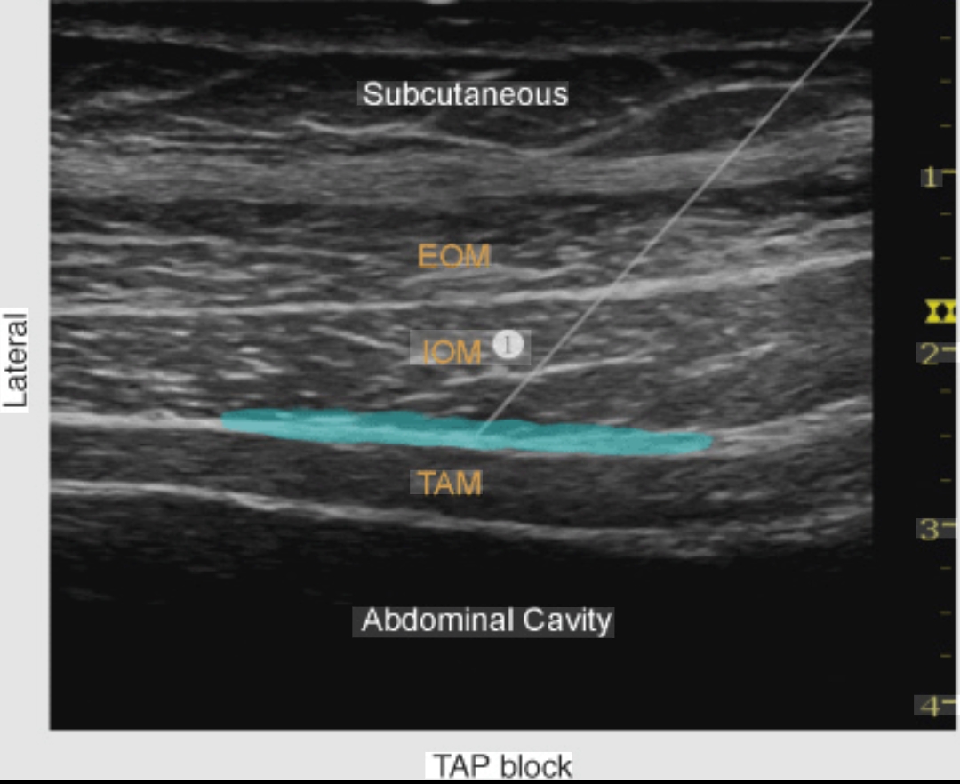
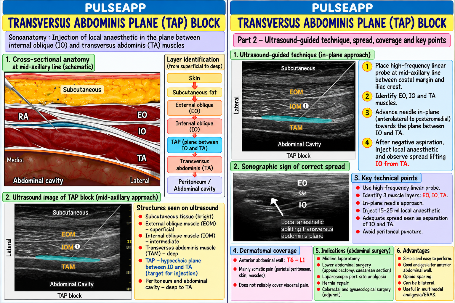
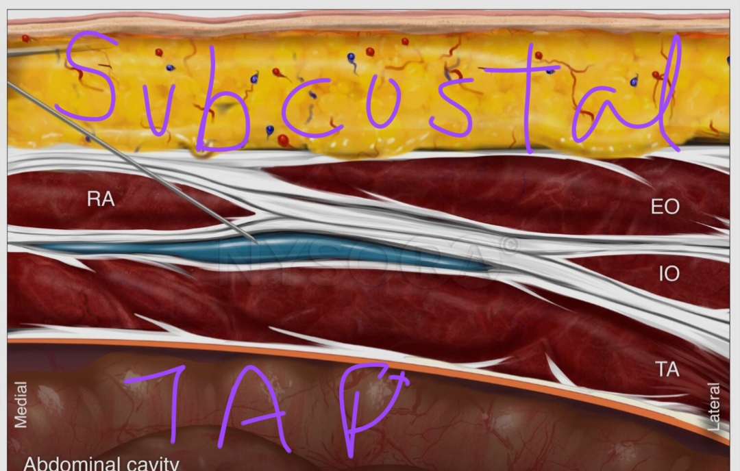
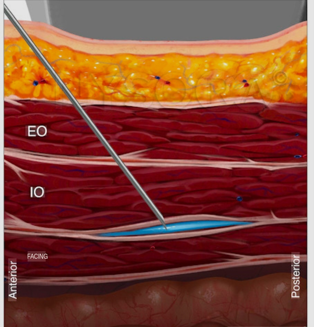
Anatomy: the lateral abdominal wall has three muscle layers: external oblique (most superficial), internal oblique (middle), and transversus abdominis (deepest, just above the peritoneum); the intercostal nerves T10–L1 (supplying the anterior abdominal wall from the umbilicus to the groin) travel within the fascial plane between the internal oblique and transversus abdominis — the TAP (Transversus Abdominis Plane); injection of LA into this plane bathes the nerve trunks as they fan out across the abdominal wall Approaches:

Lateral TAP (midaxillary approach): probe in the mid-axillary line between the iliac crest and costal margin; image the three muscle layers; inject 20 mL 0.25% bupivacaine (or 0.2% ropivacaine) into the TAP layer (between IO and TA); covers T10–L1 (lower abdomen and groin); best for lower abdominal surgery (appendicectomy, hernia repair, Pfannenstiel incision)

Subcostal TAP: probe obliquely from the xiphoid toward the ASIS; targets T6–T9 nerves in the TAP layer below the costal margin; covers upper abdomen; for laparoscopic port sites below the costal margin

Bilateral TAP: bilateral injection for midline laparotomies or bilateral laparoscopic port sites; 20 mL each side = 40 mL total (stay within LA maximum dose)

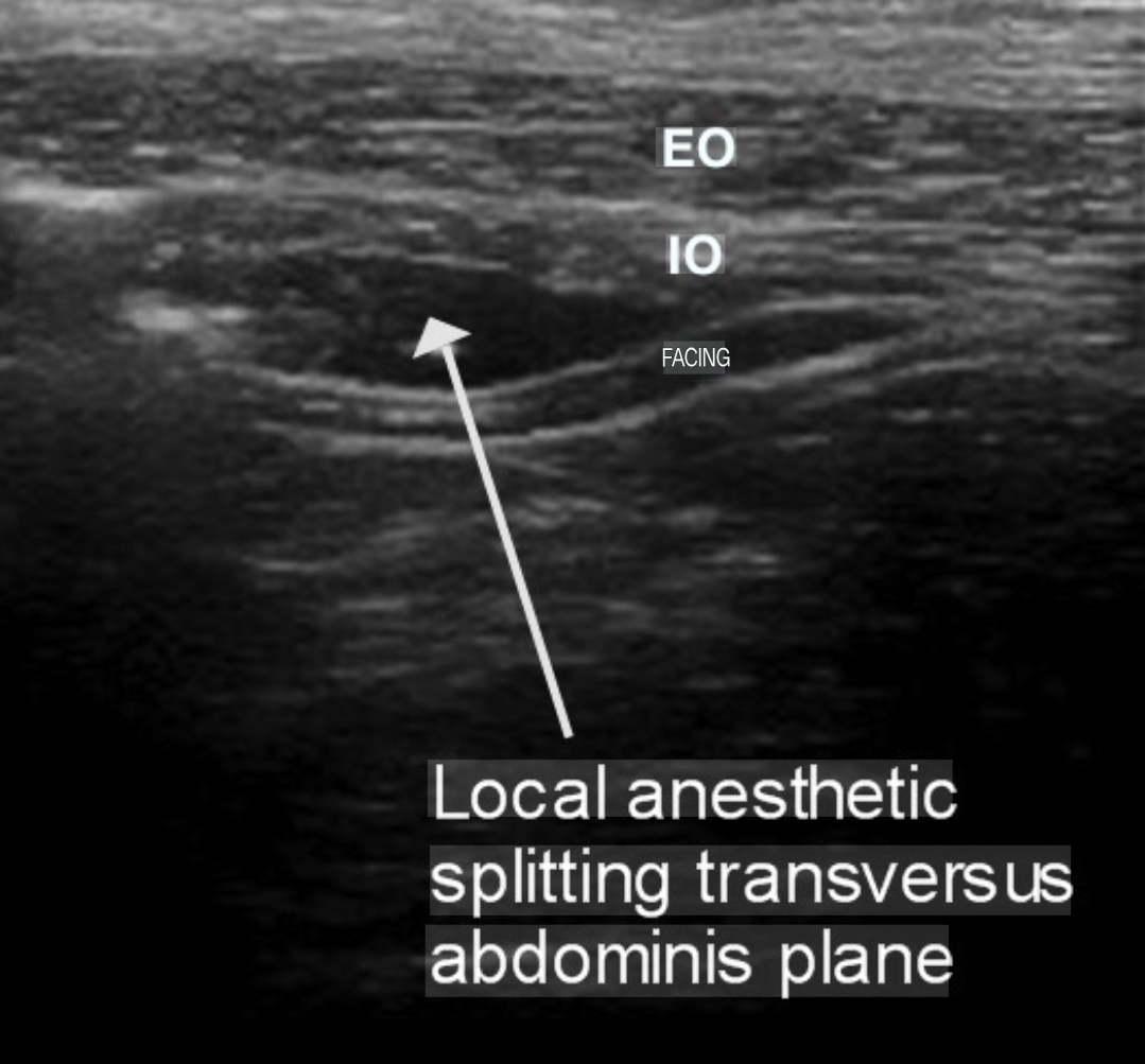
Sonoanatomy: high-frequency (12–15 MHz) linear probe; identify three muscle layers (EO = thin superficial layer; IO = middle, usually thickest; TA = deepest, most echogenic with deep fascia); the TAP lies between IO and TA; visualise the fascial plane as a bright line; inject LA to see hypoechoic spread in the plane (if "dome" appears in TA → too deep, peritoneum injected; if injection raises IO → too superficial)

B. Quadratus Lumborum (QL) Block — Types & Technique3 marks

QL Type Needle Target Coverage Indication QL1 (anterior) Anterior to the QL muscle, T10–L1 (lateral cutaneous branches); similar to lateral TAP Lower abdominal surgery, hip lateral to the transversalis surgery fascia QL2 Posterior to the QL muscle, T7–L1; better posterior cutaneous coverage than QL1; may provide Laparotomy, colectomy, renal (lateral/posterior) in the thoracolumbar fascia some visceral analgesia via epidural-like spread in the thoracolumbar surgery fascia QL3 Between the QL and psoas T5–L1; most extensive coverage; may reach the paravertebral space; Major abdominal surgery, (transmuscular) major muscles (anterior QL) provides both somatic and visceral analgesia components hysterectomy, colectomy as epidural alternative Intramuscular Within the belly of the QL Variable; less commonly used Selected cases QL muscle QL technique (lateral decubitus approach — most common): patient lateral decubitus; low-frequency curved array probe (3–5 MHz) placed on the posterior flank, lateral to the spine; identify the "shamrock sign" — the three muscles (psoas major, QL, erector spinae) meeting at the L4 transverse process like a three-leaf shamrock; place the needle tip at the correct fascial plane relative to the QL (depending on QL1, QL2, or QL3 target); inject 20–30 mL 0.375% ropivacaine or 0.25% bupivacaine; bilateral for midline surgery

C. TAP vs QL Block Comparison2 marks

Feature TAP Block QL Block Coverage T10–L1 (lateral TAP); T6–T9 (subcostal); cannot reliably T5–L1 (QL3); broader coverage including posterior abdominal wall and potentially level cover upper abdomen from a single approach visceral component via thoracolumbar fascia spread

Visceral None — purely somatic abdominal wall Some visceral analgesia from QL2/QL3 (thoracolumbar fascia spread toward analgesia paravertebral space)

Ultrasound Easier — superficial, familiar anatomy; suitable for trainees More difficult — requires identification of the shamrock sign; deeper target; more difficulty variable anatomy Duration 8–12 hours (plain bupivacaine); 24–36 hours with continuous Similar; some studies show longer duration with QL (15–16 hours) due to the fascial catheter or liposomal bupivacaine depot providing slower diffusion

Evidence Moderate — effective for port-site analgesia in laparoscopic Growing — QL3 provides superior analgesia to TAP for open colectomy in multiple for surgery; less effective than epidural for open laparotomy RCTs; increasingly used as epidural alternative when epidural is contraindicated laparotomy

D. Clinical Indications & ERAS Integration2 marks

PROSPECT recommendation 2022: bilateral TAP blocks (subcostal + lateral) recommended for laparoscopic colectomy (Grade A evidence); QL2 or QL3 block for open colectomy as alternative to epidural; single-shot TAP for Pfannenstiel CS incision analgesia (combined with intrathecal morphine)

Liposomal bupivacaine TAP: 266 mg liposomal bupivacaine (Exparel) diluted to 60 mL for bilateral TAP → provides 72-hour analgesia from a single injection; FDA-approved indication for TAP block; allows ambulatory colectomy/hysterectomy without epidural catheter management

Maximum LA dose: bilateral blocks consume twice the dose; for bilateral TAP or bilateral QL: use 0.2–0.25% ropivacaine (lower concentration × larger volume) to maintain total dose <3 mg/kg; avoid bilateral blocks with high-concentration solutions in obese patients where the total dose may approach toxic levels

🎤 Viva Corner
Q. A patient has had an open right hemicolectomy through a midline laparotomy. The surgeon asks you to perform a TAP block at the end of surgery instead of a thoracic epidural (which was declined). Would you prefer a TAP or QL block, and which specific approach?
For a midline laparotomy for right hemicolectomy, I would prefer a bilateral QL3 block over a bilateral TAP block. The reasons: the midline laparotomy incision spans from the epigastrium to the pubis, involving T6–L1 dermatomal territory. A lateral TAP block only reliably covers T10–L1 (lower abdomen); the subcostal TAP addition covers T6–T9 but requires a separate injection on each side (four injections total for complete bilateral coverage). A QL3 block on each side provides T5–L1 coverage from a single injection per side — broader somatic coverage from one needle pass. More importantly, the QL3 approach (transmuscular, between QL and psoas major) may allow some LA to spread toward the thoracolumbar fascia and potentially the paravertebral space — providing a degree of visceral analgesia that the purely somatic TAP block cannot provide; this visceral component is meaningful for a hemicolectomy where visceral pain from bowel handling and mesenteric traction contributes significantly to postoperative discomfort. Technique: bilateral QL3, with the patient in the left lateral decubitus position for the right side QL block, then repositioned for the left side; identify the shamrock sign (psoas, QL, erector spinae meeting at L4 transverse process); advance the needle through the QL muscle to position the tip between QL and psoas major; inject 25–30 mL of 0.375% ropivacaine per side (within safe dose limits); bilateral injection provides T5–L1 coverage for the entire midline incision and abdominal viscera. This would be combined with regular IV paracetamol 1 g QID, diclofenac sodium 75 mg BD (if renal function permits), and PRN oral opioid for breakthrough pain — completing the multimodal analgesia strategy.
★ Examiner's Pearl
The three muscle layers of the lateral abdominal wall (EO/IO/TA) in order from superficial to deep, with the TAP being between IO and TA, are the anatomical facts specifically tested. The QL "shamrock sign" (three muscles meeting at L4 transverse process) is the specific ultrasound landmark for QL blocks. PROSPECT 2022 recommendation (TAP for laparoscopic; QL for open laparotomy as epidural alternative) is the specific guideline evidence that demonstrates up-to-date knowledge.
Blanco R. TAP block under ultrasound guidance (Anaesthesia 2007;62:1086). El-Boghdadly K et al. Quadratus lumborum block (Reg Anesth Pain Med 2016;41:757). PROSPECT Working Group. Evidence-based recommendations for colorectal surgery 2022. Hadzic A. Hadzic's Textbook of Regional Anesthesia, 3rd Ed.
lock Protected Clinical Document • Pulse App • Screenshots Strictly Prohibited
QUESTION 74 person Asked by .
bookmark_add

Define ARDS using the Berlin 2012 criteria and the Global Definition 2023 expansion. Describe the pathophysiology of diffuse alveolar damage. Outline the evidence-based ventilation strategy including lung-protective ventilation, PEEP optimisation, prone positioning, and adjuncts.

collections Question Diagrams & Reference Images (2)
Question Reference Diagram
zoom_in View Image
Question Reference Diagram
zoom_in View Image
description Clinical Response
⚙ Core Concept
ARDS (Acute Respiratory Distress Syndrome) is a clinical syndrome of acute, bilateral, non-cardiogenic pulmonary oedema characterised by refractory hypoxaemia and reduced lung compliance. The Berlin 2012 definition standardised the classification; the 2023 Global Definition expanded it to include nonintubated patients and resource-limited settings. The ARDSNet ARMA trial (2000) — demonstrating that 6 mL/kg IBW TV reduces mortality by 22% vs 12 mL/kg — is the most important evidence in critical care of the last 25 years. (ARDS Definition Task Force — JAMA 2012; Matthay MA — Global Definition 2023; ARDSNet ARMA trial; Guérin C — PROSEVA trial; Fan E)
A. Berlin 2012 Definition2 marks

Criterion Specification Timing Within 1 week of a known clinical insult OR new/worsening respiratory symptoms Chest imaging Bilateral opacities on CXR or CT — not fully explained by effusions, lobar/lung collapse, or nodules Origin of oedema Not fully explained by cardiac failure or fluid overload; echocardiography if no risk factor for ARDS Oxygenation (on PEEP ≥5 cmH₂O) Mild: PaO₂/FiO₂ 200–300 mmHg; Moderate: PaO₂/FiO₂ 100–200 mmHg; Severe: PaO₂/FiO₂ <100 mmHg

Global Definition 2023 (Matthay et al.): expands Berlin to include patients on high-flow nasal O₂ ≥30 L/min with SpO₂/FiO₂ ≤315 (mild), and patients in resource-limited settings without PEEP-capable ventilators; acknowledges ARDS can be diagnosed without intubation

B. Pathophysiology — Diffuse Alveolar Damage (DAD)2 marks

Trigger (direct: pneumonia, aspiration, COVID-19; indirect: sepsis, trauma, pancreatitis, transfusion) → inflammatory cascade (neutrophil activation, macrophage cytokine storm: IL-1β, IL-6, IL-8, TNF-α) → alveolar-capillary membrane disruption → protein-rich fluid floods alveoli → hyaline membrane formation → Type II pneumocyte damage → impaired surfactant production → alveolar collapse → severe V/Q mismatch → refractory hypoxaemia. Simultaneously: fibroblast activation → fibrin deposition → fibrosing alveolitis in survivors (fibrotic phase after 5–7 days)

Phase Timing Pathology Clinical

Exudative Days 1– DAD, hyaline membranes, neutrophil infiltration, oedema, Refractory hypoxaemia, bilateral infiltrates, poor compliance (acute) 7 protein exudate

Proliferative Days 7– Type II pneumocyte proliferation, early fibroblast activation, Some improvement in oxygenation; risk of ventilator-induced lung injury 14 beginning repair Fibrotic >2 Collagen deposition, lung fibrosis, emphysema-like bullae Prolonged ventilator dependence; barotrauma risk; survivors: long-term weeks reduced DLCO

C. Lung-Protective Ventilation — ARDSNet Protocol3 marks
✅ ARDSNet ARMA Trial (2000) — The Most Important Ventilation Trial in History
N=861 ARDS patients; 6 mL/kg IBW vs 12 mL/kg IBW tidal volume; plateau pressure ≤30 cmH₂O in the 6 mL/kg arm; result: 28-day mortality 31% vs 39.8% — a 22% relative reduction in mortality; this is the only ventilation strategy with proven survival benefit in ARDS. Parameter Setting Rationale Tidal 6 mL/kg IBW (use IBW not actual body weight — obese Prevents volutrauma (overdistension of recruitable alveoli); small TV forces higher RR for Volume patients benefit most from correct IBW dosing) same MV but dramatically reduces plateau pressures Plateau ≤30 cmH₂O (inspiratory hold measurement) Plateau pressure >30 → barotrauma and VILI; if plateau >30 despite 6 mL/kg → reduce TV Pressure to 4–5 mL/kg IBW Driving ≤15 cmH₂O (Plateau − PEEP = Driving pressure) Driving pressure is the most predictive parameter for ARDS mortality (Amato et al. NEJM Pressure 2015); each 1 cmH₂O increase above 15 → significant mortality increase PEEP Titrate per ARDSNet higher PEEP/FiO₂ table; target SpO₂ PEEP recruits collapsed alveoli, improves oxygenation, prevents derecruitment at end88–95%; PEEP 5–20 cmH₂O depending on FiO₂ expiration; excessive PEEP → overdistension and right heart strain requirement Permissive Accept PaCO₂ up to 60–80 mmHg if plateau ≤30 cmH₂O Normalising CO₂ in severe ARDS requires dangerously high TV/RR; permissive hypercapnia hypercapnia is safer than high driving pressures Prone ≥16 hours/day for severe ARDS (PaO₂/FiO₂ <150 mmHg) PROSEVA trial (Guérin C, NEJM 2013; n=466): prone positioning significantly reduced 28positioning day mortality (16% vs 32.8%); improves V/Q matching by recruiting dependent posterior lung units
D. Adjunct Therapies3 marks

Therapy Evidence When to Use

Neuromuscular ACURASYS trial (2010): early NMBA improved 90-day mortality by 9% vs placebo in moderate-severe ARDS; Severe ARDS (P/F <150) + failure blockade ROSE trial (2019): NMBA not superior to light sedation target (contradictory); current: NMBA for severe of sedation alone to prevent (cisatracurium dyssynchrony, refractory hypoxaemia, or prone positioning dyssynchrony; facilitate prone 48h) Recruitment Sustained inflation (40 cmH₂O × 40 sec) or PEEP incremental staircase; improves oxygenation acutely; ART After ETT suctioning, accidental manoeuvres trial showed no mortality benefit and possible harm in moderate ARDS; not routinely recommended; use disconnection, or circuit change selectively in severe ARDS after derecruitment events Inhaled nitric Selective pulmonary vasodilator → improves V/Q matching → ↑PaO₂; does not improve mortality; use as Refractory hypoxaemia as bridge oxide (iNO) bridge to lung transplant or ECMO decision therapy

ECMO (VV- EOLIA trial (Combes A, NEJM 2018): VV-ECMO for severe ARDS (P/F <50–80 despite optimal ventilation); Severe ARDS (P/F <80) at ECMOECMO) significant reduction in 60-day mortality (35% vs 46%); rescue therapy when conventional management fails capable centre; bridge to recovery or transplant

Conservative FACTT trial: conservative fluid management (target CVP <4 cmH₂O vs liberal CVP <10) reduced ventilator- Haemodynamically stable ARDS — fluid strategy free days and ICU days without worsening renal outcomes; reduce fluid once haemodynamically stable switch from resuscitative to conservative fluid balance

🎤 Viva Corner
Q. An ARDS patient has PaO₂/FiO₂ of 85 mmHg (severe) on FiO₂ 1.0, PEEP 14 cmH₂O, TV 6 mL/kg IBW, plateau pressure 28 cmH₂O. SpO₂ is 84%. What are your next three management steps in order of priority?
This patient has severe refractory ARDS — SpO₂ of 84% is critically low despite optimal ARDSNet ventilation (TV 6 mL/kg IBW, plateau ≤30 cmH₂O). Three management steps in priority order: First — prone positioning immediately. PROSEVA trial evidence: prone positioning for ≥16 hours/day reduced 28-day mortality from 32.8% to 16% in severe ARDS (P/F <150 mmHg). In the prone position, the previously dependent (posterior) dorsal lung regions — which in the supine position were compressed by the heart and diaphragm and atelectatic — become non-dependent and recruit; the previously non-dependent (anterior) lung regions which were being over-ventilated become dependent and receive less ventilation; this redistribution dramatically improves V/Q matching. Oxygenation typically improves within 1 hour of proning; a non-response (<20 mmHg PaO₂ improvement) within 4–6 hours suggests severe irreversible disease. Ensure the team is trained in proning (requires at least 4 people, a proning protocol, and careful management of the ETT and all lines during the turn). Second — if not already started, initiate a cisatracurium infusion (37.5 mg/hr) for neuromuscular blockade. NMBA prevents patient-initiated tidal volumes that exceed 6 mL/kg IBW (breath stacking, dyssynchrony → P-SILI — patient self-induced lung injury); also facilitates prone positioning safety; this is most beneficial in severe ARDS with dyssynchrony. Third — consider inhaled nitric oxide (iNO) at 5–20 ppm. While iNO does not improve mortality in ARDS, it produces selective pulmonary vasodilation in ventilated lung units → improves V/Q matching → acute oxygenation improvement in 60% of patients; it buys time while the prone positioning takes effect and while ECMO referral is being considered. If all three measures fail to improve SpO₂ above 88%: refer to ECMO centre urgently (EOLIA criteria: P/F <80 mmHg for >3 hours or P/F <50 for >30 minutes despite optimal management).
★ Examiner's Pearl
ARDSNet ARMA trial (6 vs 12 mL/kg IBW; 22% mortality reduction) with the specific numbers (31% vs 39.8% mortality) is the landmark trial — cite it with data. Driving pressure ≤15 cmH₂O (Amato NEJM 2015) is newer and specifically tested as a more nuanced parameter than TV or plateau pressure alone. PROSEVA trial (Guérin NEJM 2013; prone ≥16h; 16% vs 32.8% mortality) for severe ARDS (P/F <150) is the other landmark that must be cited. Berlin definition severity (mild 200–300; moderate 100–200; severe <100 mmHg PaO₂/FiO₂ with PEEP ≥5) must be reproduced with specific numbers.
ARDS Definition Task Force. Acute respiratory distress syndrome — Berlin Definition (JAMA 2012;307:2526-2533). ARDSNet. Ventilation with lower tidal volumes as compared with traditional tidal volumes — ARMA trial (NEJM 2000;342:1301-1308). Guérin C et al. Prone positioning in severe ARDS — PROSEVA trial (NEJM 2013;368:2159-2168). Amato MB et al. Driving pressure and survival in ARDS (NEJM 2015;372:747-755). Combes A et al. ECMO for severe ARDS — EOLIA trial (NEJM 2018;378:1965-1975).
lock Protected Clinical Document • Pulse App • Screenshots Strictly Prohibited
QUESTION 75 person Asked by .
bookmark_add

Describe the physiological criteria for assessing readiness to wean from mechanical ventilation. Outline the Spontaneous Breathing Trial (SBT) protocol. Discuss the Rapid Shallow Breathing Index (RSBI), predictors of extubation failure, and post-extubation NIV strategy.

collections Question Diagrams & Reference Images (2)
Question Reference Diagram
zoom_in View Image
Question Reference Diagram
zoom_in View Image
description Clinical Response
⚙ Core Concept
Weaning from mechanical ventilation is the transition from full ventilatory support back to unassisted spontaneous breathing — a process that is as potentially harmful as ventilator initiation if done too early (extubation failure → reintubation → worse outcomes) or too late (prolonged ventilator dependence → VAP, muscle atrophy, ICU morbidity). The SBT is the gold-standard test of a patient's ability to breathe independently; the RSBI is the most validated predictor of SBT outcome. (Boles JM — ERS/ESICM Task Force 2007; Esteban A — NEJM 1995; Yang KL — RSBI; Girault C — NIV postextubation; Ely EW — SAT + SBT)
A. Readiness-to-Wean Criteria3 marks

Domain Criteria for Readiness Cause of The primary reason for ventilatory support is improved or resolved (pneumonia improved, pulmonary oedema resolved, post-operative recovery respiratory adequate) failure Oxygenation PaO₂/FiO₂ ≥150–200 mmHg on FiO₂ ≤0.4–0.5 with PEEP ≤5–8 cmH₂O; SpO₂ ≥90% on these settings Ventilation Stable spontaneous respiratory effort; pH ≥7.25; PaCO₂ at or near baseline; RR ≤35 breaths/min spontaneously

Haemodynamics Stable — MAP ≥65 mmHg without escalating vasopressor requirements; no active ischaemia; HR ≤120 bpm Neurological Patient awake and following commands (GCS ≥8 if no sedation); capable of protecting the airway (cough and gag reflex present); secretion management — can clear secretions (not requiring suctioning more than every 2 hours) Sedation SAT (Spontaneous Awakening Trial) has been performed — sedation stopped or weaned to allow patient to demonstrate neurological readiness; Ely

EW (NEJM 2008): SAT + SBT combined strategy reduces ventilator days by 3 days and 1-year mortality by 14% vs SBT alone

B. Spontaneous Breathing Trial (SBT) Protocol3 marks

Mode: T-piece (patient breathes entirely spontaneously through the ETT circuit with no ventilator support) OR low-level pressure support (PS 5 cmH₂O +

PEEP 5 cmH₂O — overcomes ETT resistance while minimising support); duration: 30–120 minutes

Success criteria (pass SBT): all of the following maintained throughout: SpO₂ ≥90% (or PaO₂ ≥60 mmHg on ≤40% O₂) RR ≤35 breaths/min No accessory muscle use or paradoxical breathing HR 50–140 bpm; MAP ≥65 mmHg; no new arrhythmia

No agitation, diaphoresis, or distress

Failure criteria (abort SBT): any of the above reversed — reinstate full ventilatory support; do NOT retry SBT for at least 24 hours (patient needs rest); identify and treat the cause of SBT failure

After passing SBT: assess for extubation readiness (additional criteria beyond ventilation ability)

C. Rapid Shallow Breathing Index (RSBI)2 marks

RSBI = f/VT where f = respiratory rate (breaths/min) and VT = tidal volume (litres) measured during 1 minute of spontaneous breathing (T-piece or minimal PS)

Normal: RSBI <105 breaths/min/L (the original Yang & Tobin 1991 threshold); values below this predict successful extubation with sensitivity 97%, specificity 64%

Practical interpretation: patient breathing at 25 breaths/min with TV 0.35 L → RSBI = 25/0.35 = 71 → favourable for extubation; patient breathing at 35/min with TV 0.20 L → RSBI = 175 → very high risk of extubation failure

Limitations: alone insufficient — high sensitivity but lower specificity; must be combined with clinical assessment; not validated in chronically ventilated patients or COPD; some guidelines now use RSBI <80 as a stricter threshold for high-confidence extubation

D. Extubation Decision & Post-extubation NIV2 marks

Additional extubation criteria beyond passing SBT: adequate cough force (peak cough flow >60 L/min); secretion burden manageable (<2-hourly suctioning); upper airway intact — cuff leak test positive (if failed cuff leak → laryngeal oedema → consider IV dexamethasone 8 mg, delay extubation 24 hours, consider airway exchange catheter before extubation); no recent high aspiration risk episode

High-risk extubation: patients with >2 risk factors for re-intubation (age >65, underlying cardiac/respiratory disease, ≥2 comorbidities, BMI >30, weak cough, excessive secretions, prolonged ventilation >7 days) should receive preventive NIV (BiPAP) immediately post-extubation for at least 24 hours — reduces reintubation rate by 20–30%

High-flow nasal O₂ (HFNO) post-extubation: alternative to NIV; delivers heated-humidified O₂ at 30–60 L/min; reduces work of breathing from washout of dead space; evidence (FLORALI trial): HFNO equivalent to NIV for post-extubation respiratory failure in non-hypercapnic patients; now widely used as default post-extubation support

Reintubation: 15–20% of extubated patients require reintubation within 48–72 hours; associated with significantly higher mortality; early reintubation (within 6 hours of extubation failure signs) is associated with better outcomes than delayed reintubation after prolonged failed rescue NIV

🎤 Viva Corner
Q. An ICU patient passes a 30-minute SBT and has RSBI of 78. However, when you inspect her, she has copious thick secretions requiring suctioning every 30 minutes. Do you extubate?
No — I would not extubate this patient despite passing the SBT and having a favourable RSBI. The SBT and RSBI assess the patient's respiratory mechanics and ventilatory capacity — whether the lungs and respiratory muscles can sustain breathing without support. They do not assess the other critical requirement for safe extubation: the ability to protect the airway and manage secretions independently after the ETT is removed. Copious thick secretions requiring suctioning every 30 minutes indicates that: the secretion burden is high; the patient's own mucociliary clearance and cough are insufficient to clear secretions at this rate; once the ETT is removed, the patient will not have the suction catheter access to the trachea, and her larynx must close rapidly enough to cough secretions to the oropharynx where they can be swallowed or expectorated. If secretion management is marginal with the ETT in place, it will almost certainly be inadequate after extubation — the likely outcome is aspiration of retained secretions, post-extubation pneumonia, and reintubation within hours. My management: defer extubation; continue mechanical ventilation with active secretion clearance: physiotherapy (manual chest percussion and vibration), mucolytics (nebulised acetylcysteine or hypertonic saline to thin secretions), adequate hydration to reduce secretion viscosity, and increase suctioning frequency if needed. Reassess for extubation at 24 hours when the secretion burden has improved. If secretion burden is intrinsic to an underlying condition (difficult-to-treat pulmonary infection, tracheobronchomalacia), consider mini-tracheostomy (allows regular tracheal suction without an ETT) or percutaneous tracheostomy for long-term airway management and secretion access.
★ Examiner's Pearl
RSBI formula (f/VT in breaths/min/L; threshold <105 — Yang Tobin 1991) with a calculated example must be reproduced. SAT + SBT combined strategy (Ely NEJM 2008 — reduces ventilator days by 3 days and 1-year mortality by 14%) is the key evidence supporting daily awakening trials before SBT. Preventive NIV postextubation in high-risk patients (reduces reintubation 20–30%) is the evidence-based post-extubation intervention specifically tested.
Yang KL, Tobin MJ. A prospective study of indices predicting weaning from mechanical ventilation (NEJM 1991;324:1445-1450). Ely EW et al. Effect of sedation and ventilator weaning protocol — SAT + SBT trial (NEJM 2008;358:1861-1869). Boles JM et al. Weaning from mechanical ventilation (Eur Respir J 2007;29:1033-1056). Esteban A et al. A comparison of 4 methods of weaning from mechanical ventilation (NEJM 1995;332:345-350).
lock Protected Clinical Document • Pulse App • Screenshots Strictly Prohibited
QUESTION 76 person Asked by .
bookmark_add

Describe the major updates in the Surviving Sepsis Campaign 2024 guidelines compared to 2021. Specifically address: fluid resuscitation debates (crystalloid type, volume), vasopressor choice, corticosteroid thresholds, and the lactate- guided resuscitation endpoint controversy.

description Clinical Response
⚙ Core Concept
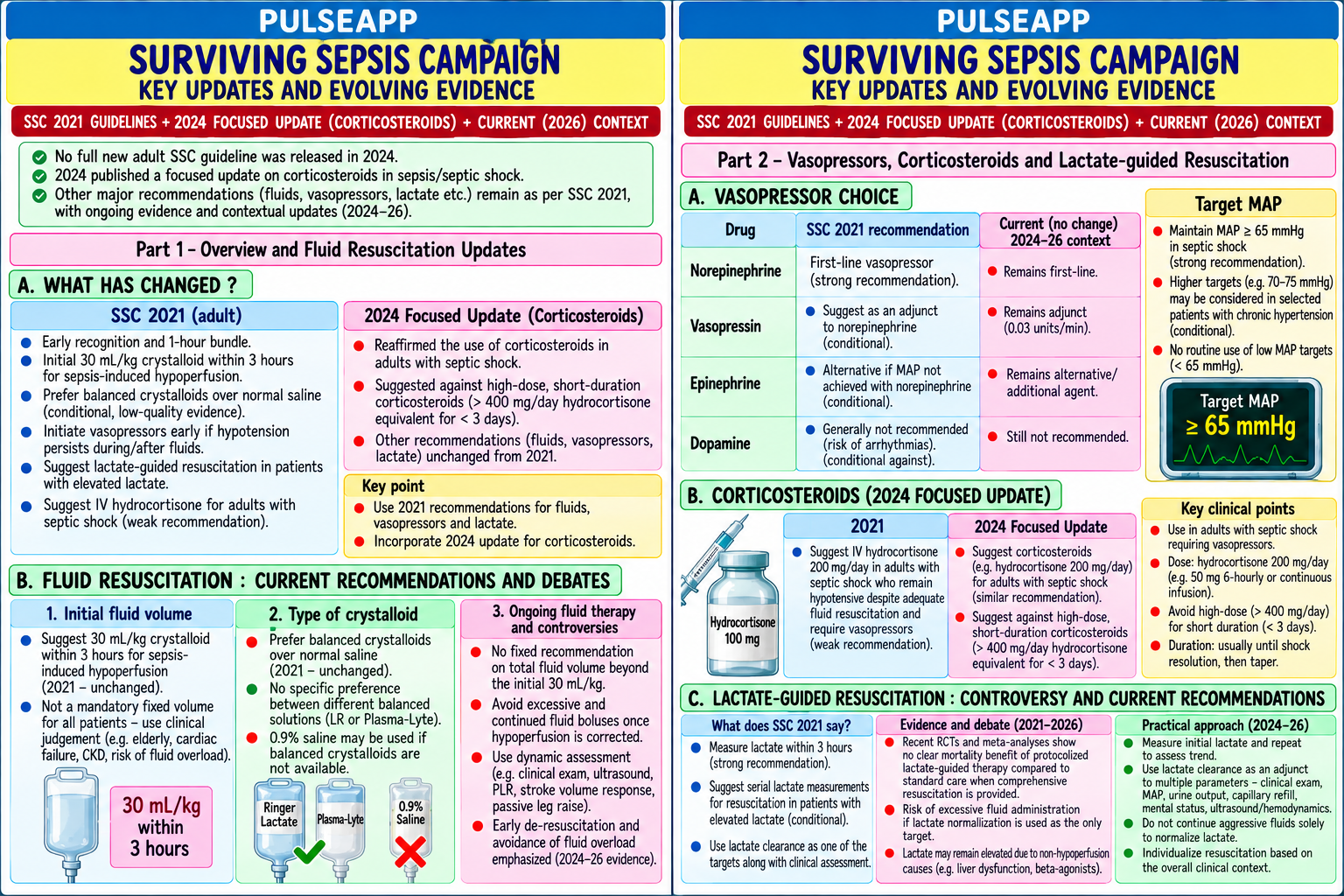
The Surviving Sepsis Campaign (SSC) guidelines undergo regular evidence-based revisions. The 2021 guidelines introduced balanced crystalloids (SMART trial), maintained noradrenaline as first-line vasopressor, and established the Hour-1 bundle. Evolving evidence from the PLUS trial (balanced vs saline), CLOVERS and CLASSIC trials (conservative vs liberal fluids), and CORTICUS-2 trial (corticosteroids) continues to reshape clinical practice. (Evans L et al. — SSC 2021; Bednarczyk JM — SSC 2024; SMART trial; PLUS trial; CLASSIC trial; CLOVERS trial)
A. SSC 2021 vs 2024 Updates — Key Changes3 marks

Domain SSC 2021 2024 Updates/Evolving Evidence Fluid type Balanced crystalloids preferred PLUS trial (NEJM 2022; n=5037): Plasmalyte-148 vs 0.9% NaCl in ICU — NO significant difference in 90-day over 0.9% NaCl (SMART trial — mortality or AKI; raises questions about the magnitude of benefit of balanced vs saline; current consensus: balanced ↓ AKI); weak balanced crystalloids remain preferred but the benefit is modest recommendation Fluid volume 30 mL/kg crystalloid bolus in CLASSIC trial (NEJM 2022; n=1554): restrictive vs standard fluid in septic shock — restrictive (boluses only if Hour-1 for hypotension/lactate severe hypoperfusion; target CVP ≤5) vs standard: NO significant difference in 90-day mortality; confirms that the ≥4; reassess after each 500 mL traditional 30 mL/kg "mandate" is not evidence-based beyond the initial resuscitation; current emphasis: titrate fluid to dynamic fluid responsiveness markers (SVV, PLR), not a fixed volume Vasopressor Noradrenaline first-line (strong); VANCS trial: vasopressin vs noradrenaline in septic shock — vasopressin first-line non-inferior, fewer arrhythmias; choice vasopressin second (weak); ongoing debate; noradrenaline remains standard first-line; vasopressin as second agent to reduce noradrenaline consider terlipressin dose (catecholamine-sparing) Corticosteroids IV hydrocortisone 200 mg/day if CAPE-COVID, APROCCHSS trials: hydrocortisone + fludrocortisone in septic shock → significant 90-day mortality noradrenaline ≥0.25 mcg/kg/min reduction; growing evidence that corticosteroids benefit a broader population than previously defined; threshold for for ≥4 hours (weak initiation may be lower than noradrenaline 0.25 mcg/kg/min recommendation; ADRENAL trial basis) Lactate-guided Measure lactate; if >2 mmol/L → LACTATES trial: lactate-guided vs usual care in septic shock — lactate-guided resuscitation did not improve resuscitation reassess every 2 hours; lactate outcomes; questions whether lactate normalisation is the appropriate endpoint; tissue oxygenation markers and clearance ≥10% per 2 hours is a organ function (creatinine, bilirubin, mental status) may be equally or more relevant endpoints target

B. Hour-1 Bundle — Current Status2 marks

The SSC 2021 Hour-1 bundle (measure lactate, blood cultures before antibiotics, broad-spectrum antibiotics within 1 hour, 30 mL/kg crystalloid if hypotensive, vasopressor if MAP <65) has been validated in multiple observational studies showing that compliance with all 5 elements is associated with reduced mortality

Controversy: the 1-hour antibiotic target for all sepsis (not just septic shock) — some evidence that for less severe sepsis without shock, the 1-hour target may lead to unnecessary broad-spectrum antibiotic use and antibiotic resistance, without clearly improving outcomes; the 2024 updates may refine the antibiotic timing based on clinical severity

Antibiotic de-escalation: increasingly emphasised in 2024 updates — broad-spectrum antibiotics started in Hour-1 must be de-escalated as soon as culture results and clinical response allow (typically at 48–72 hours); de-escalation reduces resistance, side effects (C. difficile, nephrotoxicity), and costs

C. Fluid Resuscitation in Sepsis — Practical Approach 20243 marks
✅ Modern Fluid Resuscitation — The "ROSE" Framework (SCCM 2022)
Rescue (immediate): rapid fluids for haemodynamic crisis (severe hypotension, MAP <50 mmHg, signs of shock) Optimisation (hours 2–6): dynamic fluid responsiveness assessment (passive leg raise test, SVV, SV response to 250 mL bolus) to guide further fluid therapy; STOP giving fluid when non-responsive Stabilisation (day 1–3): conservative approach; no further routine fluids; allow spontaneous diuresis or assist with furosemide once haemodynamically stable Evacuation (day 3+): active fluid removal — furosemide diuresis or CRRT ultrafiltration to achieve zero or negative fluid balance; positive fluid balance >10% body weight is independently associated with increased mortality in sepsis Passive Leg Raise (PLR) test: the patient is positioned supine; the bed is angled to raise the legs 45° (effectively autotransfusing ~300 mL from the lower limbs to the thoracic circulation); measure cardiac output/SV with an appropriate monitor before and after PLR; if CO increases ≥10% → patient is fluid responsive → a fluid bolus will benefit haemodynamics; if CO unchanged → patient is non-responsive → further fluids will only cause harm (oedema, lung injury) Key principle: Fluid is a drug — it has a therapeutic window, dose-response, and toxicity profile; over-resuscitation causes pulmonary oedema, bowel oedema (abdominal compartment syndrome), impaired wound healing, and worsened outcomes in sepsis; match fluid therapy to demonstrated need
D. Vasopressors — Advanced Considerations2 marks

Agent Role in 2024 Guidelines Specific Advantage

Noradrenaline FIRST LINE — strong recommendation; start early Best evidence base; predictable dose-response; less tachyarrhythmia than dopamine (De (norepinephrine) (before completing 30 mL/kg if MAP <65) Backer trial)

Vasopressin SECOND LINE — add when noradrenaline dose V1 receptor vasoconstriction — no catecholamine effects; may preserve renal blood flow; ≥0.25 mcg/kg/min; reduces noradrenaline reduces relative vasopressin deficiency of septic shock requirement Hydrocortisone Add when noradrenaline ≥0.25 mcg/kg/min (2021 Not a vasopressor per se but restores catecholamine receptor sensitivity (corticosteroidthreshold; 2024 may lower this) deficient state → reduced receptor responsiveness to noradrenaline; hydrocortisone restores this) Dopamine NOT recommended as first-line; alternative only if De Backer NEJM 2010: dopamine → more arrhythmias + higher mortality in cardiogenic shock noradrenaline unavailable subgroup vs noradrenaline Angiotensin II Rescue therapy for refractory shock on high-dose AT1 receptor agonist → vasoconstriction independent of catecholamine pathway; ATHOS-3 (Giapreza) noradrenaline + vasopressin trial: improved MAP in refractory vasodilatory shock; expensive, limited availability

🎤 Viva Corner
Q. An ICU patient with septic shock has received 3.5 litres of Plasmalyte over 6 hours. MAP is 62 mmHg on noradrenaline 0.3 mcg/kg/min. Passive leg raise test shows no change in cardiac output. Should you give more fluid?
No — more fluid is not indicated in this patient based on the PLR result, and giving additional fluid is likely to cause harm. The passive leg raise test is a validated, reversible method of assessing fluid responsiveness: by raising the legs 45° and tilting the bed, approximately 250–300 mL of blood is autotransfused from the lower limbs to the central circulation; this acts as a temporary "fluid bolus"; if the cardiac output increases ≥10% in response → the patient is preload-responsive and a fluid bolus would improve haemodynamics. No change in cardiac output (the result here) means the patient is NOT fluid responsive — their ventricles are operating on the flat part of the Frank-Starling curve (ventricular filling is adequate or excessive); additional preload from a fluid bolus would not increase stroke volume, but WOULD increase capillary hydrostatic pressure → cause pulmonary and peripheral oedema. The patient has already received 3.5 L over 6 hours — substantial positive fluid balance relative to likely insensible losses + urine output; the risk of fluid overload complications (pulmonary oedema impairing oxygenation, bowel oedema worsening ileus, abdominal compartment syndrome) is real. Management: DO NOT give further fluid boluses. Address the hypotension through the vasopressor pathway: increase noradrenaline from 0.3 to 0.4–0.5 mcg/kg/min; ADD vasopressin 0.03 units/min as a second vasopressor (catecholamine-sparing, V1 receptor-mediated vasoconstriction); add hydrocortisone 200 mg/day IV infusion (noradrenaline is already at 0.3 mcg/kg/min approaching the SSC 2021 threshold of 0.25 mcg/kg/min where steroids are recommended). Target MAP ≥65 mmHg through vasopressor optimisation rather than volume. Once haemodynamically stable with vasopressors, consider furosemide-assisted diuresis to achieve neutral or negative fluid balance.
★ Examiner's Pearl
PLUS trial (Plasmalyte vs saline — no mortality difference in ICU) is the key 2022 RCT that qualifies the SSC 2021 balanced crystalloid recommendation — state both trials and their conclusions. CLASSIC trial (restrictive vs liberal fluids — no difference in mortality) similarly qualifies the 30 mL/kg mandate. The ROSE framework (Rescue/Optimisation/Stabilisation/Evacuation) is the 2022 SCCM conceptual advance for fluid management in sepsis. PLR test (≥10% CO increase = responsive) with the specific mechanism (autotransfusion 250–300 mL) is the dynamic fluid responsiveness test most tested.
Evans L et al. SSC Guidelines 2021 (Intensive Care Med 2021). Zampieri FG et al. PLUS trial — Plasmalyte vs saline (NEJM 2022). Meyhoff TS et al. CLASSIC trial — restrictive vs standard fluid (NEJM 2022). Marik PE et al. The ROSE concept — fluid management in sepsis (Chest 2022). De Backer D et al. Dopamine vs norepinephrine (NEJM 2010;362:779-789).
lock Protected Clinical Document • Pulse App • Screenshots Strictly Prohibited
QUESTION 77 person Asked by .
bookmark_add

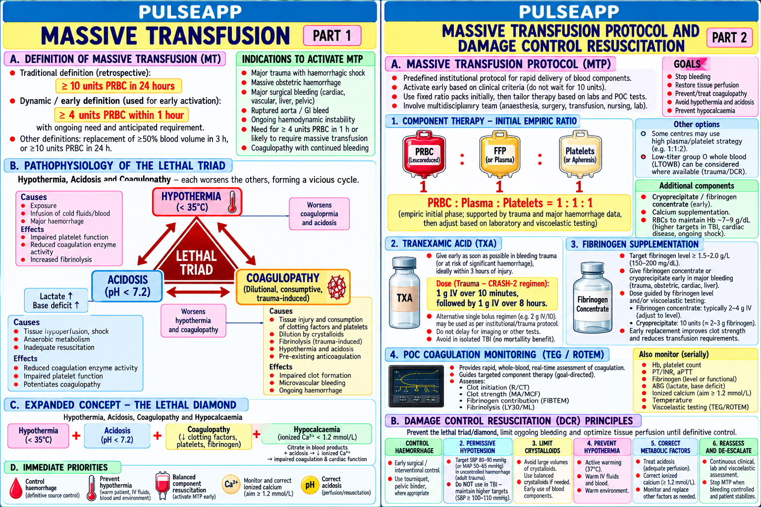
Define massive transfusion. Describe the pathophysiology of the "lethal triad." Outline the components of a Massive Transfusion Protocol (MTP) including product ratios, tranexamic acid, fibrinogen supplementation, and POC coagulation monitoring. Discuss damage control resuscitation principles.

description Clinical Response
⚙ Core Concept
Massive transfusion — defined as ≥10 units RBC within 24 hours (or ≥4 units RBC within 1 hour) — is required in approximately 1–3% of trauma patients and carries mortality of 30–40%. The "lethal triad" of hypothermia, acidosis, and coagulopathy creates a self-perpetuating cycle that kills the patient faster than the original injury unless specifically interrupted through haemostatic resuscitation. Damage Control Resuscitation (DCR) replaces the traditional large-volume crystalloid approach with early blood products in a 1:1:1 ratio and permissive hypotension until surgical haemostasis. (Holcomb JB — PROPPR trial; CRASH-2 trial; Sperry JL — PAMP trial; Borgman MA; Miller's Anaesthesia 9th Ed)
A. The Lethal Triad2 marks

Haemorrhage → hypothermia (heat loss from haemorrhage, exposure, resuscitation with cold fluids) + acidosis (tissue hypoperfusion → lactic acidosis) + coagulopathy (dilution by crystalloids + consumption of clotting factors + direct hypothermia/acidosis impairment of coagulation enzyme activity) → these three form a positive feedback loop: hypothermia impairs coagulation enzymes → worse coagulopathy → more bleeding → more acidosis → more cooling → death Component Mechanism Consequence Hypothermia Blood and tissue heat loss; cold IV fluids; OR exposure; factor activity falls 10% for Progressive coagulopathy despite "normal" laboratory (<35°C) every 1°C below 37°C; platelet function impaired at <33°C; fibrinogen activity coagulation tests (which are performed at 37°C — not impaired at <30°C reflecting the patient's actual temperature) Acidosis (pH Tissue hypoperfusion → lactate accumulation → metabolic acidosis; pH <7.2 reduces Even normal factor levels fail to generate adequate thrombin at <7.2) coagulation enzyme activity by 50%; inhibits thrombin generation low pH — coagulopathy despite normal PT/APTT Coagulopathy Factor dilution by crystalloids; factor consumption; hyperfibrinolysis (plasmin Fibrinogen is consumed first (critical threshold <1.5 g/L in activation); hypothermia/acidosis-induced enzyme failure; early trauma-induced trauma); then factor V, VIII, XI; then platelet dysfunction coagulopathy (TIC — before any resuscitation)

B. Massive Transfusion Protocol (MTP)4 marks
✅ Damage Control Resuscitation (DCR) — Core Principles
1. Permissive hypotension: target systolic BP 80–90 mmHg (MAP 50–65 mmHg) UNTIL surgical haemostasis is achieved; avoid hypertension which dislodges clots from injured vessels; exception: TBI (target MAP ≥80 to maintain CPP) 2. Haemostatic resuscitation (1:1:1): RBC : FFP : Platelets in a 1:1:1 ratio; PROPPR trial (Holcomb JAMA 2015; n=680): 1:1:1 vs 1:1:2 ratio → significantly better 24-hour and 30-day survival, better haemostasis; treats the coagulopathy simultaneously with the volume deficit 3. Early tranexamic acid (TXA): 1 g IV over 10 minutes within 3 hours of injury (CRASH-2 Lancet 2010; n=20,211: 15% relative reduction in all-cause mortality in the TXA group); if >3 hours after injury: NO benefit and possible harm; second dose 1 g IV over 8 hours if bleeding continues 4. Minimise crystalloids: crystalloids dilute clotting factors, worsen coagulopathy, cause acidosis and hypothermia (cold saline); limit crystalloids to <1.5 L for initial resuscitation; use blood products as the primary volume expander 5. Prevent/treat hypothermia: warm all blood products and IV fluids (to 37°C); external warming devices; warm blankets; increase theatre temperature; warm irrigation Product Indication in MTP Trigger/Dose RBC (pRBC) Volume replacement + O₂ carrying capacity; given early in O-negative in emergency (pending crossmatch); target Hb ≥70–80 g/L (higher in 1:1:1 with FFP + platelets TBI/IHD) FFP (Fresh Frozen All clotting factors II, V, VII, VIII, IX, X, XI, fibrinogen, protein 1:1 ratio with RBC; also for specific factor reversal (warfarin emergency — 10– Plasma) C&S; 1 unit per unit RBC 15 mL/kg) Platelets Primary haemostasis at the injury site; 1 pool per unit RBC Platelet pool (6 units) = 1 standard dose; apheresis platelets preferred if (1:1:1 ratio); target Plt >50,000/μL (higher for TBI: >100,000) available (fewer donor exposures) Cryoprecipitate / Fibrinogen is consumed first in massive haemorrhage; critical Cryoprecipitate 10 units (contains ~4–5 g fibrinogen total) OR Fibrinogen Fibrinogen threshold <1.5 g/L; replenish early concentrate 2–4 g IV; target fibrinogen >2 g/L concentrate Calcium (calcium FFP and RBC contain citrate anticoagulant → chelates ionised 10 mL of 10% calcium gluconate IV per 4 units FFP or pRBC transfused rapidly; gluconate) calcium → hypocalcaemia impairs clotting and cardiac function check ionised Ca²⁺ every 30 min in massive transfusion; target iCa >1.1 mmol/L
C. Point-of-Care Coagulation Monitoring in MTP2 marks

TEG/ROTEM: whole blood point-of-care coagulation tests; provide results within 10–20 minutes vs 60+ minutes for standard laboratory coagulation tests; guide targeted blood product replacement: TEG R time prolonged → FFP; MA reduced → platelets; LY30 elevated → tranexamic acid; fibrinogen reduced (Alpha angle low) → cryoprecipitate/fibrinogen concentrate

Standard lab tests during MTP: FBC (Hb, Plt), PT/APTT/fibrinogen, ABG (pH, lactate, calcium, glucose, temperature), TEG if available — all repeated every 30–60 minutes during active haemorrhage

Rotational thromboelastometry (ROTEM): equivalent to TEG but uses different parameter names (CT = R time; MCF = MA; LI30 = LY30); EXTEM/FIBTEM/INTEM/APTEM channels provide comprehensive haemostatic profile in <30 minutes

D. MTP Termination & Post-haemostasis Care2 marks

MTP termination criteria: haemorrhage surgically or radiologically controlled; haemodynamic stability with reducing vasopressor requirements; normal coagulation parameters (TEG normalised; pH >7.25; temperature >35°C)

Post-haemostasis care: switch from DCR (permissive hypotension, haemostatic resuscitation) to damage control surgery phase: repair injuries in planned stages; prevent abdominal compartment syndrome; reverse hypothermia aggressively; correct remaining acidosis; ICU monitoring for multi-organ failure in the subsequent days

Thromboprophylaxis after MTP: VTE risk is extremely high post-trauma; start LMWH as soon as haemostasis is secure and surgical team agrees (usually 12–24 hours after haemostasis); mechanical compression devices from admission

🎤 Viva Corner
Q. A trauma patient is bleeding massively from a pelvic fracture. The surgeon asks for "4 more litres of Hartmann's to buy time while we set up for angioembolisation." What is your response?
I would respectfully but firmly decline to administer 4 litres of Hartmann's solution in this context. The evidence clearly shows that large-volume crystalloid resuscitation in haemorrhagic shock is harmful — it represents the exact opposite of damage control resuscitation principles. The specific harms of 4 litres of Hartmann's in this patient: it would dilute all clotting factors and platelets (4 L crystalloid dilutes the blood to approximately 50% of its original concentration — essentially inducing a clinical coagulopathy equivalent to being anticoagulated); it would cause hypothermia (Hartmann's is stored at 4°C and even warming to room temperature still produces relative heat loss in the patient); it would worsen acidosis by the Stewart mechanism (large-volume Hartmann's produces a dilutional hyperchloraemic metabolic acidosis); combined, these effects directly worsen the lethal triad and increase the rate of haemorrhagic death. Instead, I would: activate the massive transfusion protocol immediately; transfuse RBC + FFP + platelets in a 1:1:1 ratio as the volume resuscitation strategy; administer tranexamic acid 1 g IV now (within 3 hours of injury); use permissive hypotension target (systolic 80–90 mmHg) — attempting to restore normal BP with crystalloids dislodges clots and increases haemorrhage; limit any crystalloid to <1.5 L as an emergency bridge while blood products are being prepared; and expedite the angioembolisation as the definitive haemostasis procedure. The surgery team needs to proceed urgently — the correct conversation is how to get to angioembolisation fastest with adequate haemostatic support, not how to fill the tank with saline while waiting.
★ Examiner's Pearl
PROPPR trial (1:1:1 vs 1:1:2 → improved 24h and 30d survival) and CRASH-2 trial (TXA within 3 hours → 15% mortality reduction; >3 hours → harm) with specific data are the two landmark MTP trials that must be cited. The lethal triad (hypothermia/acidosis/coagulopathy) with the specific effect of each on coagulation (hypothermia → enzyme failure; acidosis → thrombin generation impaired; coagulopathy → fibrinogen consumed first) is the physiological framework. Calcium gluconate per 4 units transfused (citrate chelation → hypocalcaemia → impairs clotting and cardiac function) is the specific transfusion management fact most tested.
Holcomb JB et al. PROPPR trial — transfusion of plasma, platelets and RBCs (JAMA 2015;313:471-482). CRASH-2 trial collaborators (Lancet 2010;376:23-32). Borgman MA et al. The ratio of blood products transfused affects mortality in patients receiving massive transfusions (J Trauma 2007). Sperry JL et al. PAMP trial — pre-hospital plasma in haemorrhagic shock (NEJM 2018;379:315-326).
lock Protected Clinical Document • Pulse App • Screenshots Strictly Prohibited
QUESTION 78 person Asked by .
bookmark_add

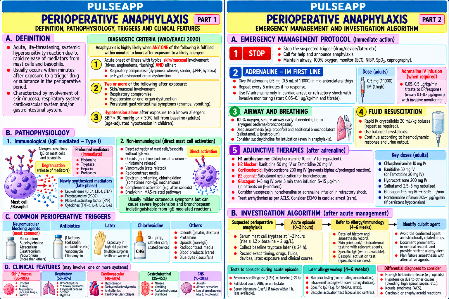
Define anaphylaxis and describe its immunological and non-immunological pathophysiology. Outline the findings of the UK National Audit Project 6 (NAP6). Describe the emergency management protocol and the systematic investigation algorithm for perioperative anaphylaxis.

collections Question Diagrams & Reference Images (1)
Question Reference Diagram
zoom_in View Image
description Clinical Response
⚙ Core Concept
Perioperative anaphylaxis occurs in approximately 1 in 10,000–20,000 general anaesthetics and carries a mortality of approximately 4–9%. NAP6 (2018) was the first large-scale national audit of perioperative anaphylaxis in the UK — revealing that neuromuscular blocking agents are the most common trigger (50%), followed by antibiotics (15%), and chlorhexidine (9%), with significant under-investigation and inadequate management in many cases. The standardised post-event investigation pathway (tryptase levels, skin testing at 4–6 weeks, RAST) is mandatory for every confirmed case. (Harper NJN et al. — NAP6 2018; Simons FER — World Allergy Organisation; Krishna MT; AAGBI guidelines)
A. Definition and Pathophysiology2 marks

Definition (World Allergy Organisation 2011): anaphylaxis is a severe, life-threatening, generalised hypersensitivity reaction; clinical diagnosis: "highly likely when ONE of the following 3 criteria is fulfilled": Acute onset with skin/mucosal involvement + either respiratory compromise OR reduced BP/end-organ dysfunction

Two or more of: skin/mucosal involvement, respiratory compromise, reduced BP, GI symptoms — occurring after EXPOSURE to a LIKELY ALLERGEN Reduced BP after exposure to a KNOWN allergen Immunological (IgE-mediated — Type I hypersensitivity): previous sensitisation → IgE antibodies bound to mast cells and basophils; re-exposure → allergen cross-links IgE → mast cell degranulation → histamine, tryptase, leukotrienes, prostaglandins, PAF → vasodilation + vascular permeability ↑ + bronchospasm + GI effects Non-immunological (anaphylactoid — no prior sensitisation needed): direct mast cell degranulation without IgE; drugs that cause this: opioids (morphine, codeine — direct mast cell degranulation; NOT IgE-mediated), hyperosmolar solutions, contrast media, aspirin (via COX pathway → ↑ leukotrienes), dextrans, protamine (complement activation)

Key mediators: histamine (H1 → bronchospasm, vasodilation; H2 → ↑ gastric acid); tryptase (the most specific mast cell marker — peaks at 60–90 min postanaphylaxis; used for diagnostic confirmation); leukotrienes C4 and D4 (potent bronchoconstrictors); prostaglandins; platelet activating factor (PAF)

B. NAP6 (2018) Findings2 marks

Finding Detail Most common NMBAs 50% (suxamethonium 26%, rocuronium 14%); antibiotics 15% (teicoplanin, co-amoxiclav, cefuroxime); chlorhexidine 9%; patent blue dye 6%; triggers latex 2%; all others 18% Incidence 1 in 10,000 general anaesthetics (estimated); true incidence uncertain due to under-reporting; approximately 266 cases identified in a 12-month period across UK Mortality 9 deaths in the audit period; estimated UK mortality from perioperative anaphylaxis: 3–9 per million GAs

Cardiovascular In perioperative anaphylaxis: cardiovascular collapse is the predominant feature (73%) in contrast to community anaphylaxis where skin and predominance respiratory symptoms predominate; skin features may be absent due to vasoconstriction/surgical drapes masking urticaria Under- Only 60% of confirmed cases were referred for formal allergy investigation; many patients discharged without follow-up testing — leaving them at risk investigation of recurrence without knowing their triggers Key practice Initial adrenaline dosing often inadequate or delayed; 25% received adrenaline >10 minutes after first symptoms; some received non-adrenaline concerns vasopressors first (noradrenaline — inadequate in anaphylaxis which requires both alpha + beta effects)

C. Emergency Management Protocol3 marks
⚠ AAGBI Perioperative Anaphylaxis Protocol
1. STOP the causative agent (if identifiable) — stop the infusion or drug administration; note the exact drug and timing 2. CALL FOR HELP — senior anaesthesiologist, anaesthetic nurse, emergency team 3. ADRENALINE — the cornerstone treatment: Adult: 0.5 mg (500 mcg) IM in the anterolateral thigh (1:1000 solution = 0.5 mL) — FIRST LINE; repeat every 5 minutes if no improvement IV adrenaline: ONLY if no pulse or experienced team; 50–100 mcg IV boluses titrated to response; IV adrenaline in inexperienced hands causes fatal cardiovascular complications Mechanism: α₁ → peripheral vasoconstriction (reverses vasodilation); β₁ → positive inotropy (reverses myocardial depression); β₂ → bronchodilation + mast cell stabilisation (inhibits further mediator release) 4. AIRWAY: 100% O₂; if bronchospasm → nebulised salbutamol 5 mg; early intubation if angioedema developing (airway oedema can progress rapidly making late intubation impossible) 5. IV FLUIDS: 500–1000 mL crystalloid bolus rapidly (treats distributive shock from vasodilation); up to 2–3 L may be needed in severe anaphylaxis; avoid excessive saline in already-acidotic patient 6. POSITION: supine with legs elevated (increases venous return to the heart in the context of vasodilatory shock); do NOT sit upright (worsens venous return) 7. SECONDARY DRUGS (AFTER adrenaline is given and haemodynamics partially restored): Chlorphenamine 10 mg IV (H1 antihistamine — reduces further histamine effects; does NOT reverse acute cardiovascular collapse) Hydrocortisone 200 mg IV (reduces late-phase biphasic reaction; onset delayed 4–6 hours; not for acute resuscitation) Salbutamol 5 mg nebulised for refractory bronchospasm Glucagon 1–2 mg IV for anaphylaxis in patients on beta-blockers (bypasses β-receptor to directly activate adenylyl cyclase → ↑ cAMP → inotropy + heart rate)
D. Post-Event Investigation Algorithm3 marks

ACUTE TRYPTASE SAMPLES — MANDATORY after every suspected anaphylaxis:

Sample 1: as soon as possible after reaction (within 30 minutes of onset)

Sample 2: 1–2 hours after reaction (peak tryptase — typically 60–90 minutes)

Sample 3: 24 hours (baseline tryptase — to exclude mastocytosis as an underlying cause) Serum tryptase >20 mcg/L (or >2× baseline + 2) confirms mast cell degranulation → supports anaphylaxis diagnosis Refer to allergy clinic at 4–6 weeks: this is the mandatory post-event investigation window; earlier testing is unreliable (residual drug/antibodies may interfere) Skin testing (SPT — skin prick test + ID — intradermal): gold standard for identifying the causative agent; each drug used during the anaesthetic is tested individually at non-irritating concentrations; a positive SPT (wheal ≥3 mm) confirms IgE sensitisation to that drug; helps identify the trigger and guide future safe anaesthesia

RAST/ImmunoCAP (specific IgE ELISA): blood test for specific IgE antibodies against drugs/allergens; less sensitive than skin testing for NMBAs but useful for penicillin allergy, latex, and chlorhexidine; complements skin testing

Future anaesthesia document: after full investigation, a written report must be provided to the patient specifying the identified trigger, safe alternative agents, and a detailed future anaesthesia plan; this document should be worn as a medical alert bracelet or kept with the patient's records

🎤 Viva Corner
Q. Why is adrenaline the drug of first choice in anaphylaxis, and why should noradrenaline NOT be given as a substitute?
Adrenaline (epinephrine) is the ONLY drug that simultaneously addresses all the major pathophysiological mechanisms of anaphylaxis through its combined alpha and beta adrenergic receptor actions. The three simultaneous beneficial effects of adrenaline in anaphylaxis: First, alpha-1 adrenergic vasoconstriction: adrenaline constricts the massively dilated peripheral vasculature → raises systemic vascular resistance → increases blood pressure → reverses the distributive shock; simultaneously, alpha-1 constriction of the bronchial mucosal vasculature reduces mucosal oedema and angioedema. Second, beta-1 adrenergic cardiac stimulation: adrenaline directly increases heart rate and myocardial contractility → increases cardiac output → further supports blood pressure in a patient who may have myocardial depression from the anaphylactic mediators. Third, beta-2 adrenergic bronchodilation: adrenaline relaxes bronchial smooth muscle → reverses bronchospasm (the second most common cause of death in anaphylaxis after cardiovascular collapse); ADDITIONALLY, beta-2 receptor stimulation on mast cells and basophils INHIBITS further degranulation — literally turning off the ongoing mediator release that is perpetuating the anaphylaxis. Noradrenaline is predominantly an alpha-1 agonist with minimal beta-2 activity: it would increase blood pressure through vasoconstriction (addressing one component) BUT it would NOT bronchodilate (severe bronchospasm would continue unabated), NOT provide adequate cardiac stimulation in the face of anaphylactic cardiac depression (beta-1 effect is weaker than adrenaline's), and critically, NOT inhibit ongoing mast cell degranulation (no beta-2 effect). Additionally, noradrenaline's intense vasoconstriction in the context of already-massive histamine-induced vasodilation can produce paradoxical and extreme cardiovascular responses. Adrenaline is therefore irreplaceable as the first-line drug — all other vasopressors are adjuncts.
★ Examiner's Pearl
NAP6 finding that NMBAs are the most common cause of perioperative anaphylaxis (50% — suxamethonium 26%, rocuronium 14%) is the specific UK epidemiological fact tested. The three adrenaline doses: IM 0.5 mg (first-line); IV 50–100 mcg boluses (experienced team only); the three tryptase sample timings (ASAP, 1–2 hours, 24 hours baseline) must be stated as a complete protocol. Cardiovascular collapse as the predominant perioperative presentation (73% vs community anaphylaxis which is predominantly skin/respiratory) is the specific NAP6 clinical finding that differentiates perioperative from community anaphylaxis.
Harper NJN et al. Anaesthesia, surgery and life-threatening allergic reactions — NAP6 2018 (BJA 2018;121:159-171). Simons FER et al. World Allergy Organisation guidelines for anaphylaxis (WAO J 2015;8:32). AAGBI. Suspected anaphylactic reactions associated with anaesthesia 2009. Ewan PW. Investigation of suspected anaphylaxis (Clin Exp Allergy 2007).
lock Protected Clinical Document • Pulse App • Screenshots Strictly Prohibited
QUESTION 79 person Asked by .
bookmark_add

Classify the causes and grading of perioperative bronchospasm. Describe the stepwise emergency management including pharmacological agents, ventilatory adjustments, and the differential diagnosis from other causes of raised airway pressure.

collections Question Diagrams & Reference Images (1)
Question Reference Diagram
zoom_in View Image
description Clinical Response
⚙ Core Concept
Perioperative bronchospasm — acute reversible bronchoconstriction during anaesthesia — occurs in approximately 2% of all GAs, rising to 9% in asthmatics and 6% in smokers. It is most commonly triggered at laryngoscopy and intubation (the highest-stimulus moments of anaesthesia) in an inadequately anaesthetised patient with reactive airways. Distinguishing true bronchospasm from other causes of raised airway pressure (ETT obstruction, pneumothorax, anaphylaxis) requires a systematic clinical assessment. (Westhorpe RN; Dewachter P; Mebazaa A; Miller's Anaesthesia 9th Ed)
A. Causes and Grading2 marks

Grade Features Management Mild Wheeze on auscultation; ↑ peak airway pressure 20–30%; SpO₂ maintained; ETCO₂ Deepen anaesthesia; salbutamol MDI via ETT adapter; upsloping plateau; patient comfortable under adequate anaesthesia continue monitoring Moderate Audible wheeze; peak airway pressure ↑30–50%; SpO₂ 90–94%; clear shark-fin ETCO₂; Salbutamol 5 mg nebulised via circuit; increase volatile agent increased work of breathing if spontaneously ventilating concentration; IV magnesium 2 g; reduce RR to lengthen expiratory time Severe Severe wheeze or silent chest (no air entry — life-threatening); peak airway pressure Emergency protocol (see below); IV adrenaline; ketamine; IV ↑>50% or cannot ventilate; SpO₂ <90%; potential auto-PEEP development and aminophylline; consider whether this is anaphylaxis cardiovascular compromise

Common triggers: intubation/laryngoscopy in light anaesthesia; aspiration of gastric contents or secretions; airway irritation (ETT irritation, suctioning); histamine-releasing drugs (atracurium, morphine, thiopentone at high doses); anaphylaxis; irritant volatile agents (desflurane — most pungent; isoflurane); cold dry gas; GORD; active smoking; uncontrolled asthma

B. Differential Diagnosis of Raised Airway Pressure2 marks

Cause Distinguishing Features Action ETT obstruction Unilateral or no breath sounds; passes suction catheter easily (if secretion plug) or does not pass (if Suction ETT; reposition; replace ETT if (kink, secretion, kinked); pass suction catheter immediately — rules out/confirms most ETT obstruction causes kinked bite) Endobronchial Unilateral breath sounds only; ↑ airway pressure; hypoxia; SpO₂ falls; confirm with fibreoptic or pull Withdraw ETT 1–2 cm; confirm bilateral intubation back ETT slowly until bilateral sounds return breath sounds Pneumothorax Absent unilateral breath sounds; hypotension; tracheal deviation; JVP raised; cardiovascular collapse if Emergency needle decompression 2nd (tension) tension; immediate clinical diagnosis; confirm with CXR or POCUS ICS MCL if tension; formal chest drain True BILATERAL wheeze; shark-fin ETCO₂; bilateral reduced air entry; responsive to bronchodilators; no Bronchodilators; deepen anaesthesia; bronchospasm sudden cardiovascular collapse (unless anaphylaxis) ventilation adjustment Anaphylaxis Bronchospasm + cardiovascular collapse + urticaria/flush (may be masked by drapes); tryptase Adrenaline IM/IV; IV fluids; full elevated; multiple drug exposures anaphylaxis protocol

C. Stepwise Emergency Management of Severe Bronchospasm4 marks

1. FiO₂ 1.0 immediately — maximum oxygenation while managing airway 2. Deepen anaesthesia: increase volatile agent to 1.5–2 MAC (sevoflurane/isoflurane — inherent bronchodilators); propofol bolus 1–2 mg/kg IV (bronchodilator properties); avoid desflurane (airway irritant) 3. Salbutamol (albuterol): 5–10 puffs MDI via ETT adapter (actuate MDI into the circuit during inspiration); or 5 mg nebulised via the inspiratory limb; the fastest and most effective initial bronchodilator; mechanism: β₂ agonist → airway smooth muscle relaxation → bronchodilation + mast cell stabilisation 4. Ventilation adjustments: reduce RR to 8–10 breaths/min (extend expiratory time → reduce auto-PEEP); I:E ratio 1:4; reduce TV if necessary (accept permissive hypercapnia rather than high airway pressures); if patient fighting ventilator → NMBA (rocuronium 0.6 mg/kg) to eliminate respiratory dyssynchrony 5. IV Magnesium sulphate: 2 g IV over 10–20 minutes — inhibits Ca²⁺-mediated smooth muscle contraction; modest bronchodilator; well tolerated; safe to give empirically 6. IV Ketamine: 0.5–1 mg/kg IV — bronchodilator via catecholamine release and direct smooth muscle relaxation; also provides additional depth of anaesthesia; particularly useful if patient is light under anaesthesia 7. IV Aminophylline: 5 mg/kg loading over 20 minutes (reduce to 3 mg/kg if on theophylline); phosphodiesterase inhibitor → ↑ cAMP → bronchodilation; narrow therapeutic index — requires ECG monitoring (tachyarrhythmia risk) 8. IV Adrenaline: if severe, life-threatening, or anaphylaxis suspected — 50–100 mcg IV boluses; activates β₂ receptors → most powerful bronchodilator; also reverses any anaphylaxis component 9. IV Hydrocortisone: 200 mg IV — anti-inflammatory; effect delayed 4–6 hours but appropriate for medium-term management; reduces airway inflammation perpetuating bronchospasm

D. Prevention in High-Risk Patients2 marks

Preoperative optimisation: ensure asthma is well-controlled (PEFR ≥80% predicted); continue all inhalers on morning of surgery; consider salbutamol nebulisation 20–30 minutes before induction; avoid elective surgery during active exacerbation

Induction: topicalise the airway (lignocaine spray) before intubation; ensure adequate depth (sevoflurane >1 MAC or propofol 2.5 mg/kg) before laryngoscopy; LMA preferred over ETT in reactive airway disease if surgically feasible (avoids subglottic stimulation); avoid histamine-releasing NMBs (atracurium → use rocuronium or cisatracurium); avoid thiopentone in high-dose (histamine release); propofol preferred induction agent

🎤 Viva Corner
Q. During appendicectomy under GA in a known asthmatic, peak airway pressure rises from 20 to 55 cmH₂O and there is bilateral wheeze on auscultation. SpO₂ falls to 88%. You have given sevoflurane 2.5 MAC and salbutamol 10 puffs MDI with no improvement. What is your next step and your differential?
Failure to respond to maximal volatile agent and salbutamol MDI in a bilateral wheezing picture requires immediate reassessment for a different underlying mechanism — true bronchospasm should improve with 2.5 MAC sevoflurane and salbutamol MDI within 2–3 minutes. At this point the differential must include anaphylaxis: during appendicectomy, multiple potential allergens have been administered (antibiotics — most likely co-amoxiclav or cefuroxime; NMB — rocuronium or suxamethonium; propofol or thiopentone; possibly latex from surgical gloves or chlorhexidine). Anaphylaxis produces bronchospasm through histamine and leukotriene-mediated bronchoconstriction, but is distinguished from pure bronchospasm by concurrent cardiovascular collapse — check the blood pressure and heart rate immediately; urticaria or flush may be present under the surgical drapes. If anaphylaxis is suspected: administer adrenaline 0.5 mg IM (anterolateral thigh) or 50–100 mcg IV immediately; stop any running drug infusions; give IV fluid bolus 500 mL rapidly; call for senior help and anaesthetic nurse. Simultaneously for the ventilation: IV magnesium 2 g over 15 minutes; IV ketamine 1 mg/kg IV (both bronchodilator and additional depth); reduce RR to 8 breaths/min and I:E to 1:4 (accommodate auto-PEEP from severe obstruction); if the patient is still fighting the ventilator despite deep anaesthesia, add rocuronium 0.6 mg/kg to eliminate respiratory dyssynchrony; accept permissive hypercapnia (PaCO₂ 60–70 mmHg) rather than dangerous peak pressures. Collect tryptase samples immediately (ASAP and at 60–90 minutes) to confirm or exclude anaphylaxis post-event. Request surgeon to expedite closure. ICU admission post-operatively for observation and investigation.
★ Examiner's Pearl
The five-step emergency protocol (FiO₂ 1.0 → deepen anaesthesia → salbutamol MDI → ventilation adjustment → escalate: MgSO₄/ketamine/aminophylline/adrenaline) must be in the correct escalating sequence. The silent chest (no wheeze) in severe bronchospasm indicating near-complete obstruction is the most dangerous clinical sign — "silence is deadly in bronchospasm." The anaphylaxis differential in refractory bronchospasm is the key diagnostic reasoning step that examiners specifically test.
Dewachter P et al. Perioperative bronchospasm (Curr Opin Anaesthesiol 2014;27:329-335). Mebazaa A et al. Practical recommendations for perioperative management of patients with possible allergic reactions (Anaesthesia 2011). Miller RD et al. Miller's Anaesthesia, 9th Ed. BTS/SIGN British Guideline on the Management of Asthma 2022.
lock Protected Clinical Document • Pulse App • Screenshots Strictly Prohibited
QUESTION 80 person Asked by .
bookmark_add

Describe the common causes of perioperative cardiac arrest and the modifications to standard ACLS required in the perioperative setting. Outline the 4 Hs and 4 Ts. Discuss specific resuscitation scenarios: local anaesthetic-induced cardiac arrest, anaphylaxis-induced arrest, and tension pneumothorax during anaesthesia.

collections Question Diagrams & Reference Images (2)
Question Reference Diagram
zoom_in View Image
Question Reference Diagram
zoom_in View Image
description Clinical Response
⚙ Core Concept
Perioperative cardiac arrest has unique features that require significant modifications to standard ACLS protocols: the cause is usually known or rapidly identifiable (unlike out-of-hospital cardiac arrest); specific antidotes exist for several causes (sugammadex for CICO-rocuronium, lipid emulsion for LAST, adrenaline for anaphylaxis); and the perioperative team has immediate IV access, airway control, and monitoring in place before the arrest. The perioperative cardiac arrest survival rate (approximately 60%) is far higher than out-of-hospital arrest (10%) — reflecting the advantage of monitored, immediate-response resuscitation. (AHA ACLS 2020; Sprung J; Newland MC — Mayo Clinic perioperative arrest; Miller's Anaesthesia 9th Ed)
A. 4 Hs and 4 Ts — Reversible Causes2 marks

4 Hs Perioperative Context 4 Ts Perioperative Context Hypoxia ETT displacement, oesophageal intubation, airway Tension Central line insertion, barotrauma, laparoscopy CO₂ tracking, obstruction, failed oxygenation; FIRST thing to exclude Pneumothorax ARDS over-ventilation; absent breath sounds + sudden — check bilateral breath sounds, SpO₂, ETCO₂ cardiovascular collapse; immediate needle decompression Hypovolaemia Surgical haemorrhage, aortocaval compression Tamponade Cardiac surgery complication, central line perforation, chest (pregnancy), fluid deficit; PEA in context of surgery and (cardiac) trauma; PEA after cardiac surgery is tamponade until proven haemorrhage; transfuse blood products immediately otherwise; pericardiocentesis or surgical drainage Hypo/Hyperkalaemia Succinylcholine in denervated patients, renal failure, Thrombosis PE during surgery (DVT, air embolism, fat embolism, amniotic fluid transfusion; ECG changes; calcium gluconate for (pulmonary) embolism); sudden hypoxia + hypotension + reduced ETCO₂; hyperkalaemia; calcium for hypocalcaemia thrombolyse if PE confirmed in arrest Hypothermia Prolonged surgery + inadequate warming; VF refractory Toxins Drug overdose (opioids, local anaesthetics), volatile agent to defibrillation below 28°C; warm the patient before overdose, anaphylaxis, medication errors; specific antidotes: lipid declaring death; "not dead until warm and dead" emulsion (LAST), naloxone (opioids), sugammadex (CICO), adrenaline (anaphylaxis)

B. Perioperative ACLS Modifications2 marks

Airway already managed: ETT usually in situ → confirm bilateral breath sounds and ETCO₂ waveform first (excludes oesophageal intubation as arrest cause); ensure 100% O₂; ventilate manually at 10 breaths/min (not continuous ventilation — pauses for CPR compression are acceptable if ETCO₂ waveform visible)

IV access established: use existing IV/central access; if fluids are running → check for medication errors or anaphylaxis-triggering drug

Abandon the surgical field: inform the surgeon immediately; control haemorrhage if possible; non-critical surgery → close and focus on resuscitation

Team roles: anaesthesiologist leads CPR and drug management; surgeon manages surgical cause if relevant; ODP/nurse circulates drugs; second anaesthesiologist for airway relief; immediate defibrillator application

Immediate defibrillation for VF/pVT: 200J biphasic immediately; do not delay defibrillation for any other intervention in shockable rhythms

Higher quality CPR target in operating theatre: sternal compression 5–6 cm depth at 100–120/min; full chest recoil; minimal interruptions; CPR feedback device if available

C. Specific Perioperative Arrest Scenarios4 marks
LAST (Local Anaesthetic Systemic Toxicity) — Cardiac Arrest

Mechanism: bupivacaine "fast in, slow out" cardiac Na⁺ channel block → VF/VT refractory to standard ACLS

Management: standard CPR; 20% lipid emulsion 1.5 mL/kg IV bolus immediately; then 0.25 mL/kg/min infusion; may repeat bolus once after 5 minutes; max 12 mL/kg total; AVOID propofol as the lipid source (not adequate lipid content for LAST at clinical doses); avoid vasopressin in LAST; adrenaline doses ≤1 mcg/kg (higher doses may worsen outcomes in LAST); consider ECMO early if LAST arrests persist despite lipid emulsion

Anaphylaxis-Induced Cardiac Arrest

Mechanism: massive vasodilation + myocardial depression + bronchospasm → PEA or VF

Management: FULL DOSE adrenaline 1 mg IV every 3–5 minutes (standard ACLS dose — do NOT use reduced dose in anaphylaxis arrest); IV fluids 1–2 L crystalloid rapidly; lie flat, legs up; stop causative drug; chlorphenamine and hydrocortisone are secondary — do not delay CPR for these; once ROSC: vasopressor infusion (noradrenaline); tryptase samples; allergy follow-up

Tension Pneumothorax During Anaesthesia

Mechanism: air in the pleural space under pressure → compresses the lung, mediastinum, and great veins → reduced venous return → obstructive shock →

PEA arrest

Recognition during anaesthesia: sudden ↑ airway pressure + sudden ↓ ETCO₂ + sudden ↓ BP → absent breath sounds unilaterally

Management: immediate needle decompression 2nd intercostal space, mid-clavicular line (anterior approach — 14G IV cannula; confirm by hiss of air under pressure and haemodynamic improvement); formal chest drain after needle decompression; do NOT delay for CXR in arrest; if bilateral pneumothorax suspected (both sides absent breath sounds, bilateral raised airway pressures in ARDS) → bilateral needle decompression simultaneously

Cardiac Arrest After Induction (Medication Error / Overdose)

Immediate check: is the correct drug drawn up? Syringe labelling error? Dose calculation correct? Weight entered correctly into TCI?

Opioid-induced arrest: naloxone 400 mcg IV; propofol overdose: supportive CPR + lipid emulsion if unresponsive

Potassium bolus error (concentrated KCl given as IV push): hyperkalaemia → VF → calcium gluconate 1 g IV immediately; sodium bicarbonate 50 mEq IV; insulin-dextrose; defibrillate for VF; dialysis

D. Post-Resuscitation Care2 marks

Targeted Temperature Management (TTM): cool to 32–36°C for 24 hours in comatose post-cardiac arrest patients (TTM2 trial 2021: 33°C vs 37.5°C — no mortality difference; maintain normothermia at minimum; avoid hyperthermia >37.5°C); continuous EEG monitoring for seizures (post-anoxic seizures are common)

Haemodynamic optimisation: MAP ≥65–70 mmHg; avoid hypotension post-ROSC; vasopressor infusion; coronary angiography if STEMI suspected as cause

Neuroprognostication: defer formal neurological assessment until 72 hours post-arrest (and 24 hours after stopping TTM); use multimodal approach: EEG, SSEP, CT brain, MRI brain, biomarkers (NSE, S100β)

🎤 Viva Corner
Q. During a thoracic epidural insertion via central line, a patient has sudden cardiac arrest with PEA. What is the most likely diagnosis and what do you do first?
Sudden PEA arrest during or immediately after central line insertion is tension pneumothorax until proven otherwise — it is the classic complication of central venous cannulation (subclavian or internal jugular approach), where the needle or guidewire can pierce the pleura, particularly in a patient with positive pressure ventilation (each breath inflates the pneumothorax). The immediate diagnostic signs to confirm: check for unilateral absent breath sounds (one side will be silent if unilateral tension pneumothorax); the airway pressure alarm will have sounded before arrest (rising peak airway pressure from the pneumothorax); ETCO₂ will have fallen just before arrest (reduced cardiac output and pulmonary blood flow). Do not wait for a chest X-ray — this is a clinical diagnosis in the arrest context. First action: while commencing CPR simultaneously, perform emergency needle decompression — 14G IV cannula inserted in the 2nd intercostal space, mid-clavicular line on the ipsilateral side (the side where the central line was being inserted); advancement through the intercostal space should result in a hiss of escaping air under pressure and dramatic haemodynamic improvement with ROSC; if no improvement after needle decompression on the ipsilateral side, decompress the contralateral side as well (bilateral pneumothorax can occur with barotrauma). Once the needle decompression relieves the tension and ROSC is achieved, insert a formal chest drain on the affected side under sterile conditions and ensure ongoing drainage. The central line procedure should be abandoned until the patient is fully stabilised. Key lesson from this scenario: anterior needle decompression (2nd ICS mid-clavicular) is now strongly supported over the lateral approach (5th ICS mid-axillary line) for emergency decompression in the supine patient — the lateral approach has better reliability in obese patients but the anterior approach is more accessible in the supine perioperative position.
★ Examiner's Pearl
The 4 Hs and 4 Ts table must be reproduced with ALL EIGHT reversible causes — partial credit only for incomplete lists. LAST cardiac arrest management (lipid emulsion 1.5 mL/kg bolus → 0.25 mL/kg/min; adrenaline ≤1 mcg/kg; avoid vasopressin) is the specific LAST protocol that differs from standard ACLS. Tension pneumothorax during anaesthesia (sudden PEA + unilateral absent sounds + raised airway pressure) → immediate needle decompression without waiting for CXR is the perioperative-specific ACLS modification most tested.
AHA/ACC. 2020 American Heart Association Guidelines for CPR and Emergency Cardiovascular Care (Circulation 2020;142:S337). Neal JM et al. ASRA Practice Advisory on LAST 2023. Newland MC et al. Perioperative cardiac arrest and its prehospitalization (Anesthesiology 2002;97:108-115). Sprung J et al. Perioperative cardiac arrests (Anesthesiology 2003;99:859-866).

Showing 7180 of 337 questions

account_tree

Subcategory Tree

Explore Anesthesia subcategories

folder_special Anesthesia
Main
lock

Category Subscription

Subscribe to Anesthesia to unlock this module and all nested subcategories.

  • check_circle Access Anesthesia & all subcategories
  • check_circle Detailed, Peer-Reviewed Answers
  • check_circle High-yield visual aids & imaging
Get Category Subscription arrow_forward
Secure 256-bit SSL Connection

Anatomical Models

Explore high-fidelity 3D visualizations included in premium modules.

Case Reviews

Real-world clinical scenarios narrated by senior consultants.

Pulse App