Anesthesia | Eklavya Medical
vaccines
medical_services Main Specialty Domain lock Subscription Required

Anesthesia

Clinical anesthesiology, local and general anesthesia delivery systems, monitoring, and perioperative care.

verified Verified Medical Faculty menu_book 337 Q&A Modules
lock Protected Clinical Document • Pulse App • Screenshots Strictly Prohibited
QUESTION 51 person Asked by .
bookmark_add

A 65-year-old with known IHD (previous NSTEMI, stent 2 years ago, NYHA II dyspnoea) requires laparoscopic colectomy. Discuss preoperative cardiac risk stratification, optimisation, intraoperative monitoring, and anaesthetic technique to minimise perioperative myocardial injury.

collections Question Diagrams & Reference Images (2)
Question Reference Diagram
zoom_in View Image
Question Reference Diagram
zoom_in View Image
description Clinical Response
⚙ Core Concept
Perioperative cardiac complications are the leading cause of death after non-cardiac surgery — approximately 2–3.5% of high-risk patients suffer major adverse cardiac events (MACE: MI, cardiac death, cardiac arrest). The ESC/ESA 2022 perioperative cardiac guidelines provide the framework for systematic risk stratification, investigation, and optimisation before elective surgery. The guiding principle is to match the procedural risk with the degree of cardiac evaluation and optimisation before proceeding. (Kristensen SD et al. — ESC/ESA 2022; Fleisher LA — ACC/AHA 2014; Lee TH — RCRI; Devereaux PJ — POISE trial; Miller's Anaesthesia 9th Ed)
A. Preoperative Cardiac Risk Stratification3 marks

Step 1: Is the surgery urgent/emergent? If emergency surgery → proceed with optimal medical management; no time for cardiac investigations; alert surgical team to cardiac risk; prepare haemodynamic monitoring plan

Elective laparoscopic colectomy → proceed with structured assessment

Step 2: Active Cardiac Conditions (ACS, decompensated HF, severe valvular disease, symptomatic arrhythmia) If present → postpone elective surgery, treat cardiac condition first; stabilise for at least 6–8 weeks after ACS before elective surgery This patient (stable IHD, NYHA II) → no active conditions → proceed to Step 3 Revised Cardiac Risk Index (RCRI / Lee's Index) RCRI Factor Present? Score High-risk surgery (suprainguinal vascular, intrathoracic, intraperitoneal) YES — laparoscopic colectomy is intraperitoneal 1 History of ischaemic heart disease YES — previous NSTEMI + stent 1 History of congestive heart failure If NYHA II = mild, evaluate carefully; if compensated → 0 0 History of cerebrovascular disease Not stated → 0 0 Diabetes on insulin Not stated → 0 0 Preoperative creatinine >2 mg/dL Not stated → 0 0 RCRI = 2 → predicted 30-day MACE rate ~7%; RCRI ≥3 → >11% MACE risk; this patient = intermediate-high risk Use ESC/ESA 2022 ACS-NSQIP or RCRI ≥3 → consider functional capacity assessment and/or further cardiac testing before proceeding

Functional Capacity — METs Assessment ≥4 METs (can climb 2 flights of stairs, brisk walk) → adequate functional reserve → proceed without further cardiac testing; perioperative MACE risk is low despite cardiac history <4 METs or unable to assess → consider non-invasive cardiac testing (stress echocardiography, MPS) to identify significant stress-inducible ischaemia

CPET (cardiopulmonary exercise test): anaerobic threshold ≥11 mL/kg/min → low perioperative risk; <11 → higher risk

B. Preoperative Investigations & Optimisation2 marks

Assessment Action ECG (12-lead) Baseline; compare with previous if available; identify LVH, bundle branch block, ischaemic changes, arrhythmia Echocardiogram Assess LV function (EF); wall motion abnormalities (prior MI territory); diastolic dysfunction; valvular disease; if EF <35% → high-risk patient requires detailed discussion and ICU plan Troponin Elevated pre-operative troponin predicts perioperative MACE; guides risk stratification; if elevated: cardiology review before surgery Beta-blockers If already on beta-blocker → CONTINUE; never stop perioperatively (rebound ischaemia); if not on beta-blocker → do NOT start de novo for noncardiac surgery within 24 hours (POISE trial: acute beta-blocker initiation increased mortality despite reducing MACE) Statins CONTINUE perioperatively; pleiotropic effects (endothelial stabilisation, anti-inflammatory) independently reduce perioperative cardiac risk; do not stop Aspirin If on aspirin alone → CONTINUE for most non-cardiac surgery (modest bleeding increase vs high cardiac benefit); discuss with surgeon for highbleeding procedures (neurosurgery, posterior eye surgery) DAPT (if DES <6 Do NOT stop clopidogrel within 6 months of DES — stent thrombosis risk 45% mortality; see Q31 management months)

C. Intraoperative Management3 marks

Element Strategy & Rationale Monitoring Standard + 5-lead ECG (leads II and V5 simultaneously — II for inferior ischaemia/arrhythmia; V5 for anterolateral ischaemia — the most sensitive lead for LV ischaemia); arterial line for continuous BP; ST-segment trend analysis; consider TOE for direct wall motion assessment in high-risk/EF<40% patients Avoid Tachycardia (HR >100) dramatically increases myocardial O₂ demand and reduces coronary filling time (coronary blood flow occurs during diastole — tachycardia shorter diastole with tachycardia → ischaemia in compromised coronary territories); target HR 60–80 bpm intraoperatively; short-acting beta-blocker (esmolol infusion) to manage intraoperative tachycardia Avoid MAP <65 mmHg → subendocardial ischaemia (particularly in hypertrophied LV); MAP target ≥65–70 mmHg; vasopressors (phenylephrine, noradrenaline) hypotension for vasodilatory hypotension (regional techniques, volatile agents); inotropes (dobutamine) for cardiogenic hypotension with low EF Anaesthetic Sevoflurane preferred over desflurane for cardiac patients — anaesthetic preconditioning (mitoKATP protection); avoids the rapid sympathetic tachycardia technique and hypertension on concentration increase that desflurane causes; TIVA (propofol) is the alternative — no ischaemic preconditioning but excellent haemodynamic stability with careful titration; combined GA + epidural for laparotomy provides superior analgesia and reduces sympathetic stress response Prevent Hypothermia → shivering → ↑ O₂ demand → ischaemia; active warming throughout; temperature ≥36.5°C hypothermia Blood Anaemia → ↓O₂ delivery → myocardial ischaemia; maintain Hb ≥80 g/L (transfuse if Hb <70 g/L in most settings, ≥80 g/L in IHD with high ischaemia risk); conservation cell salvage for anticipated major blood loss

D. Postoperative Management2 marks

POMI (Perioperative Myocardial Injury): troponin should be measured at 24 and 48 hours post-operatively in high-risk patients (RCRI ≥3, known IHD) — the VISION trial showed 18% of high-risk non-cardiac surgery patients had troponin rise without symptoms (perioperative myocardial injury) associated with 30-day mortality of 15%; most POMI is not a Type 1 STEMI but Type 2 MI (demand ischaemia from haemodynamic stress) Any symptomatic chest pain, new ECG changes, or haemodynamic instability post-operatively → urgent ECG + troponin; if STEMI → emergency cath lab activation; if NSTEMI → medical management + early cardiology review Resume all cardiac medications as soon as possible post-operatively (same day for most oral medications)

Enhanced recovery: early mobilisation, VTE prophylaxis, adequate analgesia to reduce sympathetic stress

🎤 Viva Corner
Q. Why does the POISE trial caution against starting a beta-blocker acutely before non-cardiac surgery to reduce cardiac risk?
The POISE trial (NEJM 2008, n=8,351 patients, 190 hospitals, 23 countries) randomised patients with or at risk of atherosclerotic cardiovascular disease undergoing non-cardiac surgery to metoprolol succinate 100 mg 2–4 hours before surgery (then 200 mg/day for 30 days) vs placebo. Results: the metoprolol group had significantly FEWER non-fatal MIs (4.2% vs 5.7%) but significantly MORE strokes (1.0% vs 0.5%) and MORE deaths (3.1% vs 2.3%). The net clinical harm was from two mechanisms: first, acute beta-blockade in the perioperative period caused significantly more clinically important hypotension (15.0% vs 9.7%) and significant bradycardia — both of which are recognised causes of perioperative stroke (cerebral hypoperfusion) and in some cases contributed directly to deaths; second, the dose was high (200 mg metoprolol — a relatively large acute dose in patients not previously established on this drug), administered immediately before surgery without titration, in patients with multiple risk factors for haemodynamic instability. The lesson: beta-blockers DO reduce perioperative MI in high-risk patients when established chronically (weeks to months before surgery at tolerated doses) — they should be continued in patients already taking them. However, acute initiation within 24–48 hours of surgery at high fixed doses causes net harm through haemodynamic instability. Current ESC 2022 guidelines: continue beta-blockers in patients already on them (Class I); consider initiating in patients with ≥3 RCRI risk factors if started ≥2 weeks before surgery at low starting dose with careful titration (Class IIb); do NOT initiate on the day of surgery (Class III harm).
★ Examiner's Pearl
RCRI components (6 factors: high-risk surgery, IHD history, CHF history, CVA history, insulin-dependent DM, creatinine >2 mg/dL) with MACE risk by score (0=1%, 1=1.5%, 2=7%, ≥3=11%) are numerically tested. POISE trial (beta-blocker acutely before surgery → more stroke + more death despite fewer MIs) with the specific mechanism (hypotension → cerebral ischaemia) is the landmark safety trial. The CONTINUE beta-blocker principle (never stop in patients on them) vs do NOT start acutely principle is the key perioperative medication rule.
Kristensen SD et al. ESC/ESA Guidelines on Non-Cardiac Surgery 2022 (Eur Heart J 2022;43:3826). Lee TH et al. Derivation and prospective validation of RCRI (Circulation 1999;100:1043). Devereaux PJ et al. POISE trial (Lancet 2008;371:1839-1847). VISION investigators. Myocardial injury after non-cardiac surgery (Lancet 2012;379:2233-2240).
lock Protected Clinical Document • Pulse App • Screenshots Strictly Prohibited
QUESTION 52 person Asked by .
bookmark_add

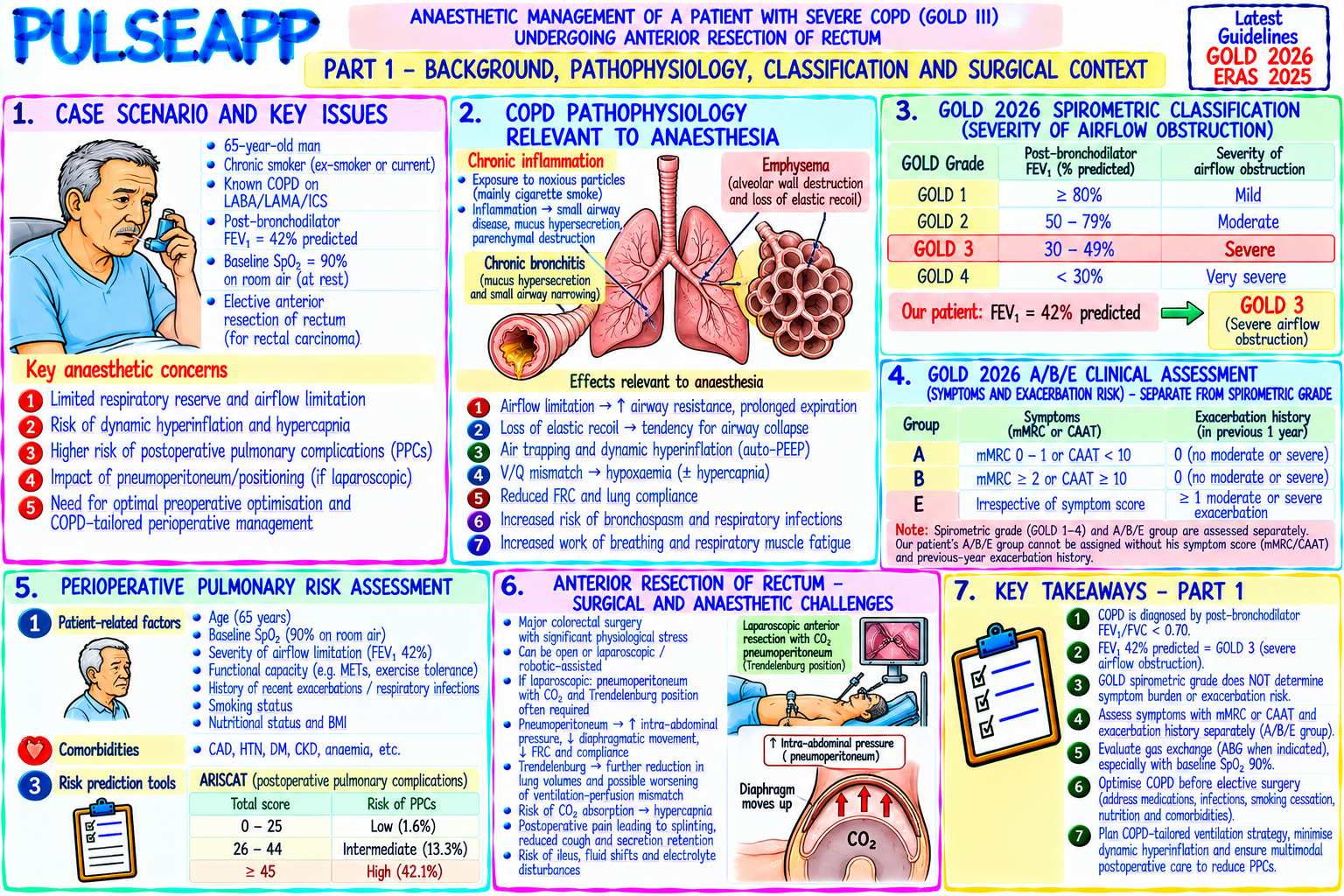
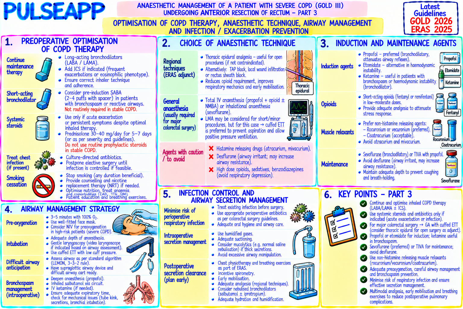
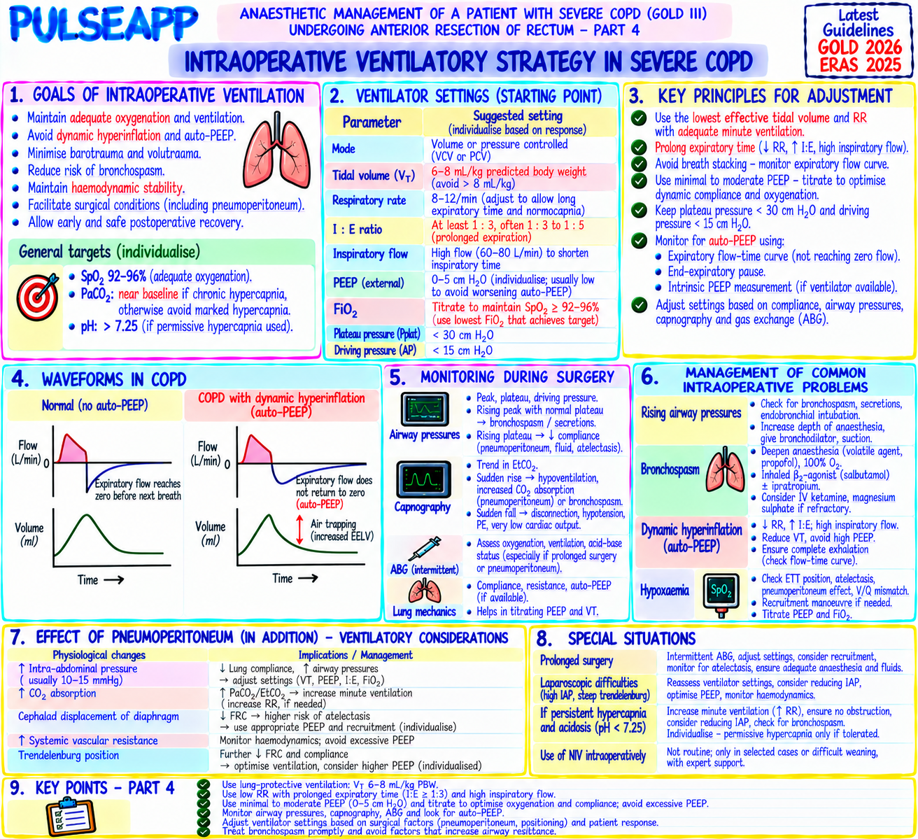
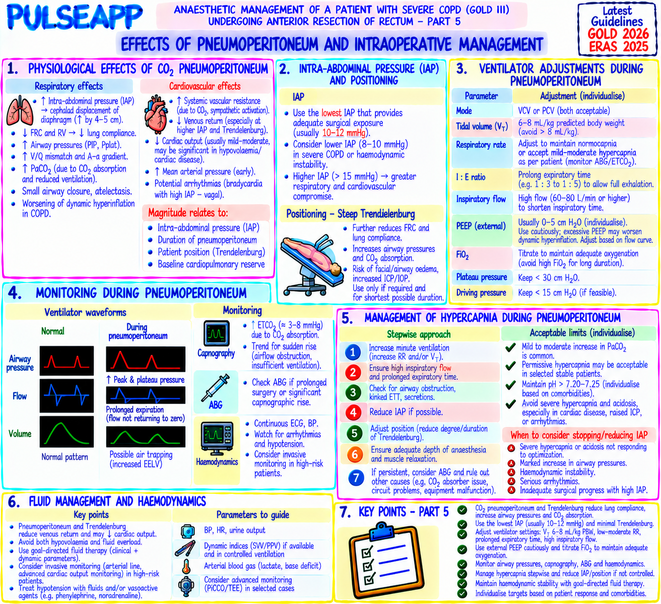
A 65-year-old male with GOLD III COPD (FEV1 42%, baseline SpO₂ 90% on room air, on home LABA/LAMA/ICS) requires elective anterior resection of rectum. Discuss preoperative optimisation, choice of anaesthetic technique, intraoperative ventilatory strategy, and postoperative pulmonary complication prevention.
collections Question Diagrams & Reference Images (2)
Question Reference Diagram
zoom_in View Image
Question Reference Diagram
zoom_in View Image
description Clinical Response
⚙ Core Concept
COPD represents a spectrum of airflow obstruction from reversible airway inflammation to fixed emphysematous destruction — but from the anaesthetic perspective, the universal consequence is increased work of breathing, air trapping (auto-PEEP), impaired gas exchange, and dramatically reduced pulmonary reserve. For major abdominal surgery, the PPC rate in GOLD III COPD exceeds 30–40%, making this one of the highest-risk perioperative respiratory scenarios. (GOLD 2023 Guidelines; Canet J — ARISCAT; Qaseem A — Ann Int Med; Miller's Anaesthesia 9th Ed)
A. GOLD Classification & Risk Assessment2 marks

GOLD Stage FEV1 Anaesthetic Risk Post-op Recommendation I (Mild) ≥80% Low — near-normal reserve Standard care; consider incentive spirometry II (Moderate) 50–79% Moderate — significant symptom burden Bronchodilators optimised; NIV plan post-op III (Severe) 30–49% High — this patient (42%); PPCs >30% Aggressive preoperative optimisation; HDU/ICU post-op; TEA analgesia IV (Very severe) <30% Very high; consider surgery alternatives MDT decision; ICU post-op; may need post-op ventilation

ARISCAT risk score: incorporates age, SpO₂ (<96% = highest risk — this patient 90%!), anaemia, upper abdominal/intrathoracic incision, duration >2 hours, emergency surgery, respiratory infection; this patient scores very high on SpO₂ alone (90% at rest) PaCO₂ >45 mmHg at rest → severe disease with CO₂ retention → very high post-op ventilatory failure risk; check ABG preoperatively

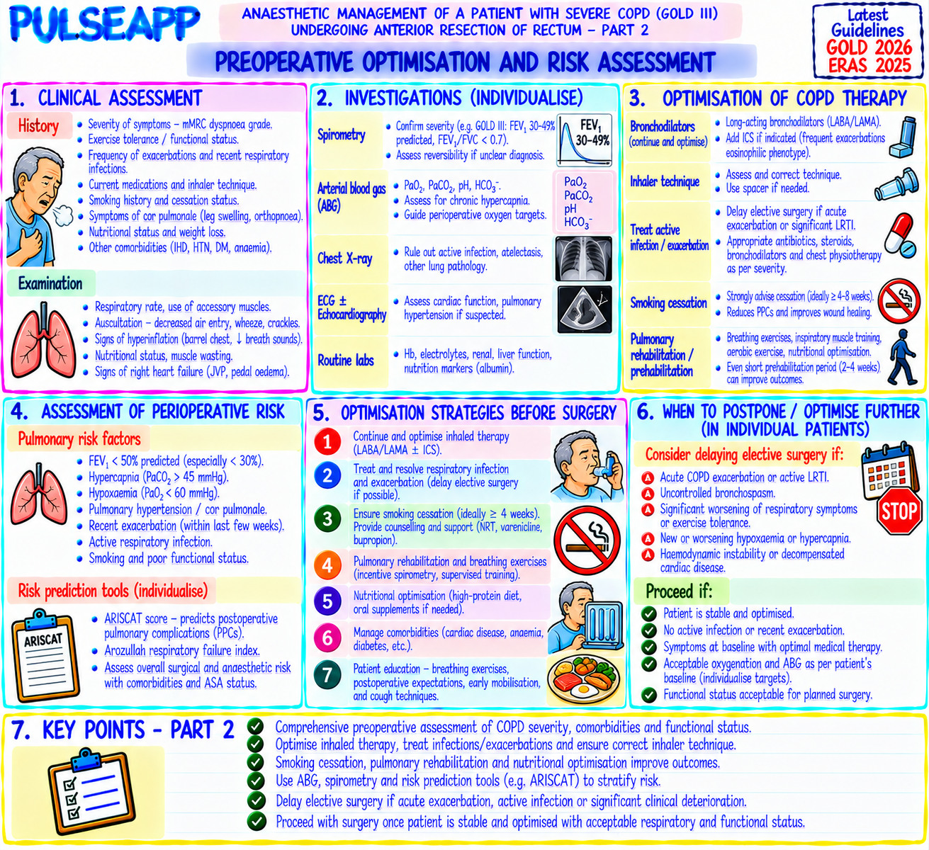
B. Preoperative Optimisation2 marks

Intervention Specific Action Smoking Absolute cessation ≥8 weeks before surgery; immediate cessation improves carboxyhaemoglobin within 12 hours and mucociliary function within 2–4 cessation weeks Bronchodilators Optimise existing LABA/LAMA/ICS regimen; add SABA (salbutamol MDI) PRN pre-operatively; continue all inhalers on morning of surgery with sip of water; spirometry re-check 4 weeks post-optimisation Physiotherapy Pre-operative breathing exercises, incentive spirometry (IS), and chest physiotherapy for secretion clearance; IS 10 repetitions hourly pre- and postoperatively reduces atelectasis rate by 30% Treat acute If purulent sputum/fever → antibiotics for ≥4–6 weeks before surgery; elective surgery during active exacerbation is contraindicated (triples PPC risk) exacerbation Pulmonary 4–8 week supervised exercise programme if FEV1 <50% and surgery is semi-elective; improves functional capacity and reduces PPCs rehabilitation

C. Intraoperative Anaesthetic Strategy3 marks

Regional Anaesthesia Preference Thoracic epidural (TEA) T6–T8: provides surgical anaesthesia ± sedation for laparotomy in selected patients; avoids GA and its airway/ventilatory consequences; reduces PPCs by 30% compared with GA; insert epidural preoperatively; combined epidural+GA preferred (epidural → superior post-op analgesia → better respiratory function post-op)

If GA Required (Laparoscopic Colorectal)

Bronchodilator pre-treatment: nebulised salbutamol 2.5 mg before induction via nebuliser

Induction: avoid histamine-releasing drugs (atracurium, morphine, thiopentone — all can cause bronchospasm); propofol (bronchodilator) preferred for induction; rocuronium for intubation

Airway: ETT preferred over LMA (secure airway needed for abdominal surgery + risk of bronchospasm); however LMA can be used for selected procedures if leak pressure >25 cmH₂O Ventilatory Settings for COPD Parameter Setting Rationale Tidal Volume 6–8 mL/kg IBW Lung-protective; avoid overdistension of already hyperinflated lung Respiratory Rate 8–12 breaths/min Lengthens expiratory time → complete exhalation → prevents gas trapping and auto-PEEP build-up (LOW) I:E ratio 1:3 or 1:4 (prolonged Standard 1:2 inadequate for obstructive physiology; prolonged expiratory time allows complete emptying through expiration) narrowed airways PEEP Minimal (3–5 cmH₂O) or Auto-PEEP already present; extrinsic PEEP >auto-PEEP level adds to air trapping; measure auto-PEEP by expiratory zero hold Permissive Accept PaCO₂ 50–60 Avoid normalising PaCO₂ at cost of excessive airway pressures; this patient may normally retain CO₂ (chronic hypercapnia mmHg hypercapnia — check preop ABG baseline) Volatile agent Sevoflurane preferred Bronchodilator properties; avoid desflurane (airway irritant, may trigger bronchospasm)

D. Postoperative Pulmonary Complication Prevention3 marks

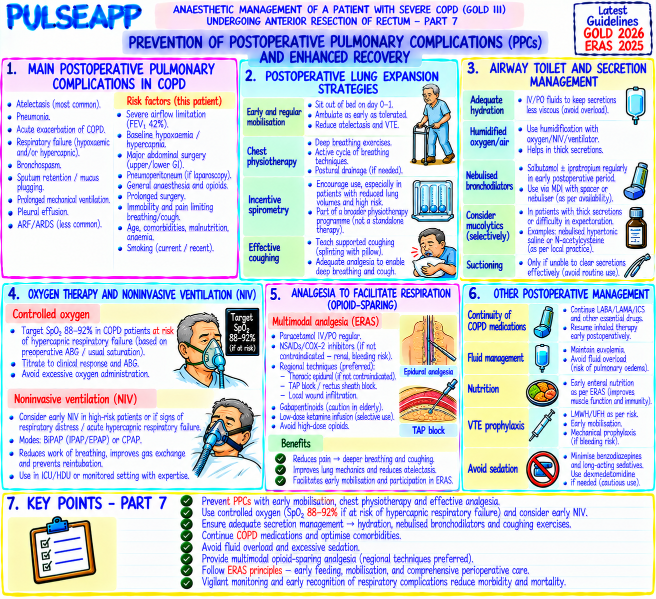
Epidural analgesia: continue thoracic epidural for 48–72 hours post-op → allows deep breathing and productive coughing → reduces splinting → reduces atelectasis and pneumonia; superior to IV morphine for COPD patients

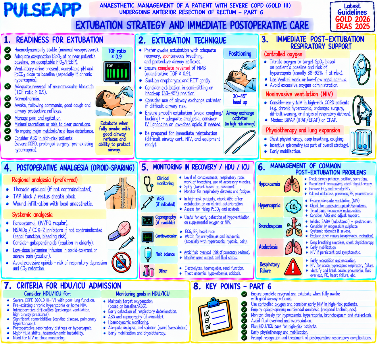
NIV/CPAP: plan for immediate BiPAP support post-extubation in GOLD III/IV patients; BiPAP settings: EPAP 5 cmH₂O + IPAP 12–16 cmH₂O; reduces reintubation rate; may be used prophylactically for first 24 hours

Early mobilisation: sitting out of bed day 1 post-op; physiotherapy-supervised ambulation; improves FRC and reduces atelectasis

Minimise opioids: opioid-sparing multimodal analgesia (TEA + paracetamol + NSAIDs); opioid-induced respiratory depression is catastrophic in this patient

HDU/ICU: book ICU/HDU bed preoperatively; plan for extended monitoring; daily spontaneous breathing trials if intubated post-op
🎤 Viva Corner
Q. Intraoperatively, auto-PEEP of 12 cmH₂O is measured. Peak airway pressure is 38 cmH₂O and the patient is becoming hypotensive. What is happening and how do you manage it?
Auto-PEEP of 12 cmH₂O with high peak pressures and hypotension in a COPD patient describes dynamic hyperinflation causing haemodynamic compromise — a complication of inadequate expiratory time allowing progressive gas trapping. The mechanism: each breath delivers a tidal volume but insufficient time is allowed for complete exhalation; the next breath starts before the previous one is fully exhaled → progressive lung hyperinflation → thoracic cage over-distension → intrathoracic pressure rises → compresses the great veins → reduces venous return → reduces cardiac output → hypotension (obstructive shock from dynamic hyperinflation — can mimic tension pneumothorax). Management: first, confirm there is no tension pneumothorax (clinical examination, immediate bilateral breath sounds — if unequal, emergency needle decompression); if bilateral breath sounds present → the picture is dynamic hyperinflation. Disconnect the patient from the ventilator and manually deflate the lungs by pressing on the chest (this allows the trapped gas to escape — an immediate dramatic improvement in haemodynamics may occur within seconds as venous return is restored). Simultaneously: give IV fluid bolus; vasopressors. Then reprogram the ventilator: reduce RR to 6–8 breaths/min; increase I:E to 1:4; reduce PEEP to zero; allow permissive hypercapnia; do not add extrinsic PEEP above the auto-PEEP level. The auto-PEEP must be reduced by lengthening expiratory time — this is the primary intervention. The specific manoeuvre of disconnecting the ventilator to confirm the diagnosis (if haemodynamics dramatically improve when the patient is off the ventilator and gas escapes → confirms dynamic hyperinflation as the cause) is a recognised emergency diagnostic and therapeutic step.
★ Examiner's Pearl
GOLD classification with FEV1 thresholds (I≥80%/II 50-79%/III 30-49%/IV<30%) must be reproduced. The ventilator settings table with specific I:E ratio (1:3 or 1:4) and low RR (8–12) are the most specifically tested numbers in COPD anaesthesia. Auto-PEEP measurement (expiratory hold manoeuvre) and the dynamic hyperinflation → obstructive shock mechanism with ventilator disconnect as the emergency treatment are high-yield viva topics.
GOLD. Global Strategy for Diagnosis, Management and Prevention of COPD 2023. Canet J et al. ARISCAT study (Anesthesiology 2010;113:1338). Qaseem A et al. Reducing risk for PPCs (Ann Intern Med 2006;144:575). Lohser J. Management of one-lung ventilation (Anesthesiol Clin 2008). Miller RD et al. Miller's Anaesthesia, 9th Ed.
lock Protected Clinical Document • Pulse App • Screenshots Strictly Prohibited
QUESTION 53 person Asked by .
bookmark_add

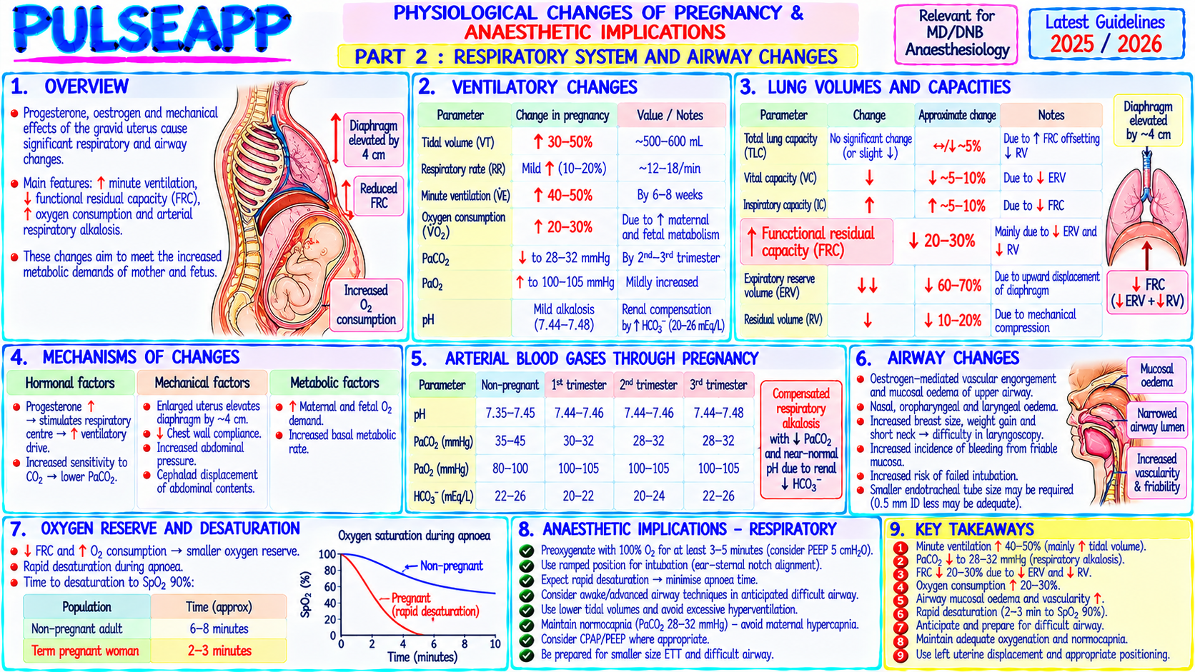
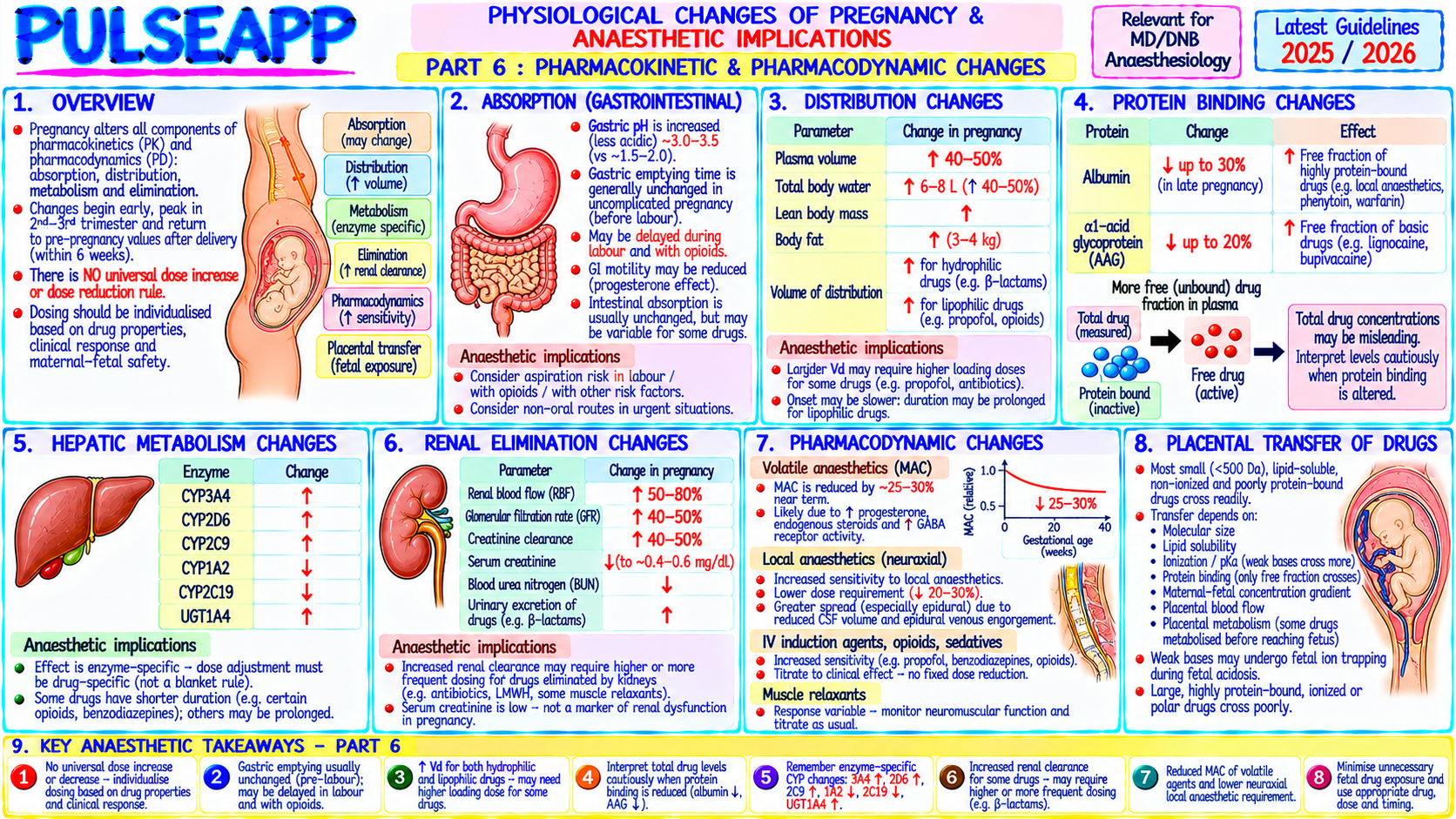
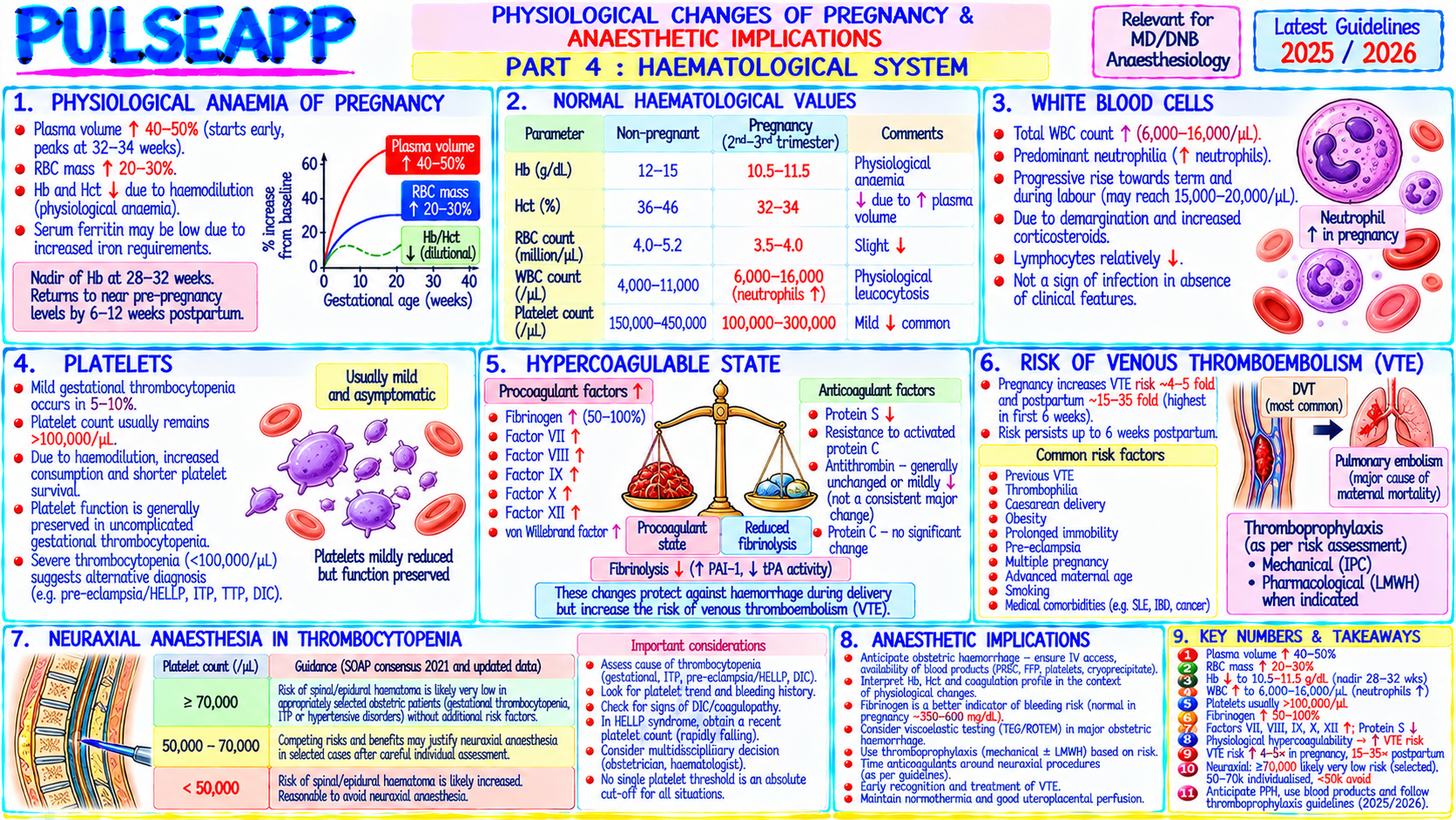
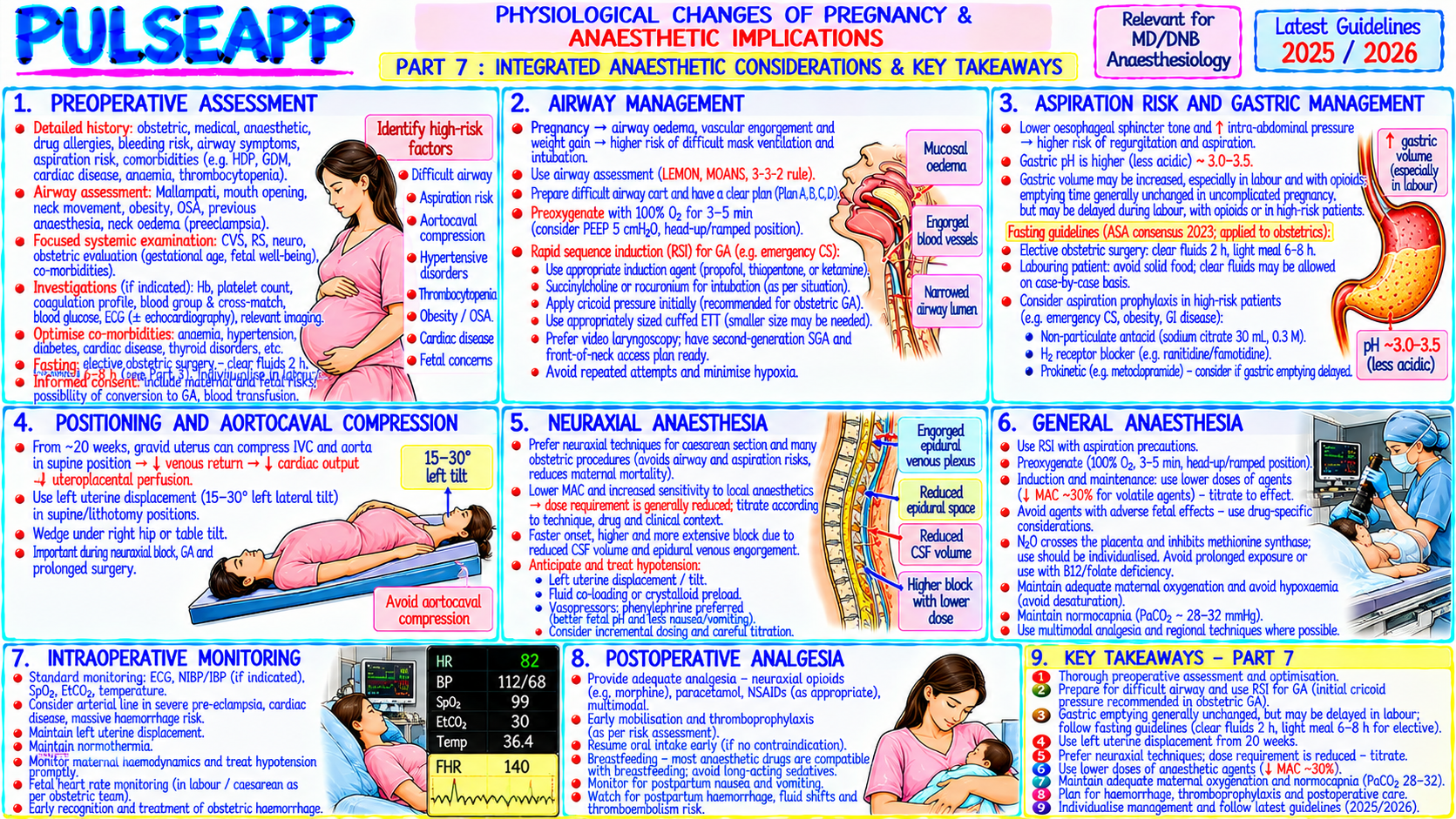
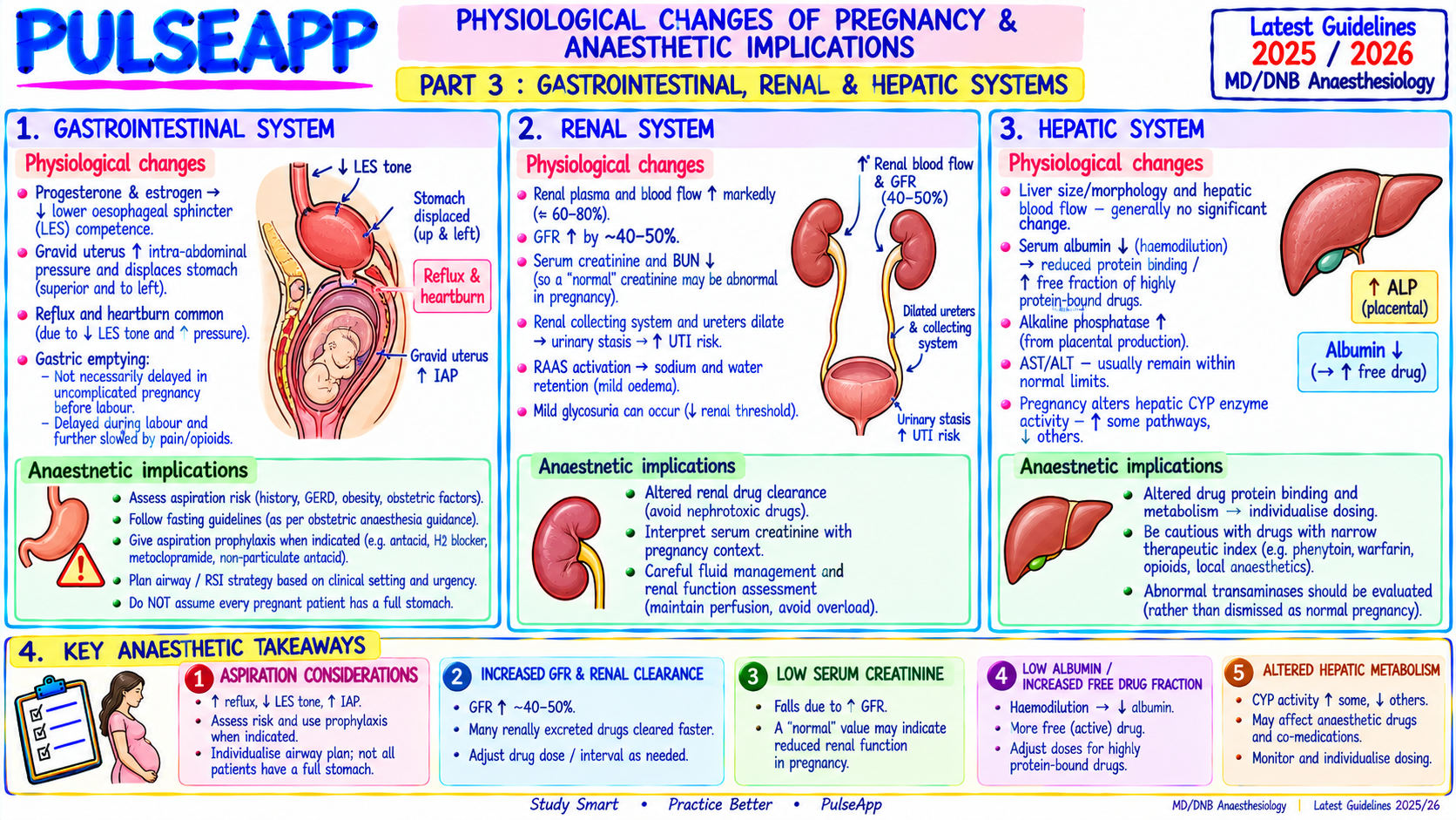
Describe the major physiological changes of pregnancy across all organ systems and their specific anaesthetic implications. Include: cardiovascular, respiratory, gastrointestinal, haematological, and pharmacokinetic changes.

collections Question Diagrams & Reference Images (1)
Question Reference Diagram
zoom_in View Image
description Clinical Response
⚙ Core Concept
Pregnancy produces profound physiological adaptations across every organ system — driven primarily by the hormonal milieu of progesterone, oestrogen, and human placental lactogen, and secondarily by the mechanical effects of the enlarging uterus. For the anaesthesiologist, these changes collectively produce: a difficult airway, rapid oxygen desaturation, aspiration risk, haemodynamic instability, and altered drug pharmacokinetics — every element of the anaesthetic challenge is affected. (Miller's Anaesthesia 9th Ed; Chestnut's Obstetric Anaesthesia; Pilkington S — physiological changes; RCOG; Capeless EL)
A. Cardiovascular Changes2 marks

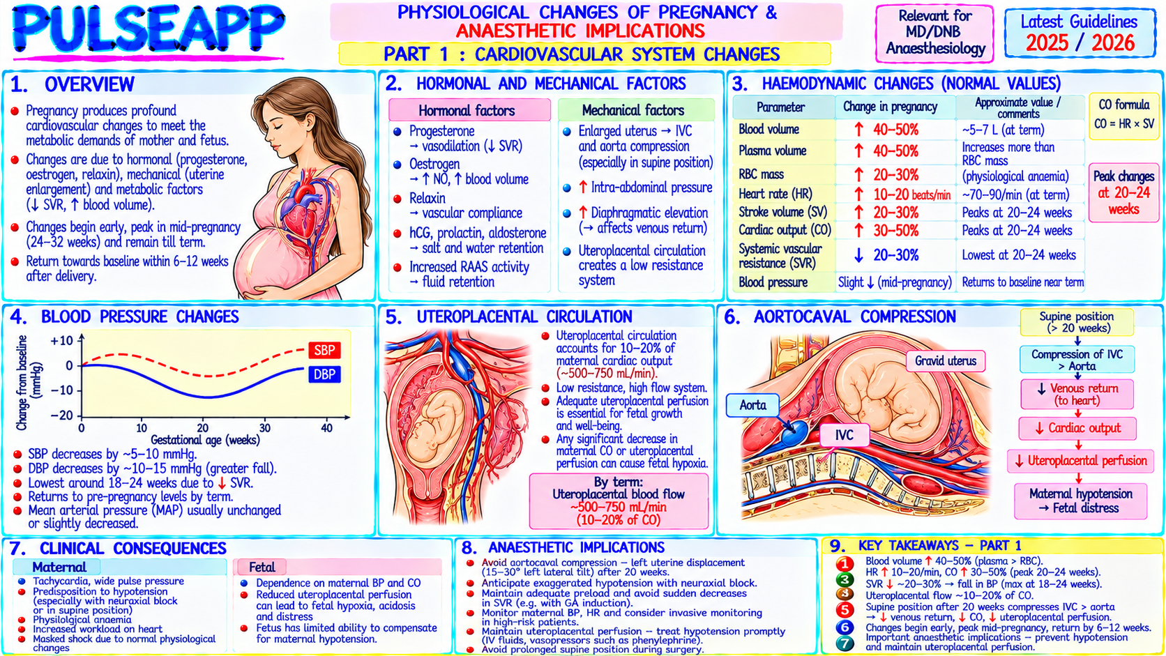
Parameter Change by Term Mechanism Anaesthetic Implication Blood ↑ 40–50% (plasma ↑45%, RBC Progesterone → aldosterone activation → Na⁺/water Physiological anaemia of pregnancy (Hb <100 = Volume ↑20%) — dilutional anaemia (Hb retention; oestrogen → renin-angiotensin; plasma pathological); increased blood volume provides falls to 105–110 g/L at term) expansion exceeds RBC production haemorrhage buffer but masks volume depletion Cardiac ↑ 40–50% by 28–32 weeks; CO ↑ Blood volume, ↓ SVR (progesterone → High CO masks underlying cardiac pathology; cardiac Output = HR × SV; HR ↑15–25 bpm; SV vasodilation), ↑ metabolic demand of the feto- disease patients may decompensate in late pregnancy; CO ↑25–30%; further ↑ 15% during placental unit peaks 32–36 weeks not at term labour contractions SVR ↓ 20% (vasodilation from Progesterone → smooth muscle relaxation; Low SVR → relative hypotension; regional anaesthesia → progesterone + prostacyclin) prostacyclin production ↑; results in low-resistance further ↓ SVR → risk of severe hypotension particularly in hyperdynamic circulation pre-existing vasoconstricted states Aortocaval Supine hypotension syndrome in Gravid uterus compresses IVC (right side) and aorta ALWAYS left lateral tilt (15°) for supine procedures after 20 Compression 10–15% at term; IVC (left lateral reduces this) when supine; collateral weeks; for cardiac arrest in pregnancy: manual uterine compression → ↓ venous return circulation (paravertebral, azygous veins) displacement + left lateral tilt during CPR; perimortem CS → ↓ CO → hypotension compensates partially in most patients within 5 minutes if no ROSC

B. Respiratory Changes2 marks

Parameter Change Anaesthetic Implication FRC ↓ 20% (400 mL) — most important respiratory ↓ Oxygen reservoir → rapid desaturation during apnoea; SpO₂ falls faster than in non-pregnant; change; diaphragm is pushed up 4 cm by gravid preoxygenation mandatory and may be less effective; closing capacity may exceed FRC in the uterus supine position → airway closure → shunt → worse oxygenation O₂ ↑ 20–30% at term (metabolic demands of fetus Rapid O₂ consumption + ↓ FRC → very short safe apnoea time; preoxygenate with 100% O₂ for 3–5 Consumption + uterus) minutes ALWAYS before obstetric RSI Tidal Volume TV ↑ 40%; RR ↑ 15%; MV ↑ 50%; driven by Hyperventilation of pregnancy: PaCO₂ falls to 30–32 mmHg (normal for pregnancy); "normal" PaCO₂ + MV progesterone stimulating the respiratory centre 40 mmHg indicates CO₂ retention in a pregnant patient; pH maintained by renal bicarbonate excretion (HCO₃⁻ falls to 18–21 mEq/L) Airway Oedema + hyperaemia of upper airway Smaller ETT (6.5–7.0 mm vs 7.5–8.0 mm standard); use nasal airways with extreme caution mucosa (oestrogen effect); capillary engorgement → (epistaxis); video laryngoscopy first-line for all obstetric intubations; Mallampati class increases as increased bleeding with instrumentation pregnancy progresses

C. Gastrointestinal Changes — Aspiration Risk2 marks

Gastric emptying: progesterone relaxes lower oesophageal sphincter (LOS) → ↓ LOS tone → regurgitation risk; mechanical displacement of stomach by uterus changes gastric axis; opioids during labour dramatically slow gastric emptying → a labouring woman who received opioids should be treated as having a full stomach regardless of fasting time

Gastric acid: ↑ gastric acid production (gastrin from placenta); the combination of ↓ LOS tone + ↑ gastric acid = "at risk for aspiration" from 16–20 weeks gestation

Aspiration prophylaxis: ranitidine 150 mg oral or 50 mg IV (↓ gastric acid pH); sodium citrate 30 mL oral (non-particulate antacid — immediate neutralisation of gastric acid); metoclopramide 10 mg IV (↑ gastric emptying, ↑ LOS tone); RSI protocol for any general anaesthesia after 16–20 weeks; clear liquid fasting 2 hours, light meal 6 hours (but treat as full stomach if in active labour with opioids)

D. Haematological Changes2 marks

Physiological anaemia: dilutional (plasma ↑45%, RBC ↑20%); Hb 105–115 g/L normal at term; true anaemia in pregnancy = Hb <100 g/L

Hypercoagulability: Virchow's triad complete in pregnancy — hypercoagulability (↑ clotting factors I, VII, VIII, X, XII; ↓ protein S; acquired resistance to protein C), venous stasis (↑ venous pressure in legs; ↓ venous tone from progesterone), and endothelial injury (from placental trauma, delivery); DVT risk ↑5× vs nonpregnant; PE is leading cause of maternal mortality in the UK

VTE prophylaxis: LMWH for high-risk pregnancies (immobility, thrombophilia, previous DVT); graduated compression stockings; early ambulation; timing of neuraxial anaesthesia relative to last LMWH dose (unfractionated 4 hours; LMWH 12 hours prophylactic or 24 hours therapeutic dose)

Platelet count: mild thrombocytopenia of pregnancy (gestational thrombocytopenia — Plt 70–150 × 10⁹/L at term) — benign; does NOT contraindicate epidural; contraindication threshold for neuraxial anaesthesia: Plt <70–80 × 10⁹/L (institution-specific) with normal platelet function

E. Pharmacokinetic Changes2 marks

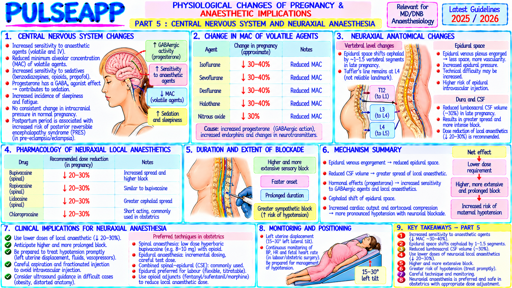
Parameter Change Drug Effect Plasma Albumin ↓ 20–30% → less drug protein-bound → more free (active) Propofol, thiopentone, local anaesthetics: increased free fraction → greater protein drug effect per dose; reduce doses accordingly binding Renal GFR ↑50% → creatinine falls to 0.5–0.6 mg/dL (normal pregnant); Renally-cleared drugs (aminoglycosides, digoxin) have shorter t½ → may need clearance renal tubular secretion ↑ higher/more frequent doses MAC MAC ↓ 25–40% from early pregnancy (high progesterone → Standard MAC concentrations produce deeper anaesthesia in pregnant (volatile GABAergic CNS depression) patients; doses should be reduced; risk of cardiovascular depression agents) Epidural LA Epidural veins engorged (↓ epidural space volume) → same LA volume Reduce epidural and spinal doses by 25–30%; a "standard" dose produces a dose spreads further → blocks are 25–30% more extensive higher block than expected in pregnancy

🎤 Viva Corner
Q. Why does a pregnant patient at 36 weeks desaturate faster than a non-pregnant adult during apnoea, and what specific preoxygenation strategies maximise the safe apnoea time?
Two simultaneous physiological changes conspire to produce rapid desaturation during apnoea in the near-term pregnant patient. First, FRC is reduced by approximately 20% (400 mL) due to the diaphragm being displaced cranially by the gravid uterus — this reduces the oxygen reservoir stored in the lungs at endexpiration. Second, oxygen consumption is increased by 20–30% to meet the metabolic demands of the fetus, placenta, and increased maternal cardiac and respiratory work. The rate of oxygen removal from the alveolar gas during apnoea is proportional to consumption; with 30% higher O₂ consumption depleting a 20% smaller reservoir, SpO₂ falls approximately 2–3 times faster than in a non-pregnant adult of equivalent size. Additionally, in the supine position (required for intubation), closing capacity may exceed FRC in pregnant women → small airway collapse → further impaired gas exchange even during preoxygenation. Strategies to maximise safe apnoea time: First, preoxygenate with 100% O₂ for 3–5 minutes via a tight-fitting face mask — this denitrogenates the FRC, maximising the oxygen store (replaces 78% N₂ with 100% O₂, dramatically increasing the available oxygen per litre of FRC). Second, use high-flow nasal O₂ (HFNO) at 30–60 L/min simultaneously during preoxygenation and maintained through the apnoeic intubation period (apnoeic oxygenation via transnasal high-flow) — this technique can extend safe apnoea time significantly by providing continuous O₂ delivery via nasal turbinates even when the patient is apnoeic and the airway is being instrumented, potentially extending safe apnoea to 5–10 minutes vs 3–4 minutes without it. Third, 20° head-up positioning during preoxygenation — this reduces diaphragmatic splinting by the uterus, increases FRC toward a more vertical position compared to the traditional supine for intubation, and is increasingly used as the standard preoxygenation position for obstetric RSI.
★ Examiner's Pearl
The FRC reduction (↓20%, 400 mL) combined with ↑O₂ consumption (↑20–30%) is the specific two-factor explanation for rapid desaturation — state both components, not just FRC. MAC reduction in pregnancy (↓25–40% from progesterone) means standard volatile concentrations are excessive — this is the most commonly missed pharmacological change. The epidural dose reduction (25–30% less because engorged epidural veins reduce space) explains why obstetric blocks are more extensive than expected and is specifically tested.
Pilkington S et al. Increase in Mallampati score during pregnancy (BJA 1995). Capeless EL, Clapp JF. Cardiovascular changes in early phase of pregnancy (Am J Obstet Gynecol 1989). Chestnut DH et al. Chestnut's Obstetric Anesthesia, 6th Ed. Miller RD et al. Miller's Anaesthesia, 9th Ed, Chapter 77. RCOG Green-top Guidelines.
lock Protected Clinical Document • Pulse App • Screenshots Strictly Prohibited
QUESTION 54 person Asked by .
bookmark_add

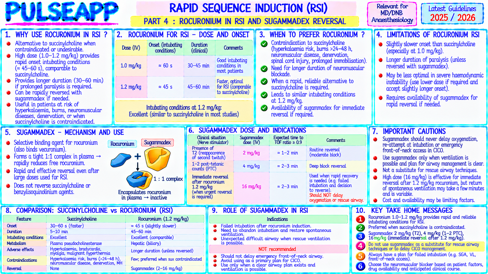
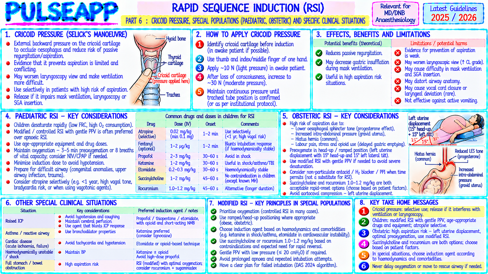
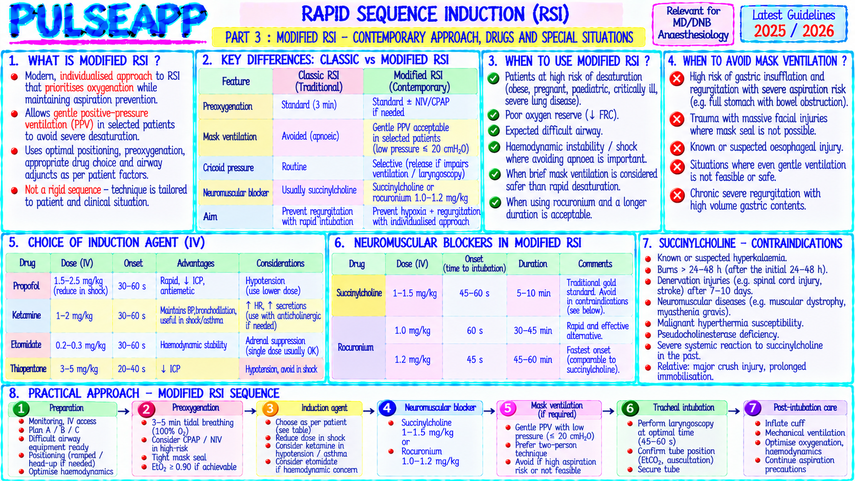
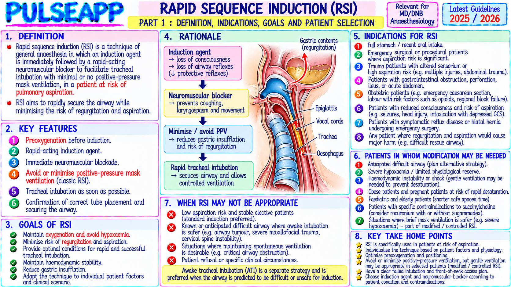
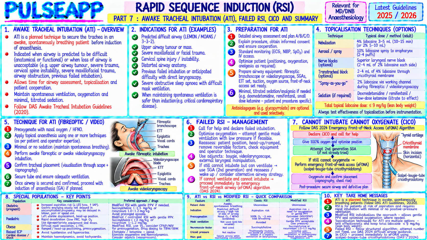
Define RSI and list its indications. Describe the classic RSI technique (cricoid pressure, thiopentone + succinylcholine). Outline modifications including: rocuronium-sugammadex RSI, preoxygenation strategies, sellick's manoeuvre controversy, and special situations (paediatric RSI, awake RSI).

collections Question Diagrams & Reference Images (1)
Question Reference Diagram
zoom_in View Image
description Clinical Response
⚙ Core Concept
RSI is the technique for securing the airway in patients at risk of pulmonary aspiration of gastric contents — where the usual practice of ventilating the patient by face mask between induction and intubation is abandoned to minimise the interval between induction and definitive airway control. Every component of the technique — the choice of drugs, the application of cricoid pressure, the management of failed intubation — is designed to reduce this critical interval while maximising the chance of successful intubation on the first attempt. (Higgs A et al. — DAS RSI Guidelines 2018; Morris J — cricoid pressure controversy; El-Orbany M; Miller's Anaesthesia 9th Ed; Sajayan A)
A. Indications for RSI2 marks

Category Examples Full stomach / known Emergency surgery (<6 hours from last solid meal); trauma (pain + opioids delay gastric emptying); inadequate fasting (non-compliant aspiration risk patient, unknown time of last meal); post-pyloric obstruction (ileus, bowel obstruction); hiatus hernia + severe GORD with symptoms at rest Physiological/anatomical Pregnancy (>16–20 weeks); opioid administration within 4 hours; diabetic gastroparesis (particularly insulin-dependent); renal failure; acute delay in gastric abdominal emergency (pain reflex slows gastric emptying) emptying Raised intra-abdominal Morbid obesity; ascites; peritoneal dialysis; bowel obstruction pressure

B. Classic RSI Technique3 marks

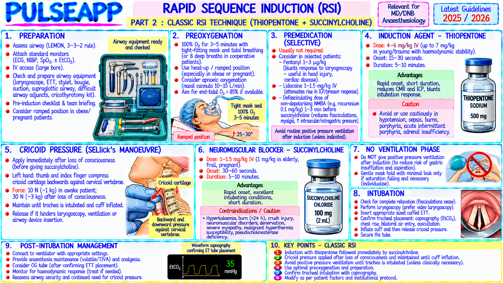
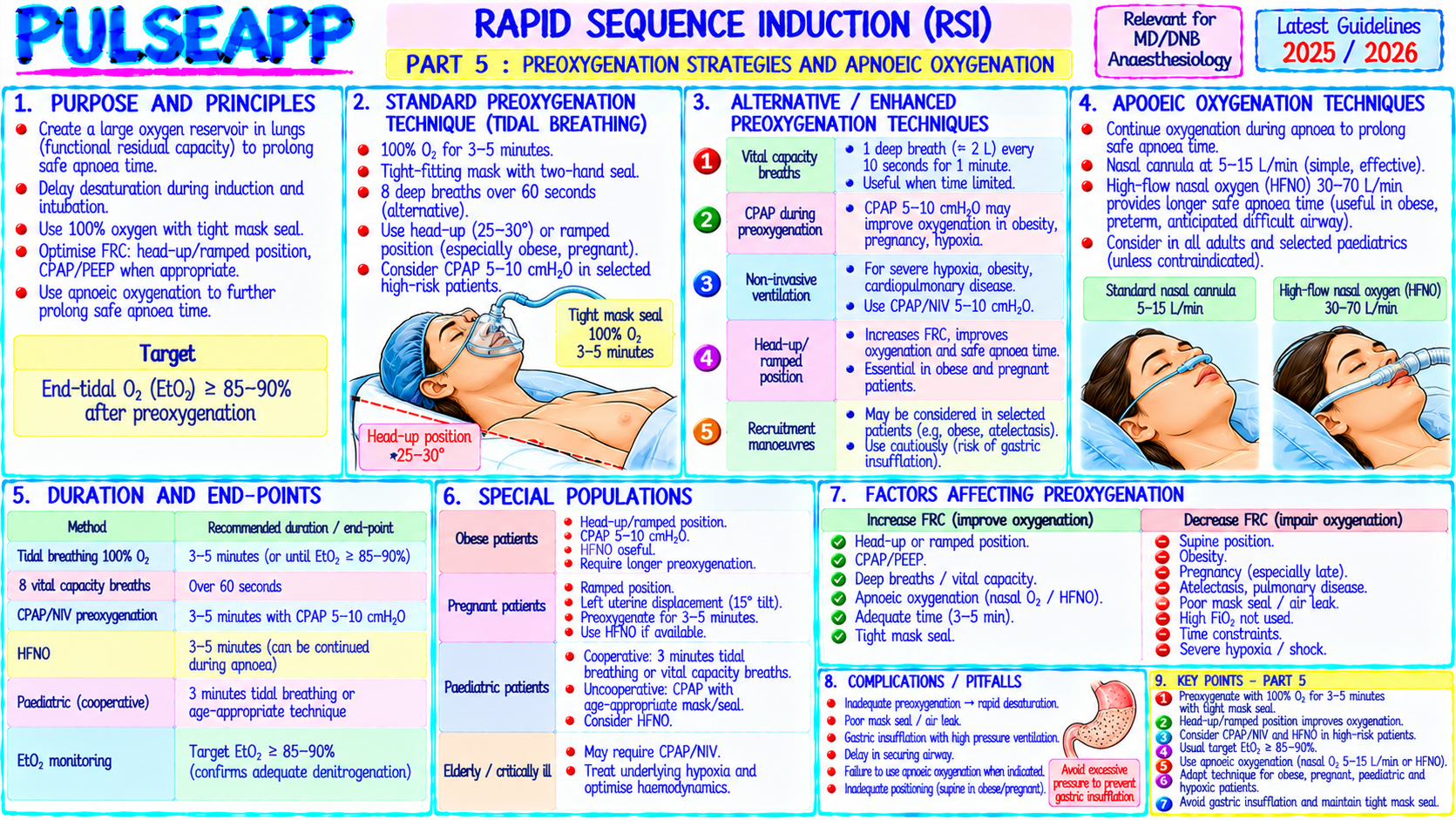
1. Preoxygenation: 3–5 minutes 100% O₂ via tight-fitting face mask (SpO₂ ≥99%); 4 vital capacity breaths as alternative if time critical; head-up 20° position increases FRC and tolerance of apnoea 2. Pre-oxygenation assessment: confirm difficult airway assessment completed; DAS algorithm available; team briefed; sugammadex 16 mg/kg drawn and ready if rocuronium used 3. Induction agent (rapid IV): thiopentone 4–5 mg/kg IV (classic choice — extremely rapid and reliable LOC in <30 sec; reduces ICP; propofol 2–2.5 mg/kg is the modern alternative — more cardiovascular depression but associated with less PONV and lower aspiration risk from earlier return of reflexes) 4. Cricoid pressure (Sellick's manoeuvre): applied simultaneously with induction drug injection by a trained assistant; 10 N (cricoid just visibly displaces) during awake phase → 30 N after LOC; maintained until ETT position confirmed; specific technique: steady downward pressure on the cricoid cartilage (not thyroid) compresses the cricoid ring against the vertebral body → occludes the upper oesophagus → prevents passive regurgitation of gastric contents into the pharynx 5. Succinylcholine 1.5 mg/kg IV: the classic RSI NMB; fastest onset (60 seconds) of any NMB; OR rocuronium 1.2 mg/kg (equivalent intubating conditions at 60 sec; sugammadex 16 mg/kg available for reversal if CICO) 6. No bag-mask ventilation between induction and intubation in classic RSI (avoids gastric insufflation); if SpO₂ falls <93% during apnoea → gentle bag-mask ventilation acceptable (modern modification) with cricoid pressure maintained 7. Intubation at 60 seconds: laryngoscopy + intubation at the time of peak NMB effect; no waiting for response to painful stimuli; direct or video laryngoscopy based on anticipated airway 8. Confirm ETT position: ETCO₂ waveform (sustained over 6 breaths) + bilateral breath sounds + SpO₂ maintenance; inflate cuff; release cricoid pressure once position confirmed; secure ETT

C. Cricoid Pressure — Evidence & Controversy2 marks

Sellick's Manoeuvre — What the Evidence Shows

Arguments FOR: Reduces regurgitation in cadaveric studies; still recommended by RCOA, DAS 2018, and most obstetric guidelines as standard of care

Arguments AGAINST: Reduces LOS competence at 30 N (paradoxically may allow regurgitation); impairs laryngoscopic view in up to 20% of cases; incomplete protection (only works against passive regurgitation, not active vomiting); no randomised controlled trial demonstrating reduced aspiration morbidity; may cause oesophageal rupture if the patient vomits against closed cricoid

DAS 2018 guidance: Cricoid pressure should be applied but must be released immediately if it impairs laryngoscopic view or interferes with intubation attempt; the airway takes priority over aspiration prevention

D. Rocuronium-Sugammadex RSI — Modern Alternative2 marks

Rocuronium 1.2 mg/kg provides equivalent intubating conditions to succinylcholine 1.5 mg/kg at 60 seconds; sugammadex 16 mg/kg immediately available for reversal if CICO

Advantages over succinylcholine: no contraindications in burns, denervation, MH susceptibility, pseudocholinesterase deficiency, hyperkalaemia risk; longer clinical duration (30–45 min vs 10 min) which can be an advantage (no recurarisation) or disadvantage (if airway fails and spontaneous recovery is needed)

CICO scenario: rocuronium RSI + CICO → sugammadex 16 mg/kg → spontaneous ventilation returns in 2–3 min → "wake and manage awake" strategy; this converts a catastrophic situation into a recoverable one

Where succinylcholine still preferred: where briefest possible duration is specifically needed (very brief intubation, hyperkalemia acceptable, no CICO concern with established airway plan)

🎤 Viva Corner
Q. A scrub nurse applies cricoid pressure during your RSI. You cannot see the glottis at laryngoscopy — only Cormack-Lehane Grade III. What do you do with the cricoid pressure?
Release cricoid pressure immediately. The DAS 2018 RSI guidelines explicitly state that cricoid pressure must be released if it impairs laryngoscopy or intubation. The rationale: cricoid pressure is applied to reduce the risk of passive aspiration of gastric contents — a potential harm. But the primary risk in an RSI scenario is failed intubation and inability to oxygenate — a certain, immediate, life-threatening harm if not managed promptly. When cricoid pressure converts a Grade I/II view to a Grade III view (Cormack-Lehane classification based on best glottic structures visible), it is actively making the most critical airway management step harder and increasing the risk of failed intubation. At this point, the theoretical aspiration protection benefit of cricoid pressure is outweighed by the concrete harm of impaired intubation. After releasing cricoid pressure: immediately attempt to optimise the view by applying BURP (Backward, Upward, Rightward Pressure on the thyroid cartilage applied by the intubating anaesthesiologist's assistant) — this is different from cricoid pressure; BURP moves the larynx to improve glottic visualisation rather than occlude the oesophagus; use a bougie as the primary intubating adjunct if view is C-L Grade III; consider videolaryngoscopy (most modern RSI protocols use videolaryngoscopy as first line anyway for difficult airway expected or emergency context). If still cannot intubate after releasing cricoid: move to DAS Plan B (SAD insertion) following the failed intubation algorithm — the priority is oxygenation.
★ Examiner's Pearl
The eight-step RSI sequence must be presented in the exact clinical order — preoxygenation → pre-assessment → induction agent → cricoid pressure → NMB → no mask ventilation → intubation at 60 seconds → confirm + release cricoid. The DAS 2018 guidance on cricoid pressure release if impairing view is the specific guideline update examiners test — the answer "release cricoid if it worsens view" must be stated with the rationale (airway takes priority). Rocuronium RSI dose (1.2 mg/kg — double the standard intubating dose) is specifically tested as different from the routine 0.6 mg/kg intubating dose.
Higgs A et al. DAS guidelines for RSI 2018 (Anaesthesia 2018;73:369-401). Sajayan A et al. Current practice of RSI in adults in UK (BJA 2016;117(S1):i69-i74). El-Orbany M, Connolly LA. RSI — Current appraisal (Anesth Analg 2010;110:1318-1325). Morris J, Cook TM. RSI: a national survey of practice (Anaesthesia 2001;56:1090-1115). Miller RD et al. Miller's Anaesthesia, 9th Ed.
lock Protected Clinical Document • Pulse App • Screenshots Strictly Prohibited
QUESTION 55 person Asked by .
bookmark_add

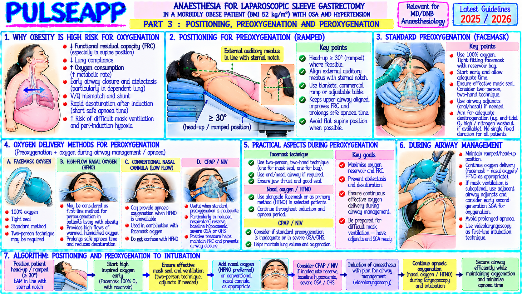
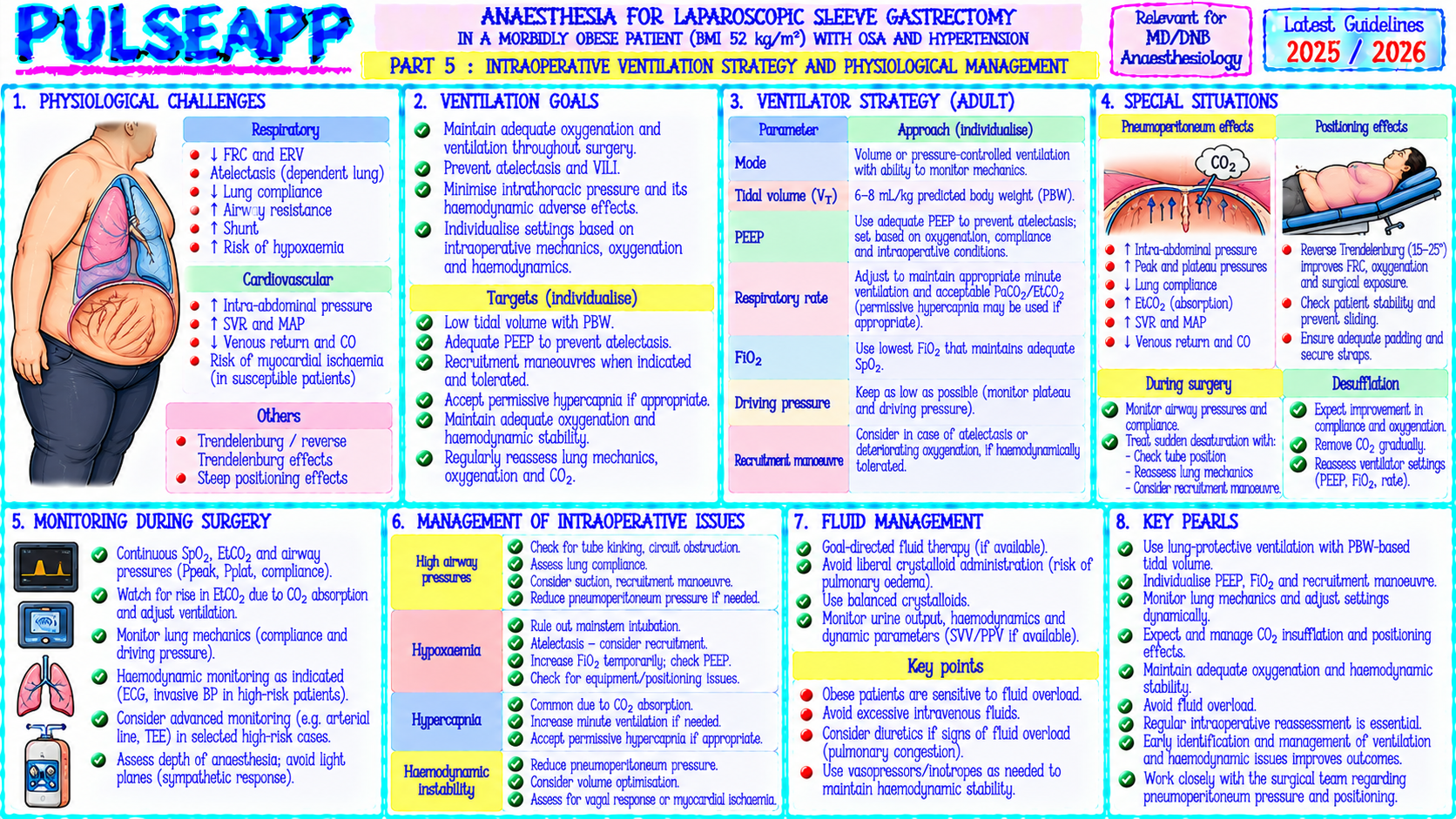
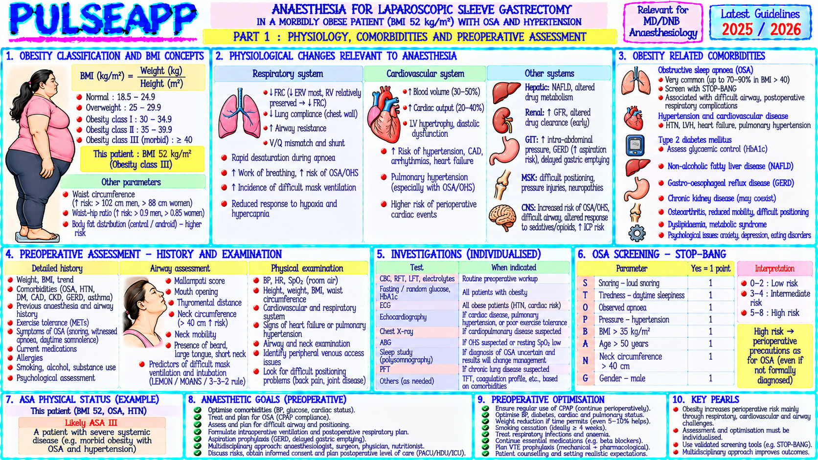
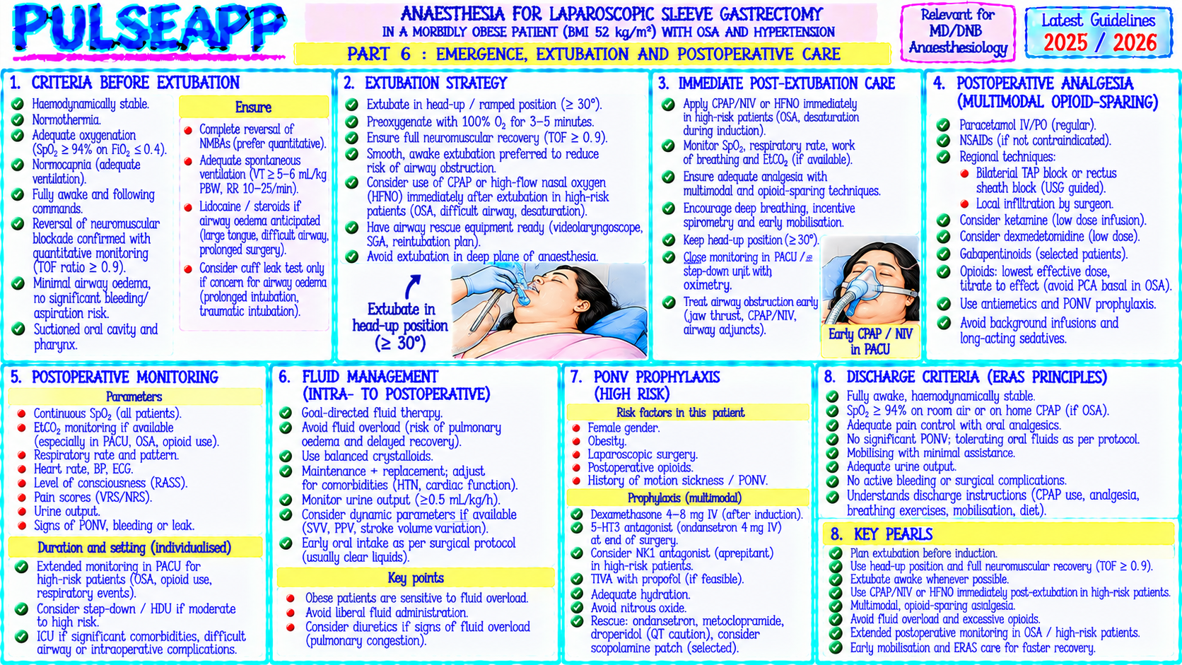
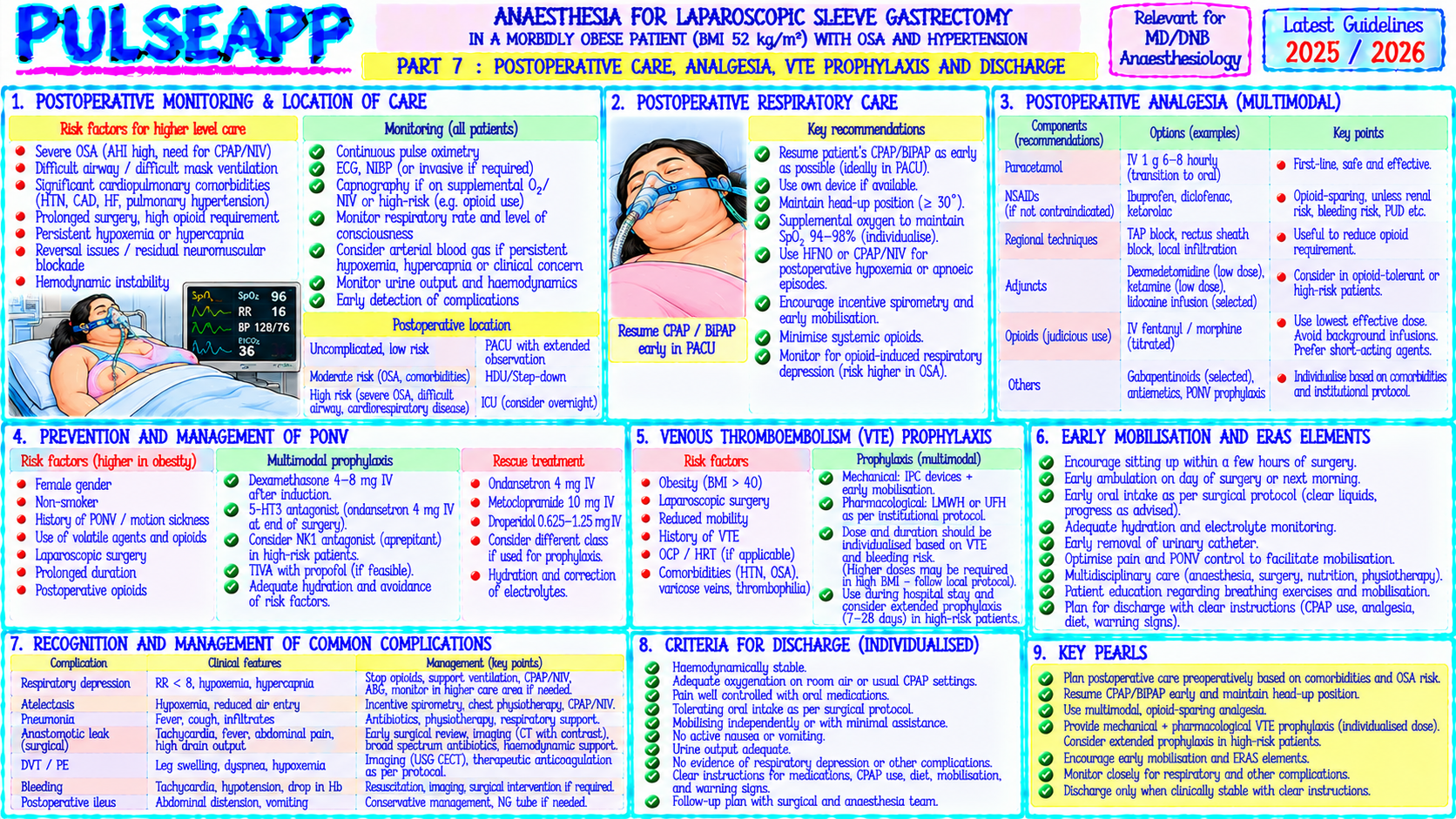
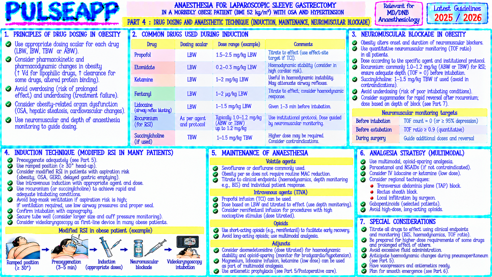
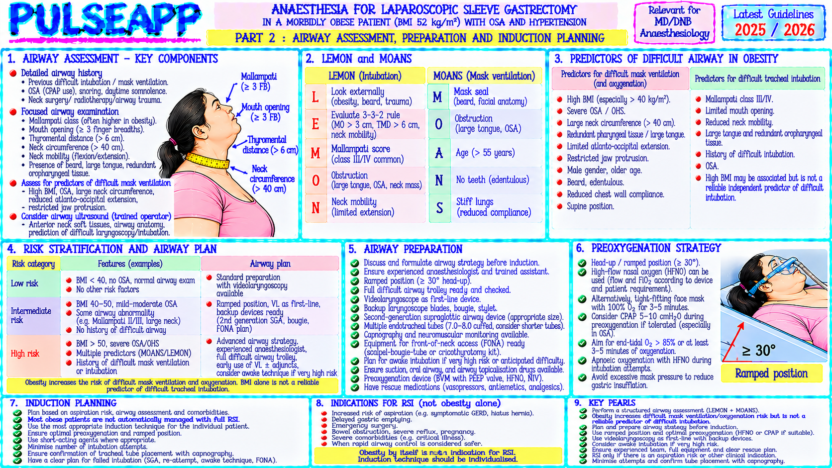
A 45-year-old female, BMI 52 kg/m², with OSA and hypertension, requires laparoscopic sleeve gastrectomy. Discuss: airway assessment and management, preoxygenation, positioning, ventilation strategy, drug dosing, and postoperative complications.

description Clinical Response
⚙ Core Concept
Morbid obesity (BMI ≥40) creates anaesthetic challenges across every phase of care — the most immediately dangerous being the difficult airway combined with markedly reduced safe apnoea time, and the most insidious being the desaturation that occurs rapidly in the supine position from loss of FRC. Yet with systematic preparation — ramped positioning, preoxygenation optimisation, appropriate drug dosing, and planned airway management — outcomes for morbidly obese patients can equal those of normal-weight patients. (STOP-BANG; El-Orbany; Neligan P — Thorax 2009; Miller's Anaesthesia 9th Ed; Nightingale CE — Anaesthesia 2015)
A. Preoperative Assessment2 marks

STOP-BANG for OSA: Snoring (loud enough to be heard through closed door?), Tired (tired/fatigued during daytime?), Observed apnoeas (by partner?), blood Pressure (hypertension?), BMI >35, Age >50, Neck circumference >40 cm, Gender male; ≥5 positive = high risk for severe OSA; this patient with BMI 52 + hypertension + female = at minimum moderate risk

Airway assessment: Mallampati class (higher in obese); neck circumference >40–45 cm is the strongest predictor of difficult intubation in obese patients (independently of Mallampati); limited neck extension; LEMON score; plan for video laryngoscopy as first-line tool

Investigations: ECG (RVH, LVH from chronic hypoxaemia/hypertension); echocardiogram (pulmonary hypertension, RV function); spirometry (restrictive pattern — ↓ FVC, ↓ FRC, ↓ ERV, normal FEV1/FVC; severe OSA with pulmonary hypertension = very high risk); ABG if suspected daytime CO₂ retention (obesity hypoventilation syndrome)

CPAP pre-admission: if OSA diagnosed — patient should be on home CPAP; bring CPAP machine to hospital; use throughout recovery and post-op
B. Preoxygenation & Positioning2 marks

Ramped position (HELP position — Head Elevated Laryngoscopy Position): pillows/foam wedge under head and shoulders to align the external auditory meatus with the sternal notch; this optimises the laryngoscopic view (brings glottis into line of sight) in obese patients and simultaneously maximises the FRC benefit of head-up positioning; the HELP position reduces failed first-attempt intubation rates in obese patients by ~50%

Preoxygenation: 3–5 minutes 100% O₂; consider non-invasive ventilation (BiPAP/CPAP 5–10 cmH₂O) during preoxygenation — applies positive pressure to stent open collapsed small airways in obese patients, increasing FRC and O₂ reserve; high-flow nasal O₂ (HFNO) 60 L/min through apnoeic period extends safe apnoea time; target ETO₂ >90% at end of preoxygenation to confirm denitrogenation Safe apnoea time is markedly reduced in morbid obesity — SpO₂ may fall to critical levels within 90 seconds of apnoea vs 8+ minutes in a normal-weight patient; this is the most important practical implication of FRC reduction in obesity

C. Intraoperative Anaesthetic Management3 marks

Drug Dosing in Obesity Drug Dose Basis Rationale Propofol Lean Body Weight (LBW) for induction dose; LBW for induction — obese patients have proportionally less increase in central compartment (induction) Total Body Weight (TBW) for maintenance volume vs LBW; TBW for maintenance — larger volume of distribution for fat-soluble propofol infusion Succinylcholine Total Body Weight (TBW) NMBs distribute into extracellular fluid which increases in proportion to TBW in obesity / Rocuronium Sugammadex Actual Body Weight (ABW/TBW) Must encapsulate total circulating rocuronium based on TBW dosing; underdosing causes incomplete reversal Fentanyl / Lean Body Weight (LBW) Opioids should be dosed conservatively in obese patients — increased sensitivity to respiratory Morphine depression; avoid boluses; prefer remifentanil infusion for precise titration Local Ideal Body Weight (IBW) LA does not distribute into fat; TBW dosing would cause LAST; reduce dose as epidural space anaesthetics smaller (fatty infiltration) (regional)

Ventilation Strategy (Laparoscopic — Trendelenburg + Pneumoperitoneum)

Lung-protective ventilation: TV 6–8 mL/kg IBW (NOT TBW — obese lungs are not proportionally larger); using TBW for TV in obesity = massive overventilation = VILI PEEP 10–12 cmH₂O: morbidly obese patients develop rapid atelectasis; higher PEEP than standard needed to prevent alveolar collapse in dependent lung regions; Trendelenburg + pneumoperitoneum → severe FRC reduction → PEEP 10–12 required

Recruitment manoeuvre: 30 cmH₂O sustained inflation for 30 seconds at position change and after pneumoperitoneum then maintain PEEP 10–12; reduces atelectasis significantly

FiO₂: avoid 100% O₂ unnecessarily — absorption atelectasis; target SpO₂ ≥95% with minimum necessary FiO₂

D. Postoperative Complications3 marks

Complication Risk in Obese Prevention Hypoxaemia / Very high; OSA + opioids + supine positioning + upper Semi-recumbent position (30–45° head-up) in recovery; CPAP immediately postObstructive abdominal incision extubation; supplemental O₂; nurse 1:1 in recovery; SpO₂ monitoring ≥12 hours apnoea post-op; avoid opioids DVT/PE ↑ VTE risk 2–3× vs normal weight (venous stasis + LMWH (dose based on TBW for adequate anti-Xa levels — standard prophylactic hypercoagulability + immobility) dose often insufficient); pneumatic compression stockings; early mobilisation; consider extended LMWH prophylaxis for 28 days post-bariatric surgery Wound Impaired wound healing (poor oxygenation of subcutaneous Laparoscopic preferred to open (fewer SSIs); adequate subcutaneous closure; complications fat); SSI 2–3× higher antimicrobial sutures; glycaemic control (<10 mmol/L) Rhabdomyolysis Specific to morbidly obese patients positioned on standard Adequate padding under all pressure points; limit case duration; CK monitoring operating tables; pressure necrosis of dependent muscle post-operatively for cases >3 hours; IV fluids to maintain UO ≥1 mL/kg/hr groups (particularly buttocks in lithotomy, back in supine)

🎤 Viva Corner
Q. Why do you use Ideal Body Weight (not Total Body Weight) for tidal volume calculation in morbid obesity?
Tidal volume in ventilated patients is calculated based on Ideal Body Weight (IBW, also called predicted body weight) rather than Total Body Weight because the functional lung volume — the actual amount of alveolar tissue available for gas exchange — does not increase proportionally with weight gain in obesity. IBW is calculated from height and sex: IBW (male) = 50 + 2.3 × (height in inches above 5 feet); IBW (female) = 45.5 + 2.3 × (height in inches above 5 feet). The lung parenchyma, alveolar surface area, and respiratory muscle force are determined primarily by height and body frame, not by the additional adipose tissue that defines obesity. A morbidly obese person at 170 cm, 130 kg has approximately the same amount of actual alveolar tissue as a lean person of the same height — the excess 70 kg is primarily adipose tissue which has NO alveolar tissue but DOES compress the chest wall and diaphragm, reducing FRC and increasing airway resistance. If we calculated TV based on TBW (130 kg × 8 mL/kg = 1040 mL), we would be attempting to deliver this large volume into a lung with the functional capacity of a lean person (perhaps 70 kg × 8 mL/kg = 560 mL adequate volume) → massively excessive TV → overdistension of the available alveoli → ventilator-induced lung injury (VILI); barotrauma; volutrauma. Using IBW (e.g., 70 kg for this patient's height) → TV = 70 × 6–8 = 420–560 mL → appropriate for the actual alveolar capacity. This IBW/TV principle is one of the most important and frequently violated aspects of ventilation management in obese patients in intensive care and in the operating room.
★ Examiner's Pearl
The drug dosing table (LBW for propofol induction; TBW for succinylcholine/rocuronium/sugammadex; LBW for fentanyl; IBW for local anaesthetics) is the most tested obesity pharmacology content. TV = 6–8 mL/kg IBW (NOT TBW) with the mechanistic explanation (obese lungs are not larger) is specifically tested in ARDS and obesity questions. PEEP 10–12 cmH₂O in obese patients (vs standard 5–8 cmH₂O) with recruitment manoeuvre is the ventilation strategy modification for obesity.
Nightingale CE et al. Peri-operative management of morbidly obese patient (Anaesthesia 2015;70:859-876). Neligan PJ et al. Continuous positive airway pressure via HFNO before RSI in morbidly obese patients (Thorax 2009;64:175-177). Chung F et al. STOP-BANG questionnaire (Anesthesiology 2008;108:812-821). Miller RD et al. Miller's Anaesthesia, 9th Ed. Brodsky JB. Positioning the morbidly obese patient (Anesthesiol Clin 2009).
lock Protected Clinical Document • Pulse App • Screenshots Strictly Prohibited
QUESTION 56 person Asked by .
bookmark_add

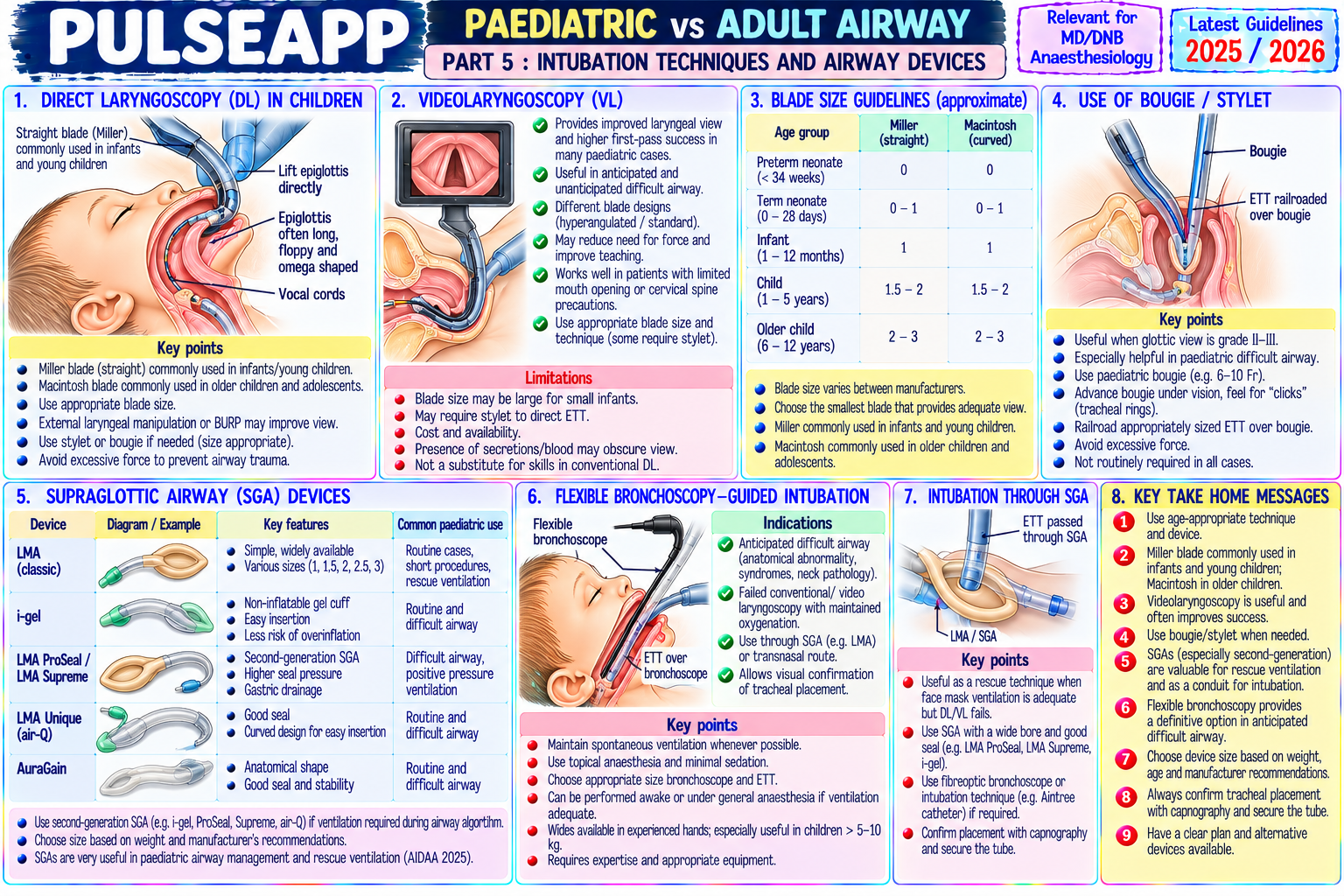
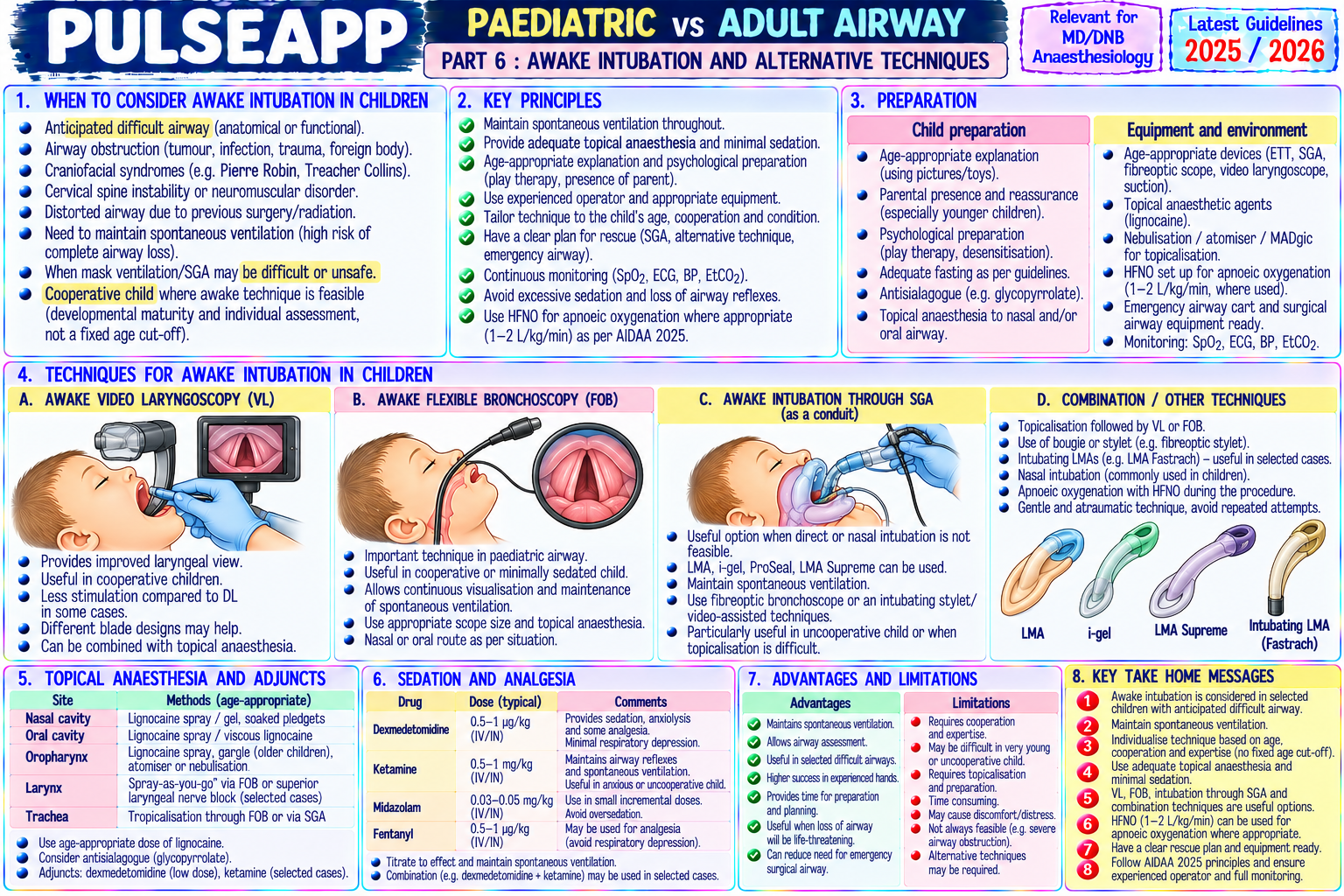
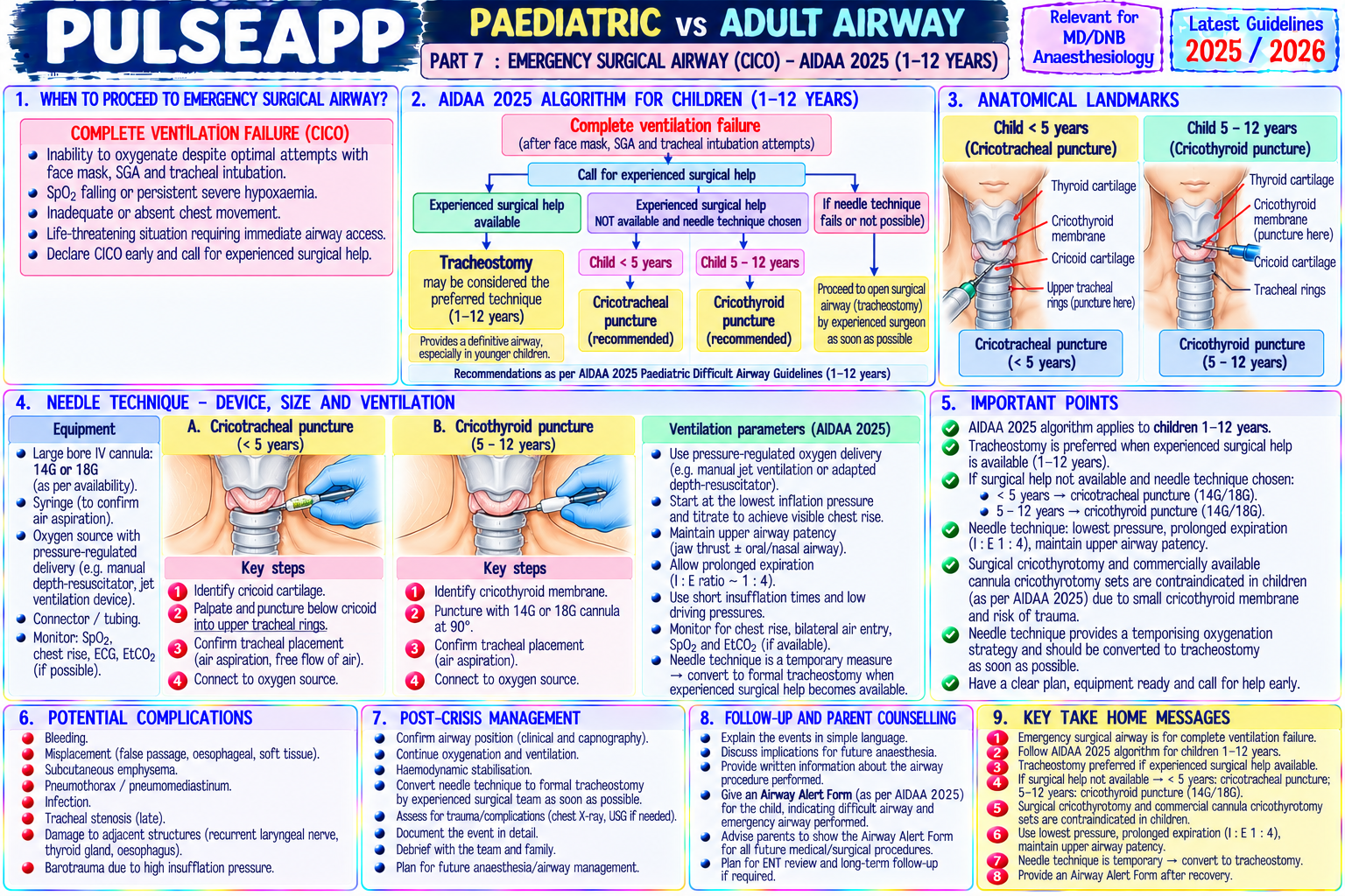
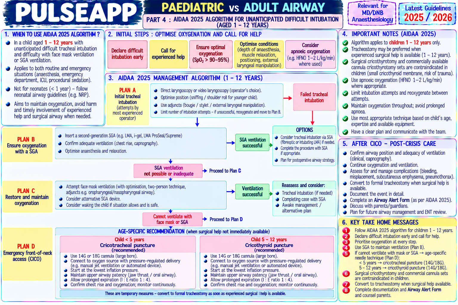
Describe the anatomical and physiological differences between the paediatric and adult airway. State the formulae for ETT sizing. Outline the management of a child with an unexpected difficult airway including: awake intubation alternatives, video laryngoscopy, and surgical airway as last resort.

collections Question Diagrams & Reference Images (5)
Question Reference Diagram
zoom_in View Image
Question Reference Diagram
zoom_in View Image
Question Reference Diagram
zoom_in View Image
Question Reference Diagram
zoom_in View Image
Question Reference Diagram
zoom_in View Image
description Clinical Response
⚙ Core Concept
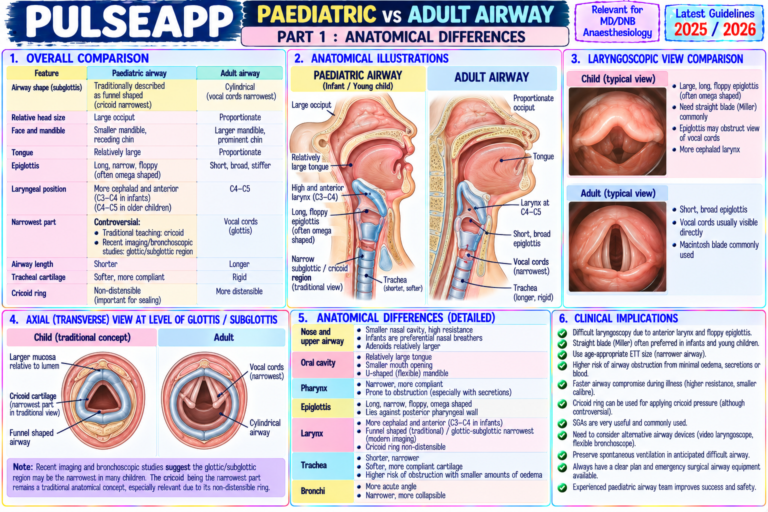
The paediatric airway is not a scaled-down adult airway — it is anatomically distinct in ways that fundamentally change the mechanics of intubation, the choice of equipment, and the consequences of airway difficulty. In neonates and infants, the narrowest point is the subglottis (cricoid ring — funnel-shaped airway), not the glottis; this is why uncuffed tubes traditionally provided an airtight fit. However, modern evidence now supports cuffed ETTs in children ≥1 year with careful cuff pressure monitoring. (Cote CJ, Lerman J — Paediatric Anaesthesia; DAS Paediatric Guidelines 2021; Black AE; Weiss M)
A. Anatomical Differences — Paediatric vs Adult Airway3 marks

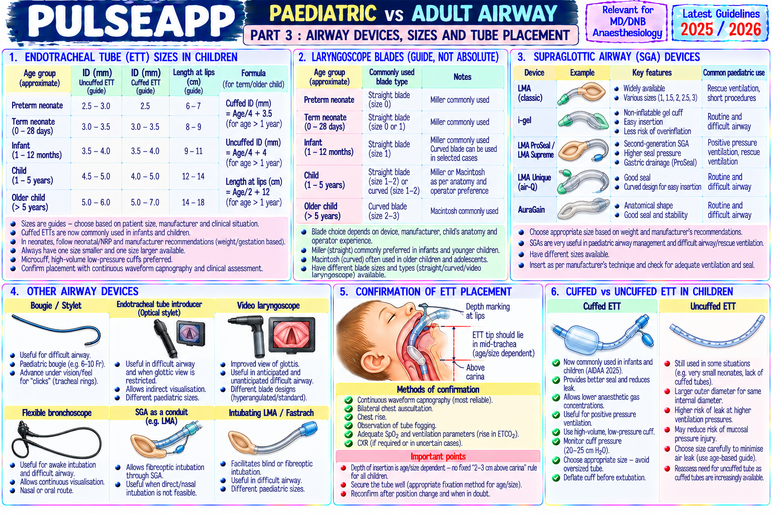
Feature Infant/Child Adult Clinical Consequence Laryngeal C3–C4 in neonates (high and anterior) C4–C5 More difficult to align larynx with the line of sight during laryngoscopy; straight position Miller blade preferred in neonates (lifts the epiglottis directly) Epiglottis Long, stiff, omega-shaped (Ω); floppy Short, flat; flexible Miller blade (straight) more effective for direct lift of epiglottis; Macintosh blade (curved, fits vallecular) less reliable in infants Narrowest Subglottis (cricoid ring) — funnel-shaped airway; Glottis (true vocal ETT that passes through the cords may still be too tight at the subglottis; too point the cricoid ring is the non-distensible narrowest cords) large a tube → subglottic oedema → post-extubation stridor; a small air leak at point 15–20 cmH₂O is the correct fit indicator for uncuffed tubes Head and Large occiput → passive neck flexion in supine Smaller occiput; In neonates: a small shoulder roll under the shoulders (not the head) brings the occiput position → relative anterior laryngeal position neck extension larynx into the line of sight; neutral "sniffing" position or slight extension; NO needed for pillow under the head intubation Tongue Relatively large tongue for the oral cavity size → Proportionally Higher aspiration and obstruction risk; jaw thrust more important; LMA insertion more easily obstructs airway; more likely to fall smaller may be more challenging back in unconscious child Rib cage Horizontal ribs → FRC-dependent breathing; Downward- Neonates desaturate extremely rapidly (≤60 seconds in a neonate vs 8+ higher closing capacity relative to FRC → small sloping ribs allow minutes in healthy adults); keep preoxygenation time adequate; work fast during airway closure at FRC → rapid desaturation active increase in apnoea FRC

B. ETT Sizing Formulae2 marks

Parameter Formula Example (5-year-old) Uncuffed ETT internal diameter (Age/4) + 4 = ID in mm (5/4) + 4 = 5.25 → use 5.0 mm uncuffed Cuffed ETT internal diameter (Age/4) + 3.5 = ID in mm (5/4) + 3.5 = 4.75 → use 4.5 mm cuffed ETT length at lips (oral) (Age/2) + 12 cm (5/2) + 12 = 14.5 cm at lip ETT length at nostril (nasal) (Age/2) + 15 cm (5/2) + 15 = 17.5 cm at nostril LMA size <5 kg = size 1; 5–10 kg = 1.5; 10–20 kg = 2; 20–30 kg = 2.5; 30–50 kg = 3; >50 kg = 4 20 kg child = LMA size 2.5

C. Management of Paediatric Difficult Airway3 marks

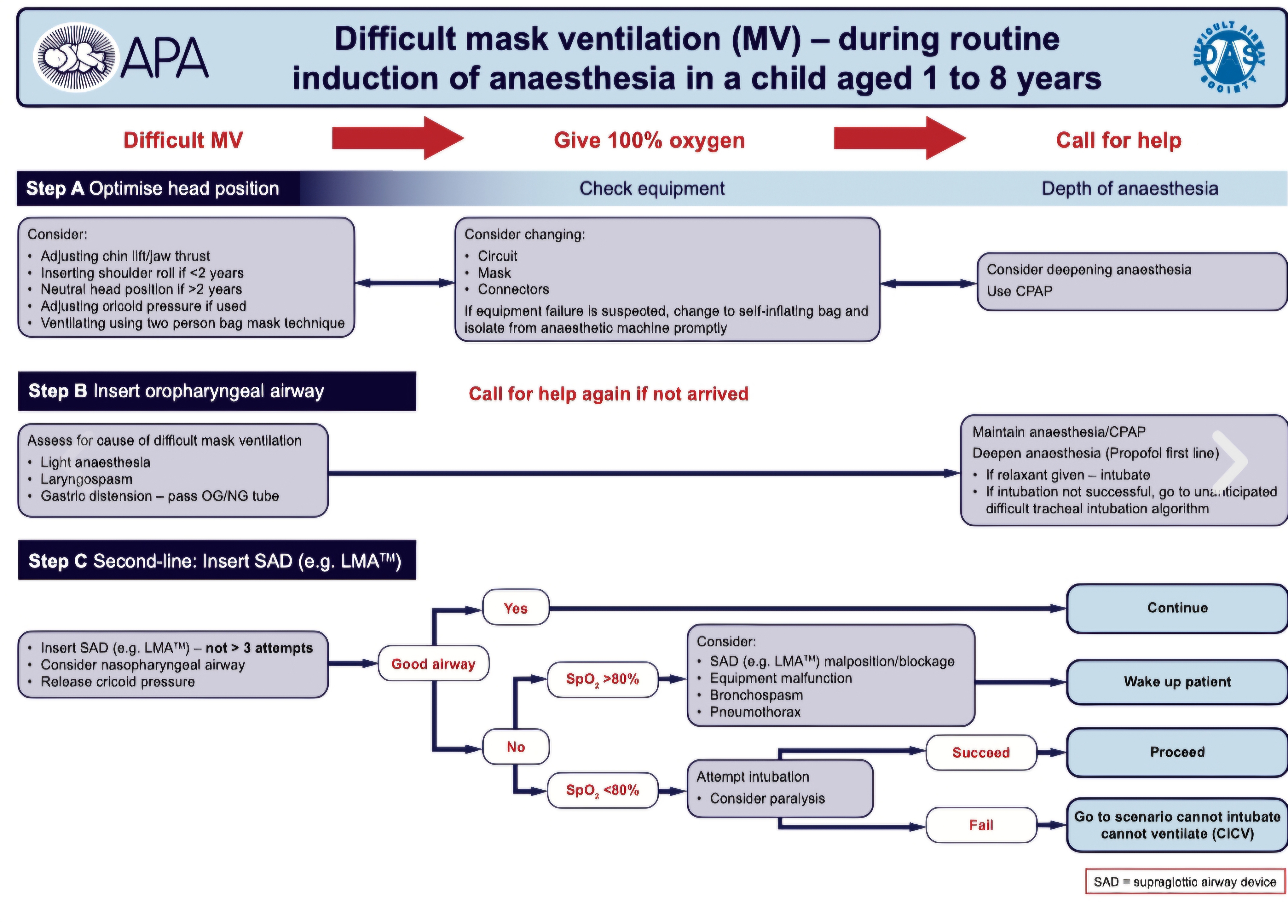
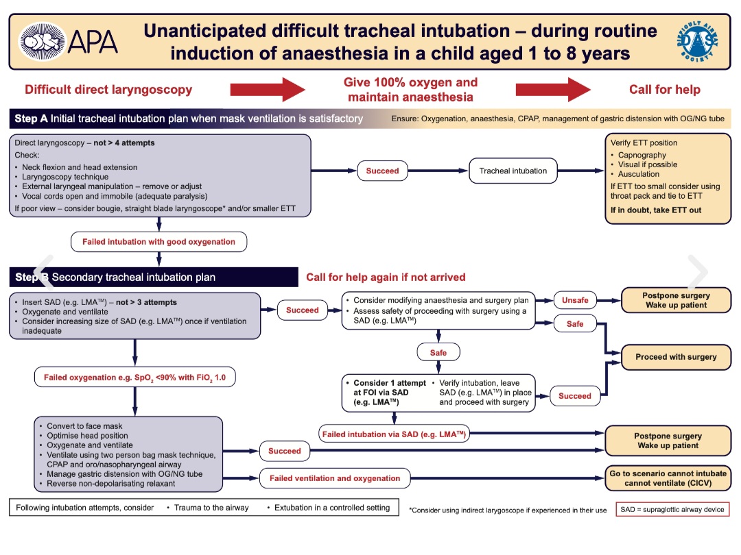
Anticipated difficult airway: plan in advance; assemble the paediatric difficult airway trolley (range of LMAs, paediatric video laryngoscope [Karl Storz DCI or CMAC paediatric blade], paediatric fiberoptic bronchoscope [2.8 mm minimum for ETT loading], paediatric cricothyrotomy kit); experienced paediatric anaesthesiologist; ENT surgeon on standby for possible surgical airway

Inhalational induction (spontaneous breathing maintained): the safest technique for anticipated difficult airway in children — induction with sevoflurane in 100% O₂, maintaining spontaneous ventilation throughout; once adequately anaesthetised, direct laryngoscopy ± video laryngoscopy attempted; if fails → LMA; if LMA → use as conduit for FOI; if all fails → wake up (spontaneous breathing maintained throughout → safe to allow the child to recover)

Video laryngoscopy in paediatrics: paediatric-sized blades (C-MAC size 0, 1, 2; Karl Storz DCI Miller size 0, 1); improving first-attempt intubation success vs direct laryngoscopy; increasingly recommended as first-line for anticipated difficult airway in children

Fibreoptic intubation (awake FOI in paediatrics): rarely practical in young children (unlike adults); infants do not cooperate for topical airway anaesthesia; preferred approach: sedated (dexmedetomidine infusion) or lightly anaesthetised FOI with spontaneous ventilation; technique: 2.8 mm or 3.5 mm paediatric fibrescope through a face mask or LMA while child breathes spontaneously; ETT railroaded over the scope once in trachea

Front-of-Neck Access (FONA) in children: true cricothyrotomy is technically challenging in infants (small cricothyroid membrane); needle cricothyrotomy (14G IV cannula) with jet ventilation is the primary emergency technique in children <8 years; surgical cricothyrotomy and tracheostomy for older children/adolescents

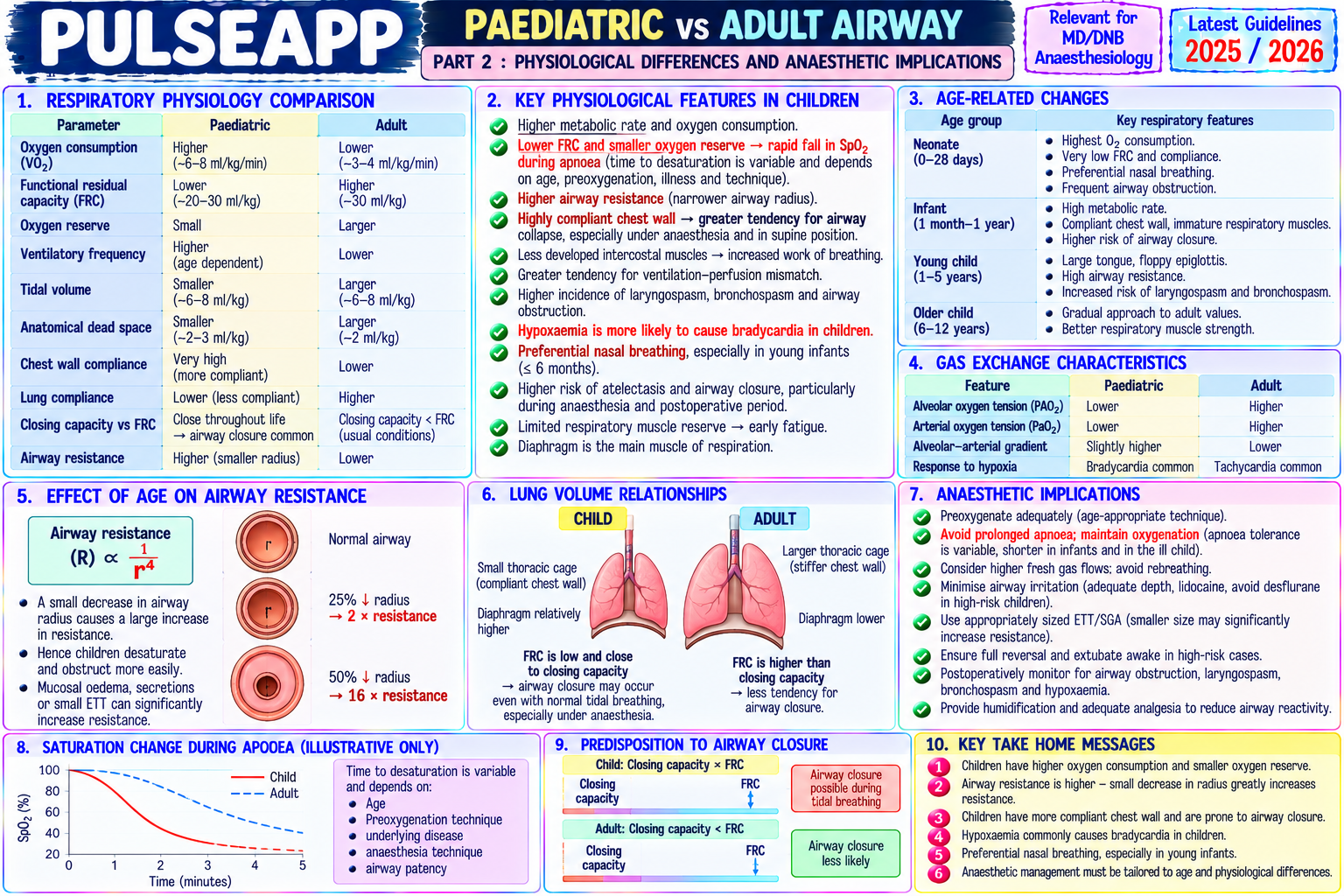
D. Physiological Differences Affecting Anaesthesia2 marks

Higher O₂ consumption per kg (6–8 mL/kg/min vs 3 mL/kg/min adults); very small functional residual capacity → rapid desaturation; safe apnoea time in a neonate ≈ 60–90 seconds Higher heart rate (normal neonatal HR 120–160 bpm; infants 100–120 bpm); cardiac output is rate-dependent in neonates (cannot increase SV significantly — immature Frank-Starling mechanism) → bradycardia = fall in cardiac output; ALWAYS give atropine 20 mcg/kg before laryngoscopy in neonates/young infants

Temperature regulation: high surface area:body mass ratio → rapid heat loss; all fluids warmed; warming blanket; theatre temperature 28°C for neonates; temperature monitoring mandatory

🎤 Viva Corner
Q. After inhalational induction of a 3-year-old with suspected subglottic stenosis, you cannot pass an ETT beyond the vocal cords. The SpO₂ is now 88%. What do you do?
SpO₂ of 88% in a child requires immediate action — oxygen reserve is already critically depleted. Critically, this child was induced with inhalational sevoflurane maintaining spontaneous ventilation, which means the child should still be breathing spontaneously despite failed intubation attempts. Immediate first action: ensure 100% O₂ continues to be delivered via the face mask; allow the child to ventilate spontaneously; apply gentle jaw thrust and oral airway to maintain airway patency and oxygenation while SpO₂ recovers to ≥95%. Do NOT continue intubation attempts until SpO₂ is restored. Once oxygenated: the fact that the ETT cannot pass beyond the vocal cords suggests subglottic stenosis — the tracheal lumen is narrowed. I would try a smaller ETT (down one size: if I was using 4.0 mm, try 3.5 mm; if 3.5, try 3.0 mm) — a tube that can pass through the narrowed subglottis provides gas exchange even if smaller than ideal. Alternatively: a supraglottic airway (LMA size 2 for a 3-year-old, approximately 12–15 kg) as a bridge device to provide ventilation and oxygenation while a more senior paediatric anaesthesiologist and an ENT surgeon are called. If LMA also fails to ventilate: needle jet cricothyrotomy (14G IV cannula through the CTM with high-flow O₂ jet ventilation); this is the paediatric emergency surgical airway while ENT performs tracheostomy. The overarching principle: "Can't intubate" does NOT mean "Can't oxygenate" if spontaneous breathing is maintained and the LMA can ventilate; the spontaneous breathing throughout inhalational induction was the safety net that makes this situation recoverable.
★ Examiner's Pearl
The ETT sizing formulae (uncuffed = age/4 + 4; cuffed = age/4 + 3.5; oral length = age/2 + 12) must be reproduced — they are tested with specific age calculations. The anatomical distinction that the narrowest point is the SUBGLOTTIS (not the glottis) in children <5–8 years is the most frequently tested paediatric airway anatomical fact. The atropine-before-laryngoscopy rule in neonates (20 mcg/kg minimum 100 mcg — prevents vagal bradycardia) is a specific neonatal safety fact tested in written papers.
Cote CJ, Lerman J, Anderson BJ. A Practice of Anaesthesia for Infants and Children, 6th Ed. Black AE et al. Management of the difficult paediatric airway — DAS guidelines 2021 (Anaesthesia 2021;76:536-556). Weiss M et al. Paediatric airway management (BJA Education 2012;12:57-63). Miller RD et al. Miller's Anaesthesia, 9th Ed, Chapter 93.
lock Protected Clinical Document • Pulse App • Screenshots Strictly Prohibited
QUESTION 57 person Asked by .
bookmark_add

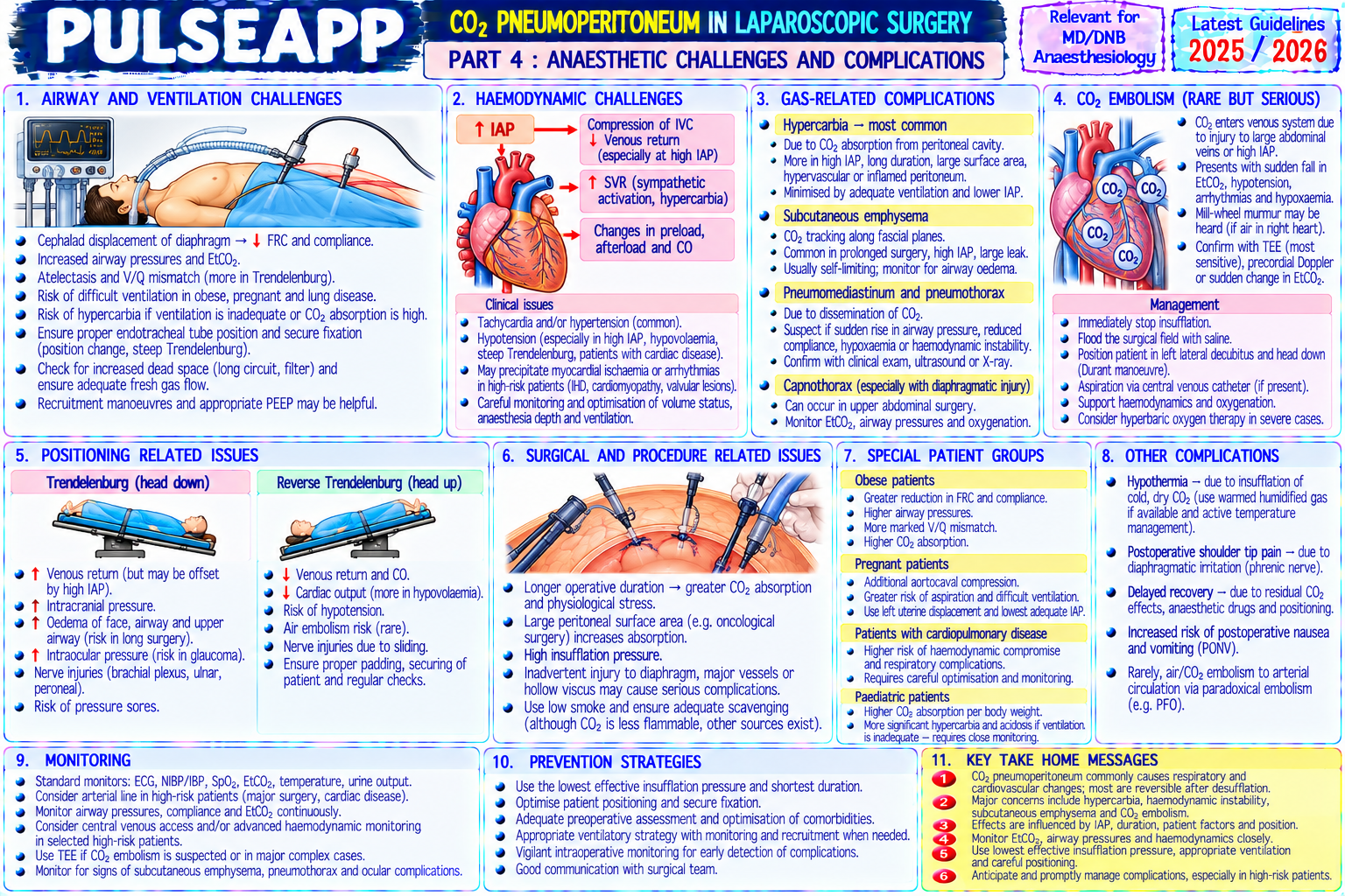
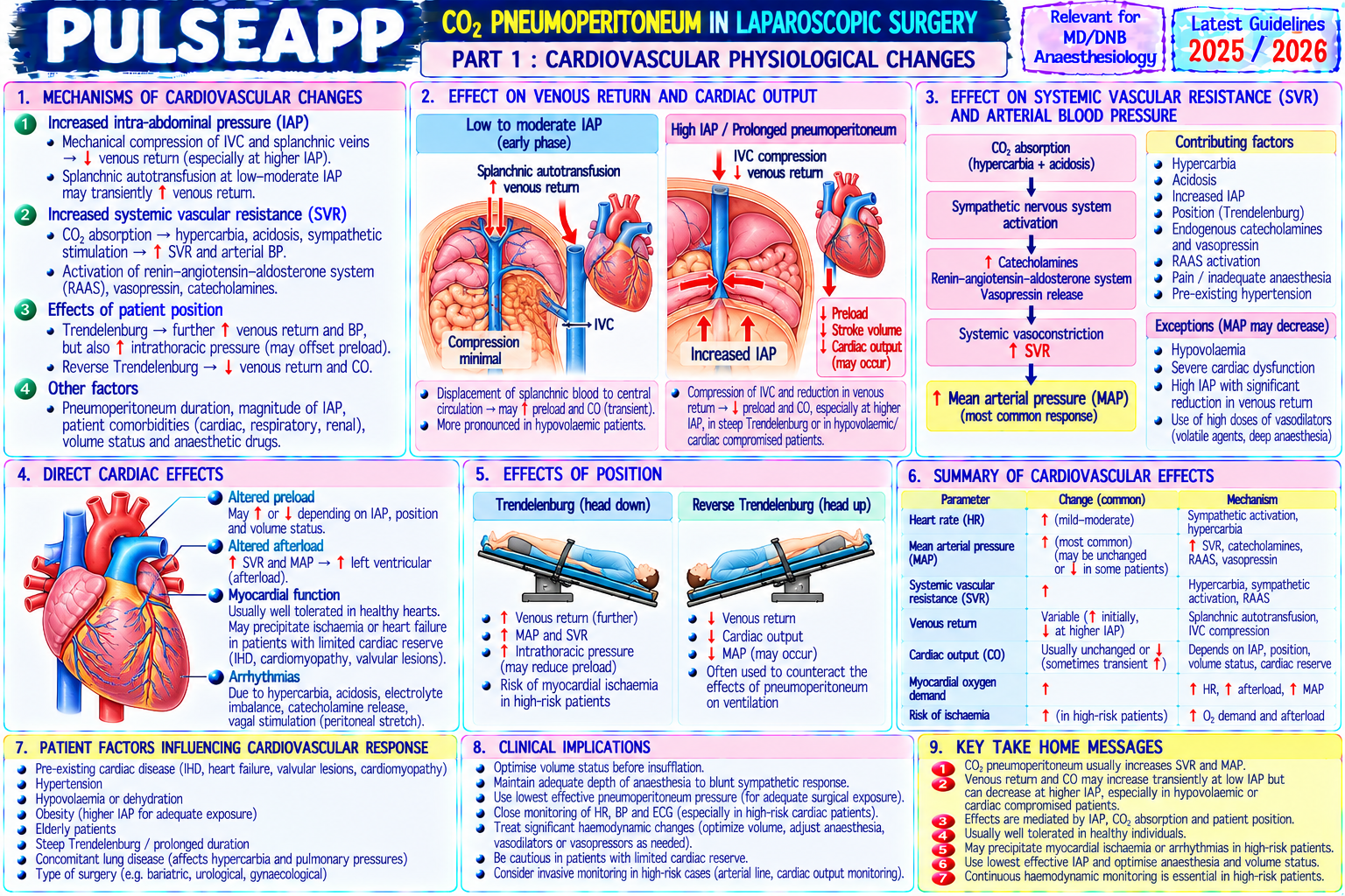
Describe the cardiovascular, respiratory, and systemic physiological changes caused by CO₂ pneumoperitoneum during laparoscopic surgery. Outline the specific anaesthetic challenges and ventilatory management for laparoscopic procedures.

collections Question Diagrams & Reference Images (3)
Question Reference Diagram
zoom_in View Image
Question Reference Diagram
zoom_in View Image
Question Reference Diagram
zoom_in View Image
description Clinical Response
⚙ Core Concept
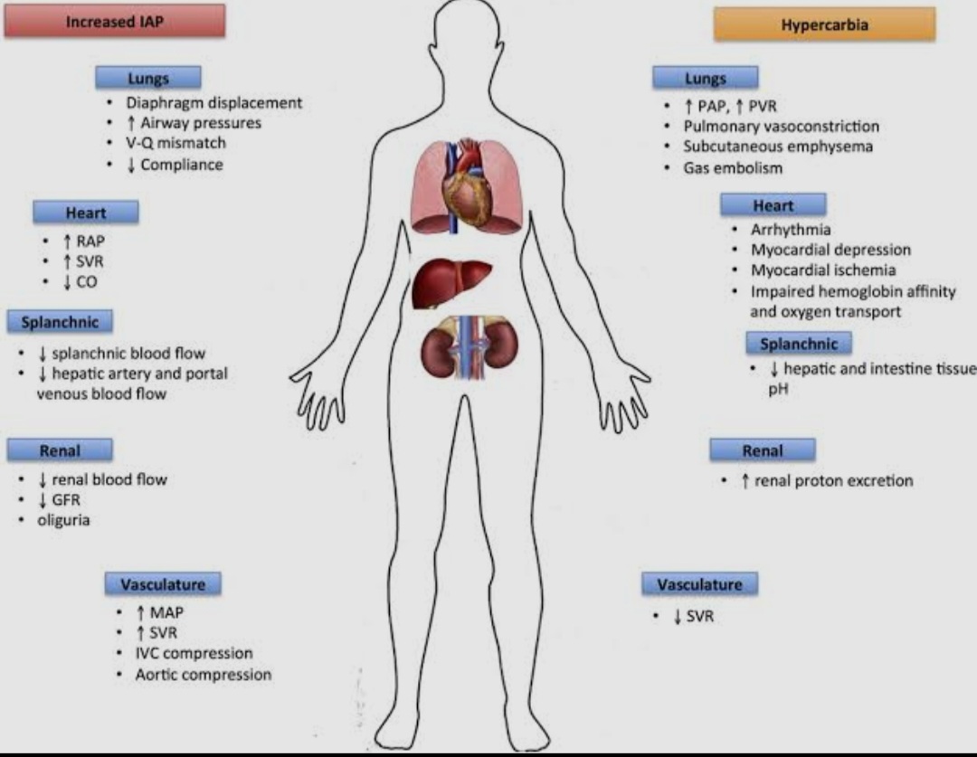
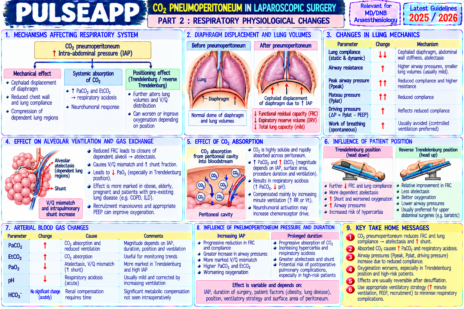
CO₂ pneumoperitoneum creates a controlled physiological perturbation: rising intra-abdominal pressure compresses the diaphragm (reducing FRC and lung compliance), compresses the inferior vena cava (reducing venous return), and absorbs CO₂ across the peritoneal surface (requiring 15–25% increase in minute ventilation). These effects are manageable in most patients but require specific ventilatory adjustments and monitoring — particularly in patients with pre-existing cardiorespiratory disease. (Miller's Anaesthesia 9th Ed; O'Malley C — BJA 1999; Joris J — Anaesthesiology 1997; Nguyen NT)
A. Cardiovascular Effects3 marks

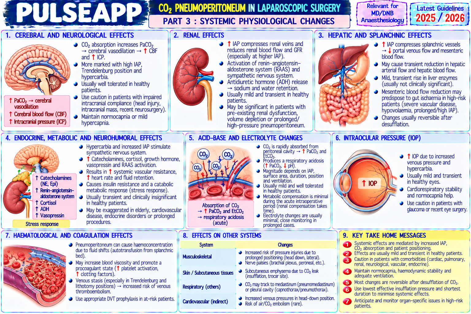
Effect Mechanism Intraoperative Management ↑ SVR (early IAP compression → mesenteric vasoconstriction; CO₂ absorption MAP rises initially; may require vasodilators in hypertensive patients; volatile and → sympathetic activation → ↑ catecholamines → vasoconstriction; agents and deeper anaesthesia help manage hypertension from CO₂ absorption sustained) IAP 12–15 mmHg → SVR ↑30–50% ↓ Venous IAP >12 mmHg → IVC compression → ↓ preload; initial ↑ CO from Adequate preloading before insufflation (250–500 mL crystalloid); vasopressors Return → ↓ leg-emptying into central circulation then falls as IVC compression for sustained hypotension; Trendelenburg position modifies venous return (↑ in CO predominates head-down; ↓ further in head-up laparoscopy) (intermediate) ↑ HR Sympathetic activation from CO₂ absorption and surgical stress; Adequate depth of anaesthesia; beta-blocker if needed for rate control (moderate) partially offset by vagal tone from peritoneal stretching Cardiac Hypercapnia (CO₂ absorption) → ↑ catecholamines → arrhythmia; Atropine available; adequate hyperventilation to prevent hypercapnia; if arrhythmia also vagal reflex from peritoneal stretching at insufflation onset → arrhythmia → check ETCO₂ and ABG; consider desufflation bradycardia → asystole (rare)

B. Respiratory Effects2 marks

↓ FRC and lung compliance: diaphragm displaced cranially by pneumoperitoneum → FRC reduced 10–20% beyond position-related reduction; peak airway pressures rise 40–50% for same TV; atelectasis in dependent lung regions

CO₂ absorption: CO₂ gas diffuses from the peritoneal cavity into systemic blood → ETCO₂ rises 10–20 mmHg above pre-insufflation baseline within 10–20 minutes; must increase MV by 15–25% to maintain normocapnia; in patients with severe COPD or very limited reserve, maintaining normocapnia may not be possible without dangerous airway pressures → accept permissive hypercapnia

Gas embolism risk: direct IV insufflation of CO₂ (rare but catastrophic) → sudden fall in ETCO₂ + cardiovascular collapse; CO₂ is more water-soluble than air → reabsorbed faster from the blood; management: stop insufflation; left lateral Durant's position; aspiration via CVP catheter; CPR

C. Other Systemic Effects2 marks

Renal: IAP >15 mmHg → renal vein compression → ↓ renal blood flow → oliguria (transient — resolves on desufflation); not associated with post-op AKI in healthy patients; ensure adequate hydration; monitor urine output; avoid NSAIDs peri-operatively if oliguric intraoperatively
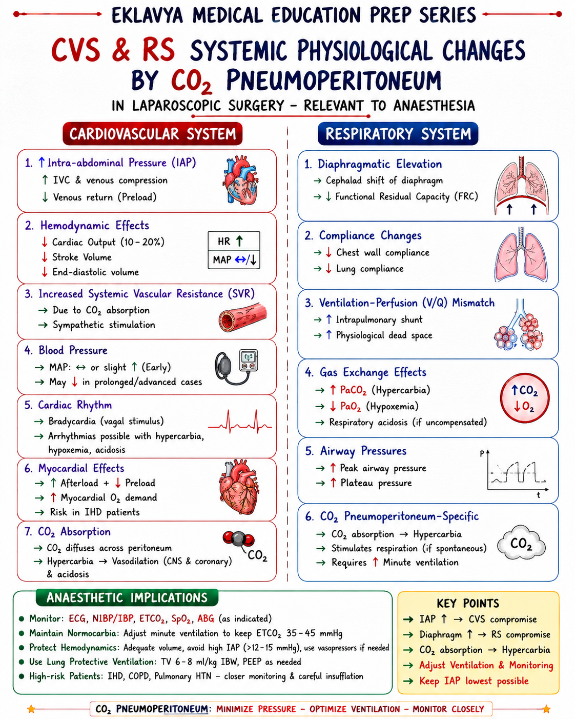
ICP rise: CO₂ absorption → hypercapnia → cerebral vasodilation → ↑ CBF → ↑ ICP; compounded by ↑ CVP from IAP raising venous pressure; head-down Trendelenburg further raises ICP; relevant in patients with pre-existing raised ICP or ventriculoperitoneal shunts

Subcutaneous emphysema: CO₂ tracks along fascial planes from the peritoneal cavity into subcutaneous tissue; presents as crepitus on palpation; may cause extensive tracking to the mediastinum and neck; results in massive CO₂ absorption → very high ETCO₂ → increase MV further; does not require specific treatment unless compromising airway or mediastinal structures

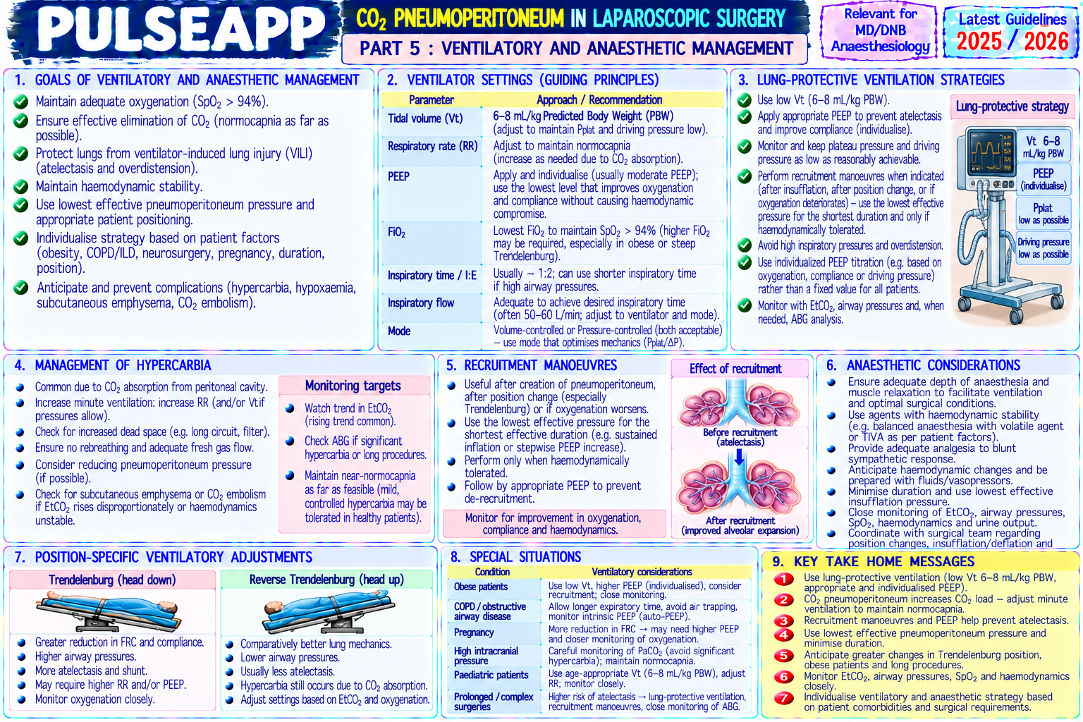
D. Specific Anaesthetic Management3 marks

Airway: ETT mandatory (positive pressure needed; aspiration risk from raised IAP; patient may need Trendelenburg); LMA is used for brief, non-obese patients in selected units with second-generation supraglottic devices (higher seal pressure)

Ventilation settings: TV 6–8 mL/kg IBW; PEEP 5–8 cmH₂O; RR adjusted to maintain normocapnia (increase by 15–25% from baseline after CO₂ insufflation); monitor ETCO₂ continuously; check ABG at 30–60 minutes if significant patient complexity; accept permissive hypercapnia in COPD patients rather than use excessive driving pressures

PONV: laparoscopy is one of the highest PONV risk procedures; full multimodal PONV prophylaxis based on Apfel score; TIVA with propofol reduces PONV incidence by 25–30% vs volatile; ondansetron + dexamethasone minimum for all laparoscopic procedures

Analgesia: port-site local anaesthetic infiltration (bupivacaine 0.5% at each port before closure); IV paracetamol + NSAIDs; opioid-sparing; TAP block for larger ports/incisions; intraperitoneal local anaesthetic instillation (bupivacaine) reduces post-operative visceral pain after laparoscopy

Desufflation awareness: sudden desufflation may cause vasovagal reaction (brief hypotension + bradycardia); anaesthesiologist should be informed before desufflation; have atropine ready

🎤 Viva Corner
Q. At ETCO₂ monitoring, the ETCO₂ suddenly rises from 38 to 65 mmHg during a laparoscopic cholecystectomy. Simultaneously, the patient develops subcutaneous crepitus in the neck. What has happened and how do you manage it?
The sudden dramatic ETCO₂ rise to 65 mmHg (27 mmHg above baseline) with neck subcutaneous emphysema indicates extensive CO₂ tracking from the peritoneal cavity into the extraperitoneal fascial planes and subcutaneous tissue. CO₂ insufflated into the peritoneal cavity has dissected along the pre-peritoneal and retroperitoneal planes, through the diaphragmatic hiatus, into the mediastinum and then subcutaneous tissues of the neck — subcutaneous emphysema. This dramatically increases the surface area from which CO₂ can be absorbed into the systemic circulation, causing massive CO₂ absorption and ETCO₂ rise. This can continue or worsen even if CO₂ insufflation is reduced or stopped. Management: first, notify the surgeon of the complication and ask them to assess the peritoneal cavity — is there any evidence of pneumomediastinum, incorrect trocar placement, or inadvertent extra-peritoneal insufflation? Increase minute ventilation substantially (increase RR to 20–22 breaths/min; increase TV if peak pressures allow) to manage the CO₂ load; target ETCO₂ 38–42 mmHg — may require MV 150–200% of baseline; FiO₂ to 1.0; the surgeon should reduce the insufflation pressure to minimum needed for surgery; subcutaneous emphysema itself does not require specific treatment beyond managing the CO₂ absorption — CO₂ is water-soluble and will be absorbed over 30–60 minutes after desufflation; check ABG (pH, pCO₂); if ETCO₂ cannot be controlled <70 mmHg despite maximum ventilation, consider desufflating and converting to open surgery. The emphysema in the neck must be assessed — if it is causing airway compression or mediastinal shift, immediate desufflation and ENT/cardiothoracic assessment is required.
★ Examiner's Pearl
The CO₂ absorption effect (ETCO₂ rises 10–20 mmHg → must increase MV 15–25%) with specific numbers is the most tested laparoscopic physiology fact. The cardiovascular effects table (↑SVR, ↓CO, ↑HR) must present the mechanisms in sequence. PONV prevention in laparoscopy (highest PONV-risk procedure → full multimodal prophylaxis mandatory) is the most practically tested anaesthesia management point for laparoscopic surgery.
O'Malley C, Cunningham AJ. Physiological changes during laparoscopy (Anesthesiol Clin North Am 2001). Joris JL. Anaesthetic management of laparoscopy (Miller's Anaesthesia). Nguyen NT et al. Respiratory changes during laparoscopic cholecystectomy (Arch Surg 1999). Miller RD et al. Miller's Anaesthesia, 9th Ed.
lock Protected Clinical Document • Pulse App • Screenshots Strictly Prohibited
QUESTION 58 person Asked by .
bookmark_add

A 45-year-old female with rheumatic mitral stenosis (MVA 1.0 cm², moderate pulmonary hypertension, AF with rate control) requires laparoscopic hysterectomy. Discuss haemodynamic goals, anaesthetic technique, and management of intraoperative complications.

description Clinical Response
⚙ Core Concept
Mitral stenosis (MS) creates a fixed obstruction between the left atrium and left ventricle — the stenotic mitral valve limits the flow of blood during diastolic filling, creating a pressure gradient that produces pulmonary hypertension, right ventricular strain, and left atrial hypertension. The anaesthetic haemodynamic goals directly oppose what the body's natural stress response tries to do (tachycardia is compensatory but in MS causes acute pulmonary oedema). (Miller's Anaesthesia 9th Ed; ACC/AHA Valvular Guidelines; Roshanov P; Stoelting — Coexisting Disease)
A. Pathophysiology of Mitral Stenosis2 marks

Normal MVA = 4–6 cm²; symptoms begin at MVA <2.5 cm²; MVA 1.0–1.5 = severe; MVA <1.0 = very severe

Mechanism: stenotic mitral valve → ↑ LA pressure → pulmonary venous hypertension → pulmonary oedema; chronic LA pressure ↑ → pulmonary arterial hypertension → RV hypertrophy and failure; fixed outflow from LA → any ↑ HR shortens diastolic filling time → less time for blood to cross the stenotic valve → sudden ↑ LA pressure → acute pulmonary oedema (the key clinical insight) AF is almost universal in significant MS (atrial dilation drives AF); loss of atrial systole ("atrial kick") removes 20–30% of ventricular filling → additional haemodynamic compromise

B. Haemodynamic Goals — "SLOW, FULL, HIGH"2 marks
✅ Haemodynamic Goals for Mitral Stenosis
SLOW heart rate (60–80 bpm): the most critical goal; tachycardia → shortened diastole → less filling time across the stenotic valve → acute ↑ LA pressure → pulmonary oedema; maintain rate with beta-blockers (esmolol infusion intraoperatively); avoid tachycardia triggers (pain, light anaesthesia, ketamine, sympathomimetics) FULL preload (maintain SR if in sinus rhythm): adequate preload supports ventricular filling through the stenotic valve; avoid excessive fluid loading (↑ LA pressure → pulmonary oedema); avoid vasodilators that cause excessive preload reduction (spinal hypotension) HIGH SVR (avoid hypotension): MS patients are dependent on adequate SVR to maintain MAP; hypotension → reduced coronary perfusion pressure → RV ischaemia in already-hypertrophied RV; phenylephrine (pure alpha-1 agonist — raises SVR without increasing HR) is the vasopressor of choice; avoid ephedrine (beta-1 agonist → tachycardia) Avoid atrial fibrillation at rapid rate: if in chronic AF, maintain rate control (digoxin, beta-blockers, calcium channel blockers continued perioperatively); if new-onset AF with rapid ventricular response → acute haemodynamic deterioration → cardiovert electrically if haemodynamically unstable
C. Anaesthetic Technique3 marks

Regional Anaesthesia (Preferred)

Spinal anaesthesia: technically simple but produces sudden sympathectomy → acute hypotension → reflex tachycardia → catastrophic in MS; spinal is RELATIVELY CONTRAINDICATED for major surgery in MS because the sympathectomy is abrupt and may cause severe tachycardia from reflex sympathetic activation; if used → titrated slowly (combined spinal-epidural with very small initial intrathecal dose + epidural top-up) or avoided

Epidural anaesthesia: preferred for regional technique — allows gradual titration of sympathectomy level; maintains more haemodynamic control than spinal; place thoracic epidural for laparoscopic hysterectomy; slow top-up in 5 mL increments; use vasopressor (phenylephrine) to maintain SVR; monitor closely for tachycardia

General Anaesthesia (for Laparoscopic Case)

Induction: etomidate (most cardiovascularly stable) or carefully titrated propofol; AVOID ketamine (↑ HR → ↑ LA pressure → pulmonary oedema); fentanyl 1–2 mcg/kg with induction to blunt laryngoscopy response

Maintenance: sevoflurane preferred (moderate rate reduction; preconditioning); avoid desflurane (sympathetic activation → tachycardia)

Intraoperative tachycardia management: esmolol 0.5–1 mg/kg IV bolus; or digoxin if not already digitalised; metoprolol 1–2 mg IV bolus

Fluid management: conservative; avoid large volume crystalloid boluses; pulmonary artery catheter or TOE for haemodynamic monitoring in MVA <1.0 cm²

D. Specific Laparoscopy Considerations2 marks

CO₂ pneumoperitoneum in MS: ↑ IAP → ↑ SVR (helps maintain MAP) and initial ↑ CO₂ absorption → sympathetic activation → tachycardia (harmful); maintain ETCO₂ strictly in normal range (increase MV to manage absorbed CO₂)

Trendelenburg position: increases venous return to an already volume-loaded right heart → may precipitate acute pulmonary oedema; head-down tilt must be gradual; monitor closely

Post-desufflation: sudden ↓ IAP → venous pooling → ↓ preload → ↓ CO; prepare vasopressor

E. Postoperative Management1 mark

HDU/ICU level monitoring for 24 hours post-op; cardiac monitoring (continuous ECG); resume rate-control medications immediately post-op; watch for AF with rapid ventricular rate; fluid balance (restrict fluids post-op; furosemide if signs of pulmonary congestion); anticoagulation for AF (warfarin or DOAC — check thromboembolism vs bleeding balance post-surgery); resume warfarin/DOAC within 24 hours if haemostasis adequate

🎤 Viva Corner
Q. Intraoperatively, this patient's heart rate suddenly rises from 78 to 138 bpm with irregular rhythm and BP falls from 110/70 to 70/45 mmHg. What has happened and what is your immediate management?
This is new-onset atrial fibrillation with rapid ventricular response causing haemodynamic collapse in a patient with severe mitral stenosis. AF with rapid rate is specifically catastrophic in MS because: the shortened diastole reduces trans-mitral flow time to a fraction of what was already inadequate through a stenotic valve; the loss of atrial kick further reduces left ventricular filling; and the rapid rate is self-perpetuating if untreated as left atrial pressure rises and perpetuates the AF substrate. The 70/45 mmHg BP indicates haemodynamic collapse. Immediate management: this requires synchronised DC cardioversion without delay — the haemodynamic compromise is severe enough to mandate immediate cardioversion rather than pharmacological rate control (which takes too long in this emergency). DC cardioversion with 150–200 J synchronised biphasic shock; the patient is already under general anaesthesia → no additional sedation needed; ensure synchronised mode is confirmed on the defibrillator (prevents shocking on T-wave → VF). While preparing defibrillator: maintain anaesthesia, 100% FiO₂; phenylephrine 100–200 mcg IV to maintain MAP while defibrillation is being prepared. If cardioversion to sinus rhythm is successful: check that the rate has normalised and haemodynamics improved; if reverting to AF: repeat cardioversion; consider amiodarone 300 mg IV over 20–60 minutes to maintain sinus rhythm after successful cardioversion. If cardioversion fails to achieve adequate haemodynamic response: consider intra-aortic balloon pump if echocardiography reveals severe LV dysfunction; emergency cardiac surgery (balloon valvuloplasty) consultation. After episode: determine the cause of AF onset (pain from light anaesthesia, hypoxia, hypercarbia, electrolyte abnormality — especially hypokalaemia); check K⁺ and Mg²⁺; correct if abnormal; document event.
★ Examiner's Pearl
The three haemodynamic goals (slow HR 60–80; full preload; high SVR) with "SLOW, FULL, HIGH" mnemonic are the core content — state each with mechanism. Tachycardia is specifically CATASTROPHIC in MS (not just undesirable) because shortened diastole → no filling time across stenotic valve → acute pulmonary oedema — this causal chain must be stated. Phenylephrine (not ephedrine) as the vasopressor of choice — because ephedrine has beta-1 effects → tachycardia — is a specific drug safety distinction tested.
Miller RD et al. Miller's Anaesthesia, 9th Ed. ACC/AHA 2014 Valvular Heart Disease Guidelines. Stoelting RK. Pharmacology and Physiology in Anaesthetic Practice, 5th Ed. Reyes G, Kshettry VR. Anaesthesia for valvular heart disease (BJA Education 2016).
lock Protected Clinical Document • Pulse App • Screenshots Strictly Prohibited
QUESTION 59 person Asked by .
bookmark_add

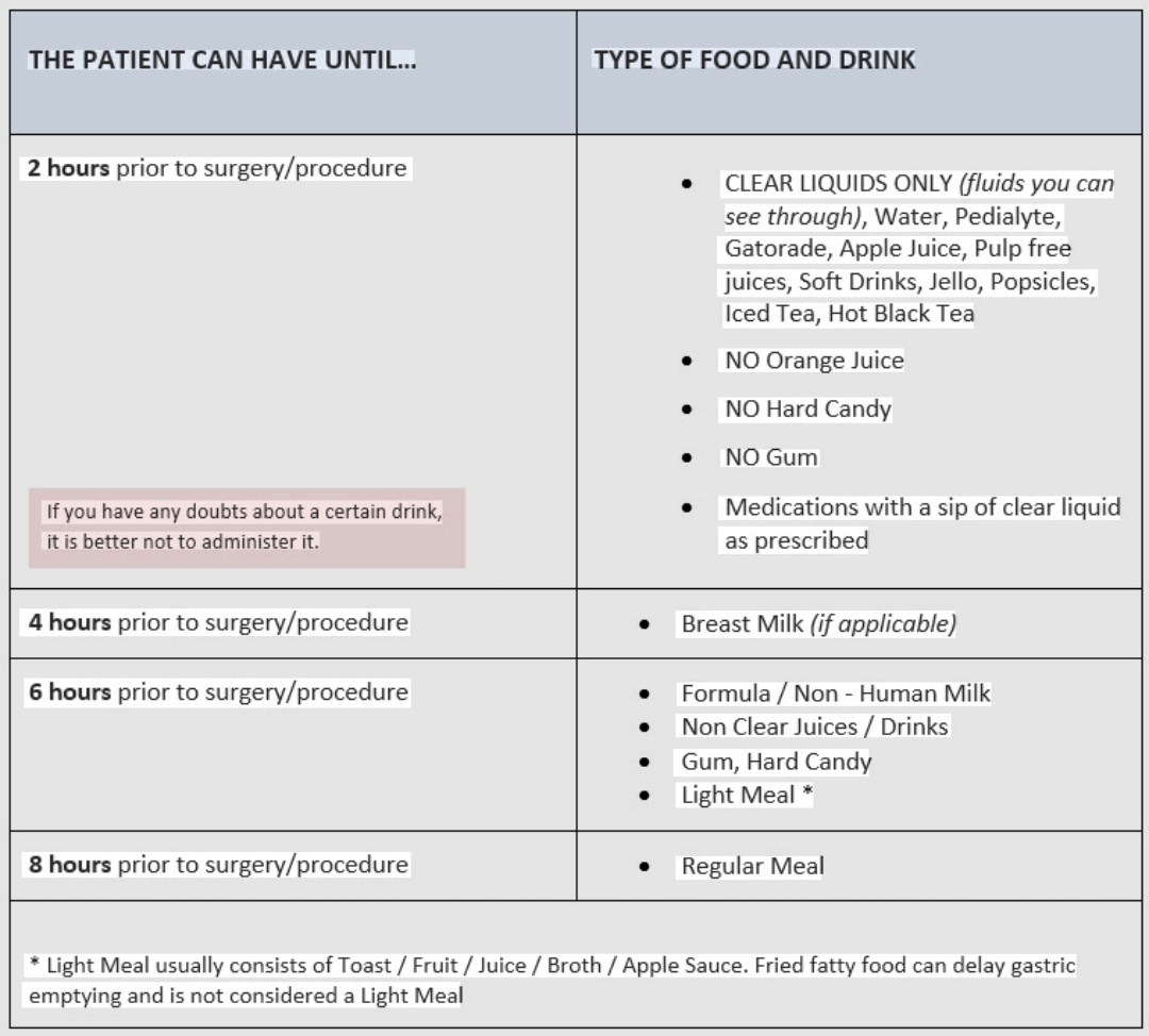
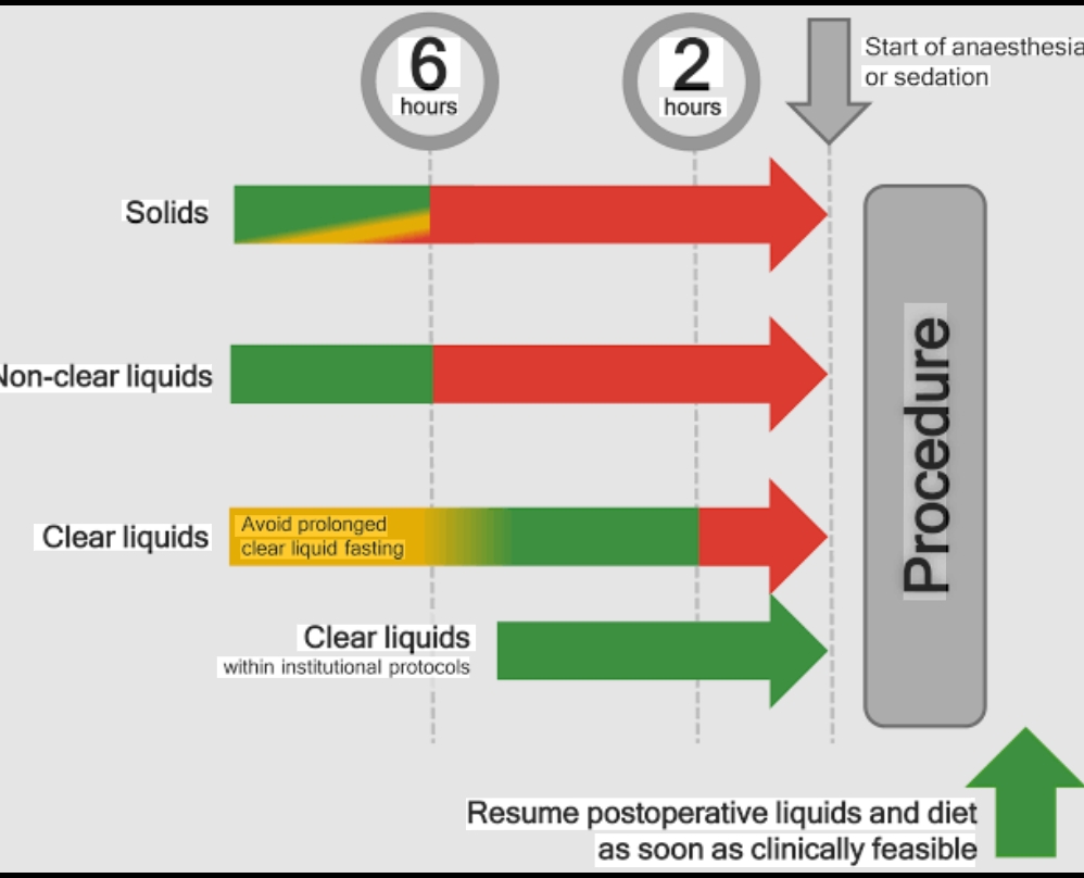
State the current evidence-based fasting guidelines for elective surgery in adults and children. Distinguish clear liquids from solids. Discuss the rationale for each fasting period and modifications for high-risk patients (pregnancy, obesity, diabetics, urgent surgery).

collections Question Diagrams & Reference Images (1)
Question Reference Diagram
zoom_in View Image
description Clinical Response
⚙ Core Concept
Traditional "nil by mouth from midnight" fasting has been replaced by evidence-based, physiologically rational guidelines that allow clear fluids up to 2 hours before elective surgery — reducing patient suffering, improving patient experience, and reducing insulin resistance and perioperative stress response without increasing aspiration risk. (ASA Practice Guidelines 2023; ESPEN; ERAS Society; Brady M — Cochrane; Smith MD)
A. Current Fasting Guidelines (ASA 2023 / ESPEN)2 marks

Item Fasting Period Rationale Clear liquids (water, black 2 hours before Clear fluids empty from the stomach within 30–60 minutes; gastric pH improves with fluid consumption (dilutes coffee, clear fruit juice without induction acid); preoperative carbohydrate drink (200–400 mL of 12.5% carbohydrate solution 2 hours before surgery) pulp, sports drinks, carbonated reduces insulin resistance and anxiety, improves postoperative outcomes — integral to ERAS protocols drinks) Breast milk (infants) 4 hours Breast milk empties from the stomach faster than formula or cow's milk due to its whey-dominant protein composition and lower fat content Infant formula / non-human milk 6 hours Higher fat and protein content → slower gastric emptying Light meal (toast, crackers, 6 hours Light meals typically empty within 4–5 hours; 6-hour fasting ensures adequate buffer simple carbohydrate meal — low fat, low protein) Full/fatty/fried meal (heavy meal 8 hours High-fat meals significantly slow gastric emptying; fat delays gastric acid secretion reduction; high-protein meals — high fat, meat, fried food) minimum are digested slowly; in practice: a large evening meal → fast from midnight (8+ hours) is appropriate (some guidelines: overnight fast)

B. High-Risk Modifications2 marks

Patient Modification Rationale Group Pregnancy Treat as full stomach regardless of fasting time (in active labour + opioids); RSI mandatory; Progesterone reduces LOS tone; opioids (>16–20 sodium citrate 30 mL oral + H₂ blocker before all GA dramatically slow gastric emptying; risk persists weeks) through full postoperative period Morbid Apply standard fasting times; however, gastric emptying may be delayed; some centres treat Obesity associated with raised intra-abdominal obesity morbidly obese patients with raised aspiration risk as "full stomach" for GA pressure and higher gastric residual volumes; GORD common Diabetes Establish surgery as first on the list (shortest fasting); monitor blood glucose hourly; variable rate Diabetic gastroparesis delays gastric emptying; mellitus insulin infusion (VRIII) if glucose >14 mmol/L; resume oral agents when eating and drinking; omit prolonged fasting → hypoglycaemia; insulin on the metformin if GA with fasting (standard glucose-containing fluids in prolonged fasting) morning of surgery without food → severe hypoglycaemia Emergency Treat as full stomach regardless; RSI if GA; aspiration prophylaxis; check last oral intake as part Emergency surgery cannot wait for fasting to surgery of pre-operative history complete; RSI is the mechanism for managing fullstomach risk when anaesthesia must proceed Gastro- Continue proton pump inhibitor (PPI) preoperatively (omeprazole 20 mg oral OD); consider PRN GORD with severe symptoms/hiatus hernia → oesophageal antacid pre-operatively; apply RSI if severe symptoms/hiatus hernia with regurgitation at rest elevated aspiration risk even with adequate fasting reflux disease (GORD)

C. Preoperative Carbohydrate Loading — ERAS Component1 mark

200–400 mL of a specialised clear carbohydrate drink (12.5% maltodextrin — e.g., PreOp, Nutricia) given 2–3 hours before induction as part of the ERAS protocol

Evidence: reduces insulin resistance by 50%; reduces preoperative thirst, hunger, anxiety; reduces length of hospital stay by 0.5–1 days in colorectal ERAS programmes

Safe: gastric emptying is complete within 90 minutes for this formulation; not associated with increased aspiration risk vs water; contraindicated in: diabetics with gastroparesis; morbid obesity with delayed gastric emptying; immediate emergency surgery

🎤 Viva Corner
Q. A patient drank a cup of black coffee with sugar (no milk) at 7 AM and is listed for a 9 AM elective knee arthroscopy. Should surgery proceed?
Yes — surgery should proceed at 9 AM, 2 hours after the black coffee with sugar. Black coffee with sugar (no milk) qualifies as a clear liquid under current ASA 2023 and ESPEN fasting guidelines: it is transparent (no particulates or milk proteins), contains no fat or protein, and will empty from the stomach within 30–90 minutes of consumption. The ASA 2023 and RCOA guidelines specify that clear liquids — including black coffee, clear fruit juices without pulp, water, clear carbonated drinks, and sports drinks — may be consumed up to 2 hours before elective surgery without clinically significant increase in gastric volume or aspiration risk. Multiple randomised studies and Cochrane reviews (Brady M et al.) have confirmed that allowing clear fluids up to 2 hours does not increase the gastric volume or acidity compared to a standard overnight fast, and significantly reduces patient thirst, anxiety, and preoperative discomfort. Important caveats for this specific patient: confirm there was NO milk added (milk contains fat and protein → 6-hour rule; adds particulates → no longer a clear liquid); confirm no food was eaten after midnight; confirm the patient is otherwise well without conditions that delay gastric emptying (diabetes, opioid use, bowel pathology). If all these are confirmed → proceed with anaesthesia as planned. Cancelling this patient for their black coffee with sugar would be a direct violation of current evidence-based fasting guidelines and would cause unnecessary harm (prolonged fasting, delayed surgery, unnecessary patient inconvenience) without any clinical benefit.
★ Examiner's Pearl
The specific fasting times (clear liquids 2h; breast milk 4h; formula/light meal 6h; heavy meal 8h) must be stated as a complete table — partial answers lose marks. The carbohydrate loading drink (12.5% maltodextrin, 2–3 hours before surgery, reduces insulin resistance 50%) is the ERAS-specific component most tested. The "black coffee = clear liquid = 2-hour rule" clinical application is specifically tested as a viva scenario.
Brady M et al. Preoperative fasting for adults to prevent perioperative complications (Cochrane 2003, updated 2010). ASA Practice Guidelines for Preoperative Fasting 2023 (Anesthesiology 2023;138:132-151). Smith MD et al. Preoperative carbohydrate loading (Cochrane 2014). ERAS Society Guidelines for perioperative care in colonic surgery (Clin Nutr 2012).
lock Protected Clinical Document • Pulse App • Screenshots Strictly Prohibited
QUESTION 60 person Asked by .
bookmark_add

Compare desflurane and sevoflurane across: physicochemical properties, blood-gas partition coefficient, MAC, cardiovascular effects, environmental impact, and specific clinical advantages and disadvantages of each agent.

collections Question Diagrams & Reference Images (1)
Question Reference Diagram
zoom_in View Image
description Clinical Response
⚙ Core Concept
Desflurane and sevoflurane represent two ends of a spectrum in modern volatile anaesthetic practice — desflurane offers the fastest recovery (lowest blood-gas λ of all volatile agents: 0.42) while sevoflurane offers patient-friendliest induction (non-pungent, suitable for gas induction) and far lower environmental impact (GWP 130 vs 2540). The growing evidence that desflurane's marginal speed advantage does not translate into clinically meaningful outcome improvements is driving its global phase-out. (Ryan SM — BJA 2010; Dahaba AA; NHS Net Zero 2020; Miller's Anaesthesia 9th Ed)
A. Comparative Properties Table3 marks

Property Desflurane Sevoflurane Blood-gas partition 0.42 (lowest of all volatile agents) 0.65 coefficient (λ) MAC (O₂, 40-year adult) 6.0% 2.0% Boiling point 23.5°C (near room temp → requires heated pressurised TEC-6 vaporiser with 58.5°C (standard draw-over TEC-7 vaporiser; no electrical power) heating required) Global Warming Potential 2540 (most potent greenhouse gas anaesthetic); atmospheric lifetime 14 years 130; atmospheric lifetime 1.1 years (GWP 100-year)

Inhalational induction NOT suitable — pungent; causes coughing, laryngospasm, breath-holding; cannot Suitable — non-pungent, pleasant odour; gold suitability be used for gas induction standard for paediatric gas induction; smooth and rapid

Cardiovascular effects — Causes marked sympathetic activation (tachycardia + hypertension) when Minimal sympathetic activation with concentration abrupt concentration concentration rapidly increased → dangerous in ischaemic heart disease; requires changes; smoother cardiovascular profile increase gradual titration Metabolism Minimal (<0.02%) — essentially no hepatic metabolism; no nephrotoxic metabolites 2–3% hepatic metabolism → inorganic fluoride + HFIP; Compound A from soda lime degradation Recovery speed Fastest of all volatile agents; significant time advantage in long cases (>6–8 hours); Rapid — marginally slower than desflurane; in (emergence) clinically meaningful in very long procedures procedures <6 hours: discharge time equivalent to desflurane Vaporiser Expensive TEC-6 heated and pressurised vaporiser required; electrically powered; Standard TEC-7 vaporiser; no power required; not portable portable; cheaper Anaesthetic Less data than sevoflurane; some evidence of preconditioning Well-documented ischaemic preconditioning effect preconditioning (mitoKATP channels); benefit in cardiac surgery

B. Clinical Implications2 marks

When desflurane might be preferred: cases expected >8 hours (morbid obesity, hepatobiliary surgery) where the faster emergence from desflurane provides meaningful time advantage; patients with high risk of prolonged emergence where rapid assessment is clinically important; very long neurosurgical cases requiring rapid wake-up testing

When sevoflurane is clearly preferred: paediatric anaesthesia (gas induction); cardiac surgery (preconditioning benefit); all routine cases (<6 hours, which is the vast majority) where recovery times are equivalent; all environmentally-conscious practice; any patient with ischaemic heart disease (desflurane sympathetic activation on concentration changes is contraindicated)

Environmental decision: UK NHS has banned desflurane from formulary (2021); ESA recommends desflurane should be reserved for exceptional clinical indications only; for the same MAC-hour, desflurane produces 19× more CO₂ equivalent emissions than sevoflurane; switching a department from desflurane to sevoflurane is among the highest-impact individual actions an anaesthesiologist can take for environmental sustainability

🎤 Viva Corner
Q. A colleague argues that desflurane must be used for bariatric surgery (8-hour case) because the faster emergence is clinically critical. Do you agree?
I partially agree with the clinical reasoning but question whether it justifies routine desflurane use even in this context. The argument in favour: desflurane's bloodgas partition coefficient of 0.42 vs sevoflurane's 0.65 means that over a prolonged 8-hour case, peripheral tissue (particularly fat) accumulation of sevoflurane will be somewhat greater, and the residual stored drug continuing to emerge from fat into blood after stopping the vaporiser will slightly extend emergence time compared to desflurane. In morbid obesity with its very large fat compartment, this theoretical difference in context-sensitive half-time behaviour is more pronounced than in normal-weight patients, and there are studies showing faster recovery with desflurane in bariatric surgery specifically. However, the counterarguments are significant: first, the actual time difference in time-to-extubation between desflurane and sevoflurane in bariatric surgery is typically 5–15 minutes — a difference that can be essentially eliminated with low-flow sevoflurane technique (reducing V3 fat accumulation), or with propofol-remifentanil TIVA (which provides comparable emergence to desflurane without any volatile greenhouse gas emissions at all); second, the post-extubation PACU stay, which is determined by OSA-related respiratory monitoring requirements and nursing care constraints rather than by anaesthetic emergence, typically far exceeds any volatile agent emergence time difference; third, TIVA with propofol-remifentanil is an established and arguably superior technique for bariatric surgery (lower PONV, lower HPV inhibition if any OLV, no volatile emissions). My practice: use propofol-remifentanil TIVA for bariatric surgery — eliminates volatile emissions entirely, provides excellent recovery profile, reduces PONV (critical in morbidly obese patients post-bariatric surgery), and avoids the desflurane sympathetic activation problem on concentration changes.
★ Examiner's Pearl
The GWP comparison (desflurane 2540 vs sevoflurane 130) with the NHS UK ban citation (2021) is the environmental policy fact most tested. The specific boiling point of desflurane (23.5°C → needs heated pressurised vaporiser) explaining why it requires a different, more expensive, non-portable vaporiser is a specific equipment fact. Desflurane sympathetic activation on abrupt concentration changes → tachycardia/hypertension → contraindicated in IHD is the cardiovascular safety fact.
Ryan SM, Nielsen CJ. GWP of inhaled anaesthetics (BJA 2010;105:760-768). NHS. Delivering a Net Zero National Health Service 2020. ESA Sustainability Task Force Statement 2021. Dahaba AA et al. Desflurane vs sevoflurane in morbidly obese patients (BJA 2004;92:209-214). Miller RD et al. Miller's Anaesthesia, 9th Ed, Chapter 26.

Showing 5160 of 337 questions

account_tree

Subcategory Tree

Explore Anesthesia subcategories

folder_special Anesthesia
Main
lock

Category Subscription

Subscribe to Anesthesia to unlock this module and all nested subcategories.

  • check_circle Access Anesthesia & all subcategories
  • check_circle Detailed, Peer-Reviewed Answers
  • check_circle High-yield visual aids & imaging
Get Category Subscription arrow_forward
Secure 256-bit SSL Connection

Anatomical Models

Explore high-fidelity 3D visualizations included in premium modules.

Case Reviews

Real-world clinical scenarios narrated by senior consultants.

Pulse App