Anesthesia | Eklavya Medical
vaccines
medical_services Main Specialty Domain lock Subscription Required

Anesthesia

Clinical anesthesiology, local and general anesthesia delivery systems, monitoring, and perioperative care.

verified Verified Medical Faculty menu_book 337 Q&A Modules
lock Protected Clinical Document • Pulse App • Screenshots Strictly Prohibited
QUESTION 61 person Asked by .
bookmark_add

Describe the pharmacology of N₂O including mechanism of anaesthesia, analgesic properties, and pharmacokinetics. Discuss its controversies: expansion of gas-filled spaces, PONV, bone marrow toxicity, and environmental impact. State its current clinical role.

collections Question Diagrams & Reference Images (1)
Question Reference Diagram
zoom_in View Image
description Clinical Response
⚙ Core Concept
Nitrous oxide is the oldest continuously used anaesthetic agent (Humphry Davy, 1800) — yet remains among the most controversial. Its unique combination of analgesic potency, rapid onset, and low cardiovascular effects made it a mainstay of anaesthesia for 200 years. But its GWP of 265, its expansion of closed gas spaces (pneumothorax, bowel obstruction), its irreversible vitamin B12 inhibition, and its association with increased PONV have led most modern anaesthesia departments to dramatically reduce or eliminate its routine use. (Miller's Anaesthesia 9th Ed; Myles PS — ENIGMA trial; Sanders RD; Ryan SM; Nunn JF)
A. Pharmacology2 marks

Mechanism: NMDA receptor antagonism (the primary mechanism for analgesia and sedation); inhibits nicotinic ACh receptors; modulates opioid receptors (μreceptor agonist — contributes to analgesia and some physical dependence); activates TREK-1 two-pore potassium channels (contributes to sedation)

Properties: colourless gas; sweet smell; no hepatic metabolism; excreted unchanged by the lungs; blood-gas partition coefficient = 0.47 (low — rapid onset and offset); MAC = 105% (requires hyperbaric conditions for surgical anaesthesia as sole agent; clinically used at 50–65% to provide MAC-equivalent of approximately 0.6 MAC)

Analgesia: significant analgesic effect at 30–50% concentrations — equivalent to moderate-dose opioids for procedural pain; the mechanism: endogenous opioid release + NMDA antagonism; Entonox (50% N₂O/50% O₂) provides effective labour analgesia, dental analgesia, and procedure analgesia

Cardiovascular: mild myocardial depression (direct) but sympathomimetic (↑ catecholamines) → net: relatively stable BP and HR; less vasodilatory than volatile agents; suitable for cardiac surgery supplementation

B. Controversies3 marks

Controversy Mechanism Clinical Evidence Expansion of N₂O is 34× more blood-soluble than N₂; diffuses into gas-filled spaces faster than Strong evidence; absolute contraindication in known gas-filled N₂ leaves → expanding gas volumes (pneumothorax, bowel obstruction, middle pneumothorax, bowel obstruction, middle ear surgery (Jobsonspaces ear, pneumocephalus, intraocular gas bubbles post-vitreoretinal surgery) Horne tympanoplasty), intraocular gas (>3 months after vitreoretinal surgery with gas tamponade), pneumocephalus, and air embolism; N₂O triples/quadruples trapped gas volume PONV N₂O activates opioid receptors in the gut → ↑ PONV; also activates vomiting ENIGMA trial (Myles PS, Lancet 2007; n=2050): N₂O-free increase centre directly; meta-analyses confirm N₂O increases PONV incidence by anaesthesia significantly reduced severe PONV; eliminating N₂O approximately 20–30% relative to N₂O-free anaesthesia from routine practice is one of the most effective PONV prevention strategies Vitamin B12 / N₂O irreversibly oxidises cobalt (Co²⁺→Co³⁺) in the active site of vitamin B12 Clinically relevant in: prolonged exposures (>6 hours); patients Methionine (cobalamin) → inactivation of methionine synthase → impaired methionine with pre-existing B12 deficiency (vegans, elderly, pernicious synthase synthesis → impaired DNA synthesis (folate-methyl trap); single anaesthetic anaemia, malabsorption); ICU patients on prolonged N₂O; patients inhibition exposure → 50% reduction in methionine synthase activity within 2–6 hours; bone on antifolate drugs (methotrexate); subacute combined marrow depression with megaloblastic changes within 24 hours after prolonged degeneration of the cord reported in N₂O abuse and chronic exposure occupational exposure Environmental GWP = 265; atmospheric lifetime 114 years; also depletes stratospheric ozone Multiple hospitals have eliminated N₂O from routine use without impact (similar mechanism to CFCs); N₂O pipeline systems leak 10–30% of delivered patient outcome impact; the ENIGMA trial in fact showed improved gas into building; healthcare N₂O accounts for approximately 5% of all healthcare outcomes in the N₂O-free arm; environmental + PONV evidence greenhouse gas emissions globally together strongly support N₂O elimination from routine anaesthesia

🎤 Viva Corner
Q. A patient post-vitreoretinal surgery with intraocular sulphur hexafluoride (SF6) gas tamponade requires emergency appendicectomy 6 weeks later. Can you use N₂O?
No — N₂O is absolutely contraindicated if SF6 intraocular gas is still present. Sulphur hexafluoride (SF6) gas, used as an intraocular tamponade after vitreoretinal surgery, has extremely low blood solubility (much lower than N₂) — it is essentially insoluble in blood and therefore takes many weeks to be absorbed from the vitreous cavity. While SF6 persists in the eye, the intraocular pressure (IOP) is determined by the gas volume within the rigid scleral shell; if N₂O is administered, it will diffuse into the SF6 bubble at a rate many times faster than SF6 can be absorbed from the eye, causing rapid expansion of the intraocular gas volume → acute rise in IOP → potentially above the ophthalmological perfusion pressure (IOP > MAP → central retinal artery occlusion → permanent blindness). The timing matters: SF6 persists for approximately 4–8 weeks; C3F8 (perfluoropropane — a longer-acting gas) persists for 6–8 weeks to 3 months. Most guidelines recommend avoiding N₂O for at least 3 months after vitreoretinal surgery with any intraocular gas regardless of the specific gas type, as the persistence of clinically significant gas volumes is patient-variable. For this patient 6 weeks post-SF6: the gas may still be present; confirm with the ophthalmologist or by clinical assessment (ask the patient if they still see the gas bubble); if any doubt → avoid N₂O entirely. Use sevoflurane or TIVA without N₂O for the appendicectomy; the surgical emergency takes priority but the anaesthetic must not worsen the ophthalmic condition.
★ Examiner's Pearl
The N₂O expansion of gas spaces mechanism (blood solubility 34× N₂ → diffuses into space faster than N₂ leaves → expands) with the specific list of absolute contraindications (pneumothorax, bowel obstruction, middle ear surgery, intraocular gas, pneumocephalus) is the most tested clinical safety content for N₂O. Methionine synthase irreversible oxidation by N₂O with the specific clinical consequence (subacute combined degeneration, megaloblastic anaemia in prolonged exposure or B12-deficient patients) is the biochemical safety fact. MAC = 105% (hyperbaric required for sole anaesthetic) is the most commonly tested N₂O pharmacological fact.
Myles PS et al. ENIGMA trial — avoidance of N₂O (Lancet 2007;369:1097-1104). Sanders RD et al. Nitrous oxide — a systematic review (Br J Anaesth 2008;101:429-435). Ryan SM. GWP of N₂O (BJA 2010). Nunn JF. Nitrous oxide. Br J Anaesth 1987;59:3-13. Miller RD et al. Miller's Anaesthesia, 9th Ed, Chapter 26.
lock Protected Clinical Document • Pulse App • Screenshots Strictly Prohibited
QUESTION 62 person Asked by .
bookmark_add

Describe ketamine's mechanism of action (NMDA antagonism), pharmacokinetics, cardiovascular and respiratory effects, clinical applications (including subanesthetic dosing), emergence phenomena, and contraindications.

description Clinical Response
⚙ Core Concept
Ketamine is unique among IV anaesthetic agents: it produces a "dissociative" state — a trance-like catalepsy with profound analgesia, amnesia, and unconsciousness while maintaining cardiovascular tone and airway reflexes (relatively). Its NMDA antagonism provides not just anaesthesia but also analgesia, anti-hyperalgesia (preventing opioid tolerance), and antidepressant effects. These properties explain its renaissance from an emergency drug to a key component of multimodal analgesia and even acute depression treatment. (Miller's Anaesthesia 9th Ed; White PF — Ketamine; Vadivelu N; Bowdle TA)
A. Mechanism of Action1 mark

Primary: non-competitive antagonist of the NMDA (N-methyl-D-aspartate) glutamate receptor — binds within the ion channel pore (open channel block) → prevents Ca²⁺ influx → inhibits glutamate-mediated excitatory neurotransmission; NMDA receptors mediate pain transmission in the dorsal horn, memory formation in the hippocampus, and consciousness in the thalamo-cortical circuits

Secondary: sigma receptor agonist (contributes to dissociative hallucinations); mu-opioid receptor agonist (weak analgesic contribution); muscarinic receptor antagonist (bronchodilation, tachycardia); voltage-gated sodium channel blockade (local anaesthetic-like effect at high concentrations)

Dissociation: ketamine produces EEG dissociation between the thalamus and limbic system — the thalamus (which normally relays sensory information to the cortex) is blocked while the limbic system (emotion and memory) continues activity; this produces the unique "dissociative" state: the patient appears conscious (open eyes, preserved nystagmus, intact airway reflexes) but is profoundly analgesic and amnestic and does not respond purposefully to stimuli

B. Pharmacokinetics1 mark

Highly lipid-soluble → rapid CNS penetration (peak brain concentration within 1 minute of IV administration); Vd = 3 L/kg (large — widely distributed); protein binding = 27% (low)

Hepatic metabolism: CYP3A4 → norketamine (an active metabolite — 20–30% of parent potency; contributes to prolonged clinical effect); norketamine → hydroxynorketamine metabolites (water-soluble, renally excreted) t½ = 2–3 hours; clinical duration of single bolus induction dose: 10–15 minutes (recovery from redistribution, not elimination); sub-anaesthetic infusion duration variable

Routes: IV (1–2 mg/kg induction; 0.5–1 mg/kg for procedural sedation); IM (4–6 mg/kg — useful when IV access unavailable; onset 5–15 minutes); oral, nasal, rectal (subanesthetic analgesic doses in paediatrics and chronic pain)

C. Cardiovascular & Respiratory Effects1 mark

System Effect Mechanism Clinical Application Cardiovascular ↑ MAP, ↑ HR, ↑ CO Inhibition of noradrenaline reuptake → ↑ circulating Ideal induction agent for haemodynamically (sympathomimetic); catecholamines + direct sympathetic stimulation → ↑ SVR + ↑ compromised patients (trauma, cardiac tamponade, myocardial depression HR + ↑ CO; in catecholamine-depleted states (severe tension pneumothorax, septic shock when directly (but masked by haemorrhagic shock), the direct myocardial depressant effect catecholamine stores are intact); provides sympathomimetic effect in may be unmasked → hypotension paradoxically anaesthesia while maintaining or improving most patients) haemodynamics Respiratory Minimal respiratory Bronchial smooth muscle relaxation from muscarinic Bronchodilator of choice for severe depression at analgesic antagonism and catecholamine release → useful in status bronchospasm/status asthmaticus; useful for doses; preserves airway asthmaticus; airway reflexes are preserved relative to other IV sedation in spontaneously breathing patients; avoids reflexes (relatively); agents but NOT completely — aspiration can still occur; do not apnoea seen with propofol/benzodiazepines bronchodilator rely on preserved reflexes for full stomach patients

D. Clinical Applications1 mark

Haemodynamically unstable RSI: induction dose 1–2 mg/kg IV; maintains cardiovascular stability when other agents would cause hypotension

Procedural sedation: 0.5–1 mg/kg IV or 4 mg/kg IM; ideal for brief, painful procedures (fracture reduction, joint manipulation, wound debridement) — maintains spontaneous ventilation and airway reflexes; use with midazolam 0.05 mg/kg to reduce emergence hallucinations

Subanesthetic analgesia: 0.1–0.5 mg/kg/hr infusion; reduces opioid consumption by 30–40% in acute pain; prevents opioid-induced hyperalgesia (NMDA antagonism blocks the central sensitisation that increases pain perception with opioid use); useful in complex regional pain syndrome, burn wound care, and multimodal analgesia

Status asthmaticus: 1–2 mg/kg IV or 0.5 mg/kg/hr infusion; bronchodilation helps while providing sedation for intubated patients with severe bronchospasm

Acute depression (S-ketamine/Esketamine): intranasal esketamine (Spravato) — FDA-approved for treatment-resistant depression; administered in clinic under observation; rapid antidepressant effect within hours (unlike conventional antidepressants — weeks); mechanism via NMDA antagonism and AMPA receptor potentiation in prefrontal cortex

E. Emergence Phenomena & Contraindications1 mark

Emergence delirium/hallucinations: unpleasant vivid dreams, hallucinations, and psychedelic experiences during recovery; more common in adults than children (>15%); can be profoundly distressing; prevented by: benzodiazepine premedication (midazolam 0.05 mg/kg IV) markedly reduces incidence; quiet, calm environment during recovery; avoid unnecessary stimulation during emergence; propofol sub-anesthetic infusion co-administered

Contraindications: severe hypertension/pre-eclampsia (raises BP further → hypertensive crisis); raised ICP/IOP (raises both — traditionally contraindicated in head injury; however, in adequately ventilated/sedated ICU patients this is disputed — recent evidence suggests ketamine does not raise ICP when used with other drugs in the context of mechanical ventilation); history of schizophrenia or acute psychosis (NMDA antagonism can exacerbate psychotic symptoms); thyrotoxicosis (catecholamine surges dangerous)
🎤 Viva Corner
Q. A trauma patient is in haemorrhagic shock (BP 70/40, HR 136) and requires emergency laparotomy. Which induction agent and why?
Ketamine is the induction agent of choice for this haemodynamically compromised trauma patient. The central pharmacological advantage of ketamine in haemorrhagic shock is its sympathomimetic mechanism — by inhibiting noradrenaline reuptake and directly stimulating the sympathetic nervous system, ketamine causes a surge in circulating catecholamines that raises heart rate, systemic vascular resistance, and cardiac output. In a healthy patient, this would be an undesirable tachycardia and hypertension; in this severely shocked patient, where catecholamine stores are partially but not completely depleted, ketamine's sympathomimetic effect offsets the direct myocardial depressant properties of the drug (which are present but masked), providing either haemodynamic stability or actually improving blood pressure and cardiac output during induction. Compare this with propofol (significant vasodilation and myocardial depression → further ↓ BP → cardiac arrest from haemorrhagic shock + propofol), thiopentone (potent vasodilator and negative inotrope → similarly dangerous in shock), or etomidate (most cardiovascularly stable alternative — no sympathomimetic, but also no myocardial depression; etomidate at 0.2–0.3 mg/kg would be the second choice in this scenario). Dose for RSI in shock: ketamine 1–2 mg/kg IV (consider using the lower end 0.5–1 mg/kg if BP is extremely low — even ketamine can cause myocardial depression if catecholamine stores are exhausted in prolonged severe shock); combine with rocuronium 1.2 mg/kg. Simultaneously: aggressive fluid resuscitation with blood products; activate massive transfusion protocol; damage control anaesthesia principles (permissive hypotension until haemorrhage control; hypoxia tolerance; hypothermia avoidance).
★ Examiner's Pearl
NMDA receptor antagonism is the primary mechanism — state this specifically with "non-competitive open-channel block of the NMDA ion channel." The haemodynamically compromised patient indication (trauma, septic shock, cardiac tamponade) is the most tested clinical application. Emergence hallucinations (>15% adults) prevented by benzodiazepine premedication (midazolam) — must state the specific prevention. Contraindication in raised ICP (traditionally) with the caveat that recent evidence disputes this in ventilated ICU patients — this nuance distinguishes thorough from superficial knowledge.
White PF et al. Ketamine — its pharmacology and therapeutic uses (Anesthesiology 1982;56:119-136). Miller RD et al. Miller's Anaesthesia, 9th Ed, Chapter 25. Vadivelu N et al. Ketamine for perioperative pain management (Curr Opin Anaesthesiol 2016;29:651-658). Bowdle TA. Adverse effects of opioid agonists and agonist-antagonists (Drug Saf 1998;19:173-189).
lock Protected Clinical Document • Pulse App • Screenshots Strictly Prohibited
QUESTION 63 person Asked by .
bookmark_add

Describe dexmedetomidine's mechanism at α₂ adrenoceptors. Outline its pharmacokinetic profile. State its clinical applications including ICU sedation, awake craniotomy, and procedural sedation. Compare it with clonidine.

description Clinical Response
⚙ Core Concept
Dexmedetomidine (Precedex/Dexdor) is a highly selective α₂ adrenoceptor agonist that produces a unique and clinically valuable sedation phenotype — patients sedated with dexmedetomidine are rousable and cooperative on verbal stimulation (they can follow commands, open their eyes, state their name) while being calm and analgesic between stimulation. This "conscious sedation" is not produced by any other class of sedative and makes dexmedetomidine specifically valuable for procedures requiring patient cooperation (awake craniotomy, awake intubation, AFOI, awake vascular surgery) and for ICU sedation where daily awakening and neurological assessment are critical. (Precedex prescribing information; Riker RR — SEDCOM trial; Reade MC — ANZICS; Miller's Anaesthesia 9th Ed)
A. Mechanism2 marks

α₂ Receptor Location Effect of Agonism Clinical Consequence Locus coeruleus (brainstem ↓ noradrenaline release → ↓ activity in the ascending arousal system → sedation The unique "co-operative sedation" — patients noradrenergic nucleus — the and hypnosis; crucially, this sedation resembles natural sleep (NREM stage 2 — can be woken easily by verbal stimulation but primary site for delta waves) rather than anaesthetic unconsciousness; the brainstem arousal return to calm sedation when stimulation stops dexmedetomidine's sedative circuits are inhibited, not the cortex directly → rousable sedation and hypnotic effects) Dorsal horn (spinal cord α₂ ↓ substance P and glutamate release from C fibre terminals → ↓ pain signal Significant analgesic effect (not as potent as receptors on presynaptic transmission → analgesia; also post-synaptically modulates dorsal horn neuron opioids but clinically meaningful); opioid-sparing nociceptive terminals) excitability (reduces morphine requirements 30–40%); reduces opioid-induced hyperalgesia Peripheral vasculature (α₂ Vasoconstriction at high doses → initially ↑ BP; at lower plasma concentrations, Bradycardia and hypotension (the most receptors on vascular smooth the presynaptic α₂ receptors on sympathetic nerve terminals are activated → ↓ common and important side effects); typically: ↑ muscle) noradrenaline release → sympatholysis → ↓ HR + ↓ BP (the predominant effect BP during loading dose followed by bradycardia at clinical infusion rates) + hypotension; atropine for severe bradycardia

B. Pharmacokinetics1 mark

Protein binding: 94%; Vd: 118 L (extensive distribution); t½: 2 hours; hepatic glucuronidation + CYP2A6 metabolism to inactive metabolites (renally excreted)

Dose: loading infusion 1 mcg/kg over 10–20 minutes (AVOID rapid loading — causes hypertension from peripheral vasoconstriction; many protocols omit loading and start at maintenance rate directly for ICU use); maintenance 0.2–1.4 mcg/kg/hr

C. Clinical Applications2 marks

Application Dose Advantage Over Alternatives ICU sedation 0.2–1.0 mcg/kg/hr infusion; no SEDCOM trial (Riker RR, JAMA 2009): dexmedetomidine vs midazolam ICU sedation — dexmedetomidine reduced (non-intubated loading dose recommended in delirium duration by 22% and time on ventilator; less respiratory depression → earlier extubation; PADIS 2018 and intubated ICU; titrate to RASS target 0 guidelines: dexmedetomidine preferred over benzodiazepines for ICU sedation patients) to −2 Awake 1 mcg/kg loading over 10 The ideal sedation for awake procedures — patient is cooperative on verbal command (can perform motor tasks for craniotomy / minutes then 0.5–0.7 awake cortical mapping; can follow commands for awake intubation), analgesic (reduces topical LA requirement), awake mcg/kg/hr maintenance and anti-anxiety; no respiratory depression (unlike benzodiazepines/propofol); dissipates rapidly when infusion fiberoptic stopped intubation (AFOI) MAC (Monitored 0.5–1 mcg/kg loading then Suitable for procedures under regional anaesthesia (prevents patient movement while maintaining cooperation when Anaesthesia 0.2–0.7 mcg/kg/hr needed); reduces supplement GA required for regional block procedure anxiety Care) sedation Attenuation of Added to propofol-remifentanil Reduces haemodynamic response to laryngoscopy, intubation, and extubation; reduces emergence agitation in sympathetic TIVA or as premedication paediatrics (reduces tachycardia and hypertension on emergence) responses Paediatric 0.5–1 mcg/kg loading over 10 Children sedated with dexmedetomidine are arousable → able to follow instructions for MRI positioning; minimal sedation min + infusion for MRI respiratory depression → safer than propofol in children without an anaesthesiologist for non-procedural imaging sedation

🎤 Viva Corner
Q. Compare dexmedetomidine and clonidine as α₂ agonists for anaesthetic use. Both dexmedetomidine and clonidine are α₂ adrenoceptor agonists but differ significantly in their receptor selectivity, potency, pharmacokinetics, and clinical applications. Receptor selectivity: dexmedetomidine is highly selective for α₂ receptors (α₂:α₁ ratio = 1600:1), whereas clonidine is less selective (α₂:α₁ ratio = 200:1); the greater selectivity of dexmedetomidine produces more potent and predictable α₂-mediated effects (sedation, analgesia, sympatholysis) with fewer α₁-related side effects (peripheral vasoconstriction, dry mouth). Potency: dexmedetomidine is approximately 8 times more potent than clonidine at α₂ receptors. Pharmacokinetics: dexmedetomidine has a t½ of 2 hours (shorter — allows more precise titration of depth of sedation); clonidine has t½ of 8–12 hours (longer-acting, less titratable — makes it suitable for oral premedication the night before or morning of surgery, not for intraoperative infusion titration). Clinical use: dexmedetomidine is used as an IV infusion for ICU sedation, awake procedures (AFOI, awake craniotomy), and procedural sedation — scenarios requiring precise control of sedation depth and rapid offset; clonidine is used as oral premedication (100–300 mcg oral 60–90 minutes before surgery — reduces anxiety, reduces MAC, provides sympatholysis to blunt surgical stress response without the IV infusion logistics); clonidine is also used as an adjuvant in neuraxial anaesthesia (15–30 mcg intrathecally — prolongs spinal block duration) and in paediatric caudal anaesthesia (1 mcg/kg — extends caudal block duration by 3–4 hours). Cost: clonidine is substantially cheaper than dexmedetomidine — relevant in resource-limited settings where dexmedetomidine is cost-prohibitive.
★ Examiner's Pearl
The three receptor locations (locus coeruleus → sedation; dorsal horn → analgesia; peripheral vasculature → bradycardia/hypotension) with specific mechanisms are the mechanistic framework the examiner tests. The "rousable cooperative sedation" distinctive phenotype — unique to dexmedetomidine among sedatives — with the specific application to awake intubation and awake craniotomy is the clinical application. SEDCOM trial (dexmedetomidine vs midazolam ICU sedation → less delirium, less ventilator time) is the specific evidence landmark.
Riker RR et al. SEDCOM trial — dexmedetomidine vs midazolam ICU sedation (JAMA 2009;301:489-499). Devlin JW et al. PADIS Guidelines for ICU sedation 2018 (Crit Care Med 2018;46:e825-e873). Miller RD et al. Miller's Anaesthesia, 9th Ed, Chapter 31. Precedex (dexmedetomidine) prescribing information, Pfizer/Hospira 2022.
lock Protected Clinical Document • Pulse App • Screenshots Strictly Prohibited
QUESTION 64 person Asked by .
bookmark_add

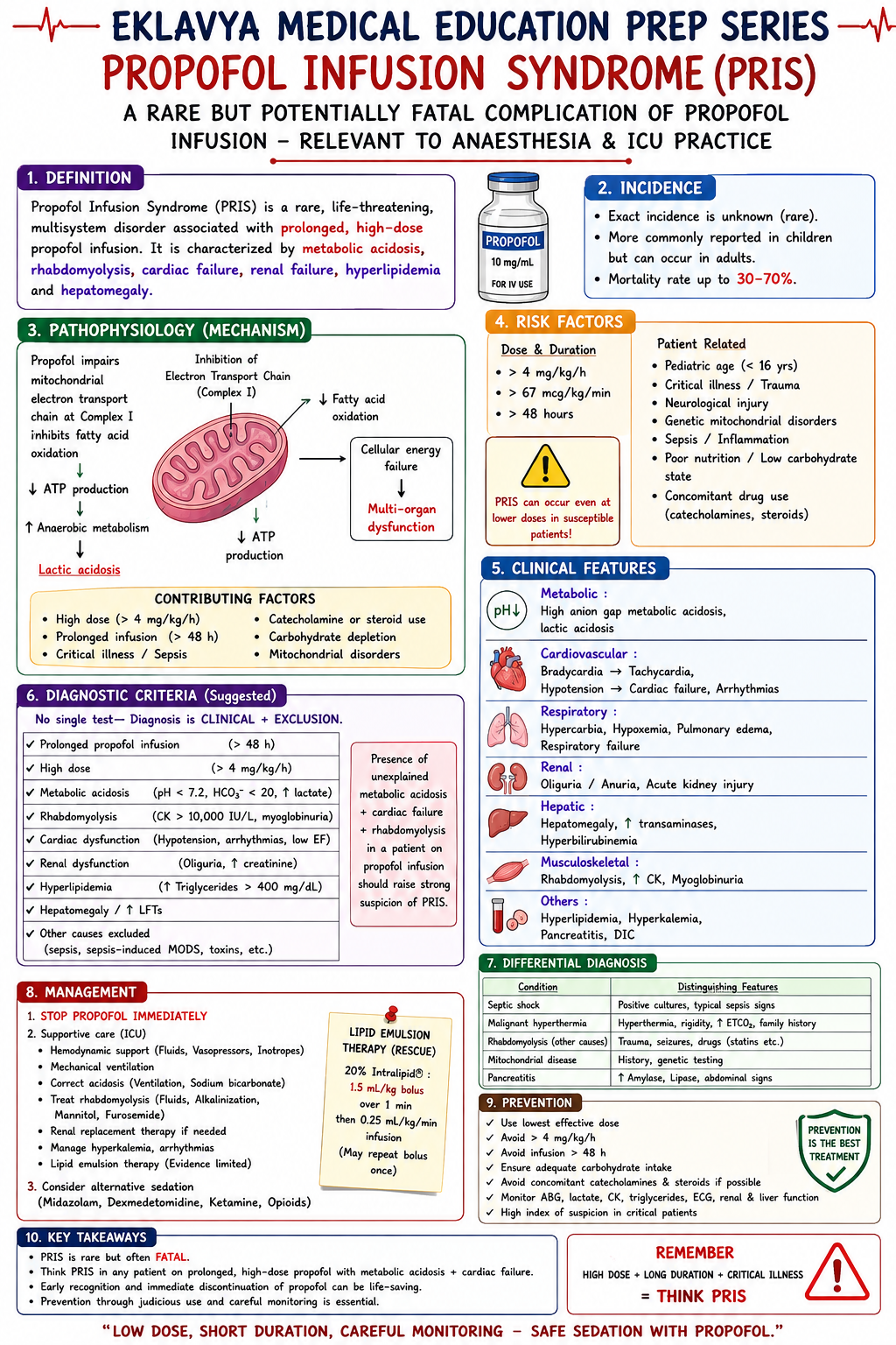
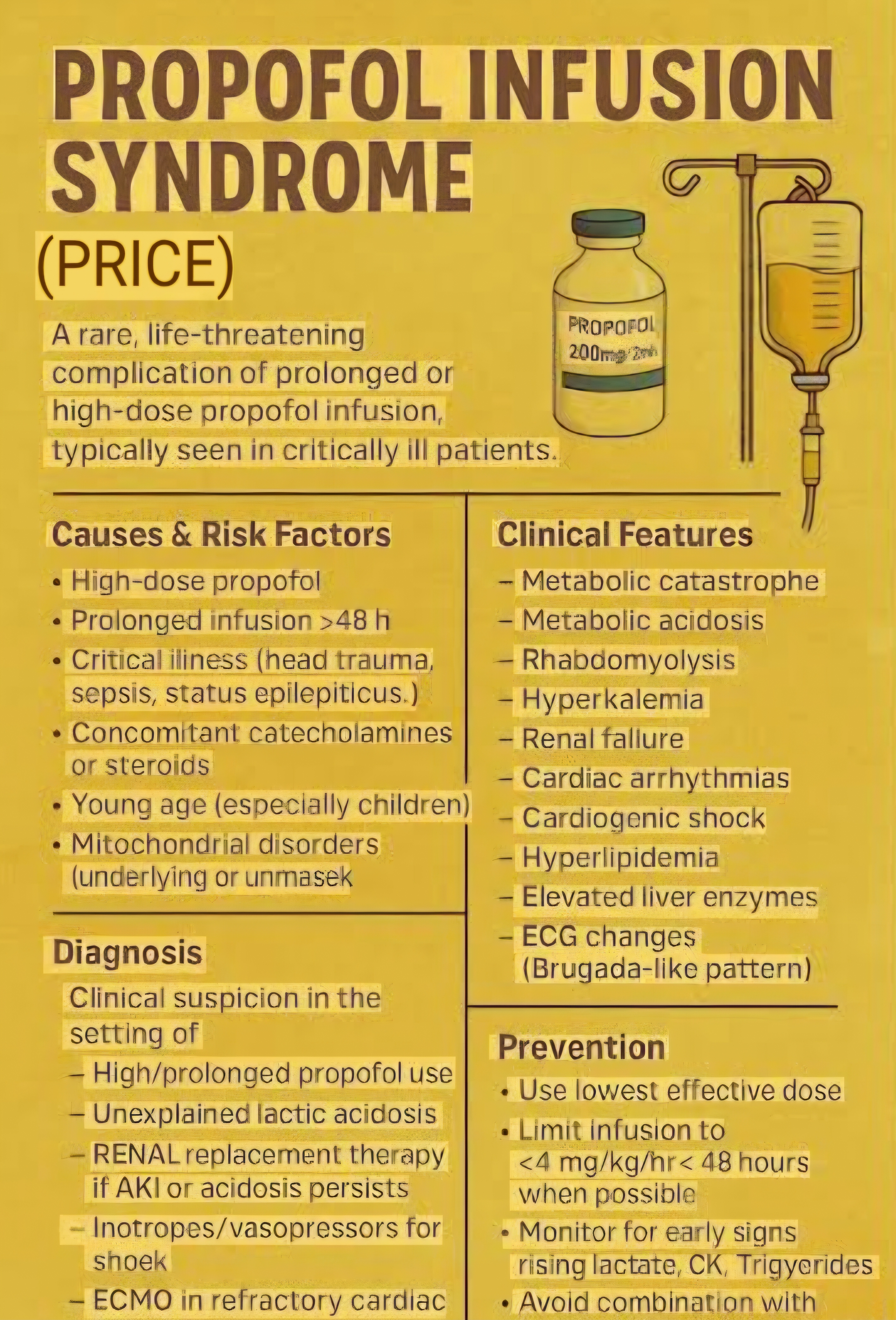
Describe Propofol Infusion Syndrome (PRIS) — its definition, molecular pathophysiology, predisposing factors, clinical features, and management. State the maximum safe infusion rates and monitoring parameters.

collections Question Diagrams & Reference Images (2)
Question Reference Diagram
zoom_in View Image
Question Reference Diagram
zoom_in View Image
description Clinical Response
⚙ Core Concept
Propofol Infusion Syndrome is a rare but frequently fatal complication of high-dose propofol infusions — characterised by metabolic acidosis, rhabdomyolysis, cardiac failure, and multi-organ dysfunction. Its molecular basis is mitochondrial respiratory chain inhibition and free fatty acid oxidation failure — the same cellular toxicity mechanism that makes it distinct from standard propofol pharmacology at therapeutic doses. Recognition is the critical challenge because its early features (rising lactate, metabolic acidosis) overlap with many other ICU diagnoses. (Cremer OL — Lancet 2001; Kam PC — Anaesthesia 2007; Roberts RJ; Miller's Anaesthesia 9th Ed)
A. Definition and Incidence1 mark

PRIS: a syndrome of new-onset metabolic acidosis (lactate acidosis — unexplained metabolic acidosis in an ICU patient on propofol infusion), rhabdomyolysis, renal failure, and cardiac failure (specifically brady-arrhythmia progressing to right bundle branch block → complete heart block → cardiovascular collapse) occurring in patients receiving high-dose propofol infusions

Incidence: approximately 1.1% of ICU patients on propofol infusions; mortality: 33–85% once the full syndrome is established; more common than recognised because early features are attributed to other causes

B. Pathophysiology2 marks

Propofol (particularly at high doses and prolonged infusions) → inhibition of mitochondrial Complex I (NADH-ubiquinone oxidoreductase) AND Complex II (succinateubiquinone oxidoreductase) of the electron transport chain → impaired oxidative phosphorylation → cells cannot use oxygen for ATP synthesis despite adequate O₂ delivery → cells switch to anaerobic metabolism → lactic acidosis. Simultaneously: propofol phenol ring → impairs mitochondrial β-oxidation of free fatty acids (the primary fuel source for cardiac muscle and skeletal muscle in critically ill patients who are frequently on carbohydrate-restricted or lipid-heavy nutrition) → free fatty acids and their acylcarnitine intermediates accumulate → directly toxic to the mitochondrial and cell membranes → rhabdomyolysis of cardiac and skeletal muscle → acute cardiac failure + skeletal myolysis → myoglobinaemia → AKI Risk Factors Risk Factor Mechanism Dose >4–5 mg/kg/hr for >48 hours Exceeds the mitochondrial capacity to process propofol metabolites; accumulation of toxic phenol metabolites High catecholamine state (severe illness, Catecholamines switch cellular metabolism toward fat oxidation → cells become MORE dependent on β-oxidation catecholamine infusions) pathways that propofol specifically impairs High-fat/low-carbohydrate intake Further increases reliance on fat oxidation; propofol vehicle (Intralipid — 1.1 kcal/mL) itself adds significant lipid load Mitochondrial disease Pre-existing mitochondrial impairment → much lower propofol dose required to tip into PRIS Paediatric patients Originally described in children; PRIS was first recognised as a distinct entity in paediatric critical care after reports of unexplained cardiac arrest in children on propofol sedation Concomitant steroids + catecholamines Steroids impair mitochondrial function; catecholamines increase fat metabolism; combination with propofol = perfect storm

C. Clinical Features and Monitoring1 mark

Feature Clinical/Laboratory Finding Metabolic lactic Unexplained metabolic acidosis (↑ lactate >4 mmol/L, ↓ pH); FIRST and most consistent early feature; check lactate every 4–6 hours in patients on acidosis propofol >4 mg/kg/hr Rhabdomyolysis Rising CK (often dramatic >10,000 IU/L); myoglobinuria (dark brown urine); muscle tenderness Cardiac New right bundle branch block (RBBB) → progressing to complete heart block → cardiovascular collapse; new-onset bradycardia refractory to dysfunction atropine; cardiomegaly on CXR; LV failure on echo Lipaemia Hyperlipidaemia (lipid load from Intralipid vehicle); visible lipaemia in plasma samples AKI Rising creatinine from rhabdomyolysis-induced renal tubular necrosis; myoglobin casts in tubules

D. Management & Safe Dosing Limits1 mark
⚠ Safe Propofol Infusion Limits
Maximum rate: 4 mg/kg/hr (67 mcg/kg/min) for ICU sedation; some sources cite 4 mg/kg/hr as the absolute upper limit beyond which PRIS risk increases significantly Maximum duration: AVOID continuous propofol >48–72 hours at doses approaching the maximum; if longer sedation needed → switch to or add alternative sedative (midazolam, dexmedetomidine, ketamine) Mandatory monitoring during prolonged ICU propofol: serum lactate every 6 hours; CK every 24 hours; triglycerides every 48 hours; urine myoglobin if CK rises; ECG continuous If PRIS suspected: STOP propofol immediately; switch to alternative sedation; supportive care: treat acidosis (sodium bicarbonate, CRRT for severe acidosis/AKI); treat cardiac failure (temporary pacing if complete heart block; inotropes for LV failure; ECMO as bridge if refractory); IV carnitine supplementation (theoretical — promotes free fatty acid oxidation via alternative pathways); renal replacement therapy; lipid rescue (20% intralipid 1.5 mL/kg bolus — same as LAST treatment — can help remove propofol from plasma)
🎤 Viva Corner
Q. An ICU patient on propofol 5 mg/kg/hr for 72 hours develops new RBBB on ECG and a lactate of 8 mmol/L. What is your immediate action?
This presentation — new right bundle branch block + severe metabolic lactic acidosis in a patient on high-dose propofol >48 hours — is PRIS until proven otherwise. Immediate action: STOP propofol infusion immediately. Do not wait for confirmation of the diagnosis — the consequences of continuing propofol in a case of PRIS are cardiovascular collapse and death; the consequences of stopping propofol and switching to an alternative are modest (brief agitation until alternative sedation is established). Switch to alternative sedation: dexmedetomidine infusion + midazolam boluses; or ketamine infusion if cardiovascularly appropriate. Simultaneously manage the existing organ dysfunction: continuous cardiac monitoring — the RBBB may progress to complete heart block; prepare for temporary transvenous pacing; have atropine and isoprenaline available for complete heart block; echocardiography urgently to assess LV and RV function. For the acidosis: arterial blood gas now to quantify; sodium bicarbonate 50–100 mmol IV for pH <7.10; initiate urgent CRRT (continuous renal replacement therapy) — provides metabolic clearance, removes lactic acid, manages any concurrent AKI from myoglobin; check CK (likely dramatically elevated); check urine for myoglobinuria; IV fluid resuscitation to maintain urine output ≥1 mL/kg/hr to prevent myoglobin precipitation in renal tubules. Check TFTs, cortisol, and cortisol stimulation test (exclude adrenal insufficiency as contributing cause of metabolic acidosis). IV lipid rescue: 20% intralipid 1.5 mL/kg IV may help sequester circulating propofol and its toxic metabolites. Inform ITU consultant and family — prognosis once PRIS is established is poor (33–85% mortality); intensive supportive care is the treatment; there is no specific antidote beyond stopping propofol.
★ Examiner's Pearl
The maximum propofol dose (4 mg/kg/hr for ICU sedation — specifically NOT the same as the intraoperative anaesthetic dose) with the duration limit (>48 hours at high dose = danger zone) are the specific clinical limits tested. The pathophysiology (mitochondrial Complex I + II inhibition → impaired oxidative phosphorylation; βoxidation failure → cardiac/skeletal myolysis) must be mechanistically stated — not just "mitochondrial toxicity." New RBBB → complete heart block progression is the most specific cardiac feature of PRIS and the most tested ECG finding.
Cremer OL et al. PRIS — long-term propofol infusion and cardiac failure (Lancet 2001;357:117-118). Kam PC, Cardone D. Propofol infusion syndrome (Anaesthesia 2007;62:690- 701). Roberts RJ et al. PRIS risk in critically ill adults (Crit Care 2009;13:R169). Miller RD et al. Miller's Anaesthesia, 9th Ed, Chapter 30.
lock Protected Clinical Document • Pulse App • Screenshots Strictly Prohibited
QUESTION 65 person Asked by .
bookmark_add

Describe the pathophysiology of phaeochromocytoma. Outline the preoperative preparation including α-blockade and β- blockade protocol. Discuss the intraoperative management of hypertensive crises during surgical manipulation, and the management of post-resection hypotension.

description Clinical Response
⚙ Core Concept
Phaeochromocytoma is an adrenaline-secreting tumour of the adrenal medulla (or extra-adrenal paraganglioma) that produces life-threatening cardiovascular crises during surgical resection — the peak intraoperative crisis (massive catecholamine release from tumour manipulation) and the postresection crash (abrupt catecholamine withdrawal with a vasoplegic circulation accustomed to massive sympathomimetic tone) represent two opposite haemodynamic extremes that must be anticipated, prepared for, and managed in real time. (Miller's Anaesthesia 9th Ed; Pacak K — endocrine reviews; Prys-Roberts C; Bravo EL; Kinney MA)
A. Pathophysiology2 marks

Phaeochromocytomas arise from chromaffin cells of the adrenal medulla (90%) or extra-adrenal sympathetic ganglia (10% — paragangliomas); secrete catecholamines (adrenaline, noradrenaline, dopamine) constitutively or in paroxysms Noradrenaline-secreting tumours → predominantly hypertension from α₁ vasoconstriction; adrenaline-secreting → mixed hypertension and tachycardia + β₂ vasodilation (can cause paradoxical hypotension); dopamine-secreting → hypertension from indirect catecholamine release

Rule of 10: 10% bilateral; 10% extra-adrenal; 10% malignant; 10% familial (MEN2A, MEN2B, VHL, SDH mutations); 10% in children; 10% discovered incidentally Chronic catecholamine excess → hypertrophied cardiovascular system with REDUCED intravascular volume (chronic vasoconstriction → pressure natriuresis + relative hypovolaemia); catecholamine cardiomyopathy (catecholamine-induced cardiac toxicity); increased sensitivity to vasodilators after tumour removal

B. Preoperative Preparation — α-blockade Protocol3 marks
✅ Standard Preoperative α-Blockade — Start 10–14 Days Before Surgery
Phenoxybenzamine (non-selective, irreversible α-blocker): start 10 mg BD; increase by 10–20 mg every 2–3 days until BP controlled (target <130/80 mmHg sitting; nasal stuffiness + orthostatic hypotension confirm adequate α-blockade); typical final dose 1–4 mg/kg/day; MUST be started before β-blockade (α-blockade first prevents paradoxical hypertension from unopposed α-stimulation if β is blocked first) Alternative: selective α₁-blockers: doxazosin (1–16 mg/day) or prazosin — shorter-acting, less orthostatic hypotension; similar efficacy to phenoxybenzamine in systematic reviews; increasingly preferred because easier to reverse intraoperatively β-blockade (add AFTER α-blockade): propranolol or atenolol — added only after ≥3 days of α-blockade; indicated for: reflex tachycardia (HR >100 from phenoxybenzamine), catecholamine-induced arrhythmia, predominantly adrenaline-secreting tumours; DO NOT start β-blocker before α-blockade (unopposed α-stimulation from catecholamine surge → severe hypertensive crisis) Volume expansion: high-salt diet + fluid intake; 2–4 L oral fluid daily in the week before surgery; IV fluid loading perioperatively; essential because chronic catecholamine excess has contracted intravascular volume — normalising volume before surgery reduces post-resection hypotension
C. Intraoperative Management3 marks

Monitoring (all before induction) Arterial line (radial artery — beat-to-beat BP monitoring throughout); large-bore IV access (2× 14G); central venous catheter (CVP monitoring, vasopressor access); TOE or PA catheter for high-risk patients (EF <40%, bilateral tumour); temperature monitoring

Induction

Avoid laryngoscopy-induced catecholamine surge: lidocaine 1.5 mg/kg IV + fentanyl 2–3 mcg/kg + esmolol 0.5 mg/kg immediately before laryngoscopy; or high-dose opioid induction (remifentanil 2–3 mcg/kg)

Avoid: histamine-releasing drugs (morphine, atracurium — can trigger catecholamine release from the tumour or direct histamine release); succinylcholineinduced fasciculations raise intra-abdominal pressure potentially triggering tumour catecholamine release; droperidol (triggers catecholamine release from phaeochromocytoma in rare cases) Hypertensive Crisis During Tumour Manipulation Drug Dose Mechanism Phentolamine (non- 2–5 mg IV bolus; repeat Competitive α₁ + α₂ blockade → acute vasodilation; rapid onset (2 min); duration 10–15 min; drug of choice for selective reversible every 2–3 minutes until BP intraoperative hypertensive crisis management α-blocker) controlled Sodium nitroprusside 0.5–8 mcg/kg/min infusion Nitric oxide → direct arterial + venous vasodilation; immediate onset; very titratable; risk of cyanide toxicity at high (SNP) doses or prolonged use; most powerful vasodilator available; useful as infusion for sustained hypertension during tumour handling Nicardipine (IV 5–15 mg/hr infusion Dihydropyridine CCB → arterial vasodilation; does not cause reflex tachycardia to the same degree as SNP; calcium channel increasingly used as an alternative first-line infusion blocker) Magnesium sulphate 2 g IV bolus Inhibits catecholamine release from the tumour; inhibits catecholamine receptor sensitivity; useful adjunct

D. Post-Resection Hypotension — The Crash2 marks
⚠ Ligation of Tumour Venous Drainage = Abrupt Catecholamine Withdrawal + Vasoplegic Shock
When the surgeon ligates the adrenal vein (or the main venous drainage of the tumour), catecholamine secretion stops abruptly; the peripheral vasculature, accustomed to massive catecholamine-mediated tone, dilates → immediate fall in SVR → haemodynamic collapse Management: WARN the anaesthesiologist before ligating the adrenal vein; stop all vasodilator infusions (SNP, nicardipine) immediately; give IV fluid bolus 500 mL rapidly; prepare vasopressor infusion: noradrenaline 0.1–0.5 mcg/kg/min (replace the lost endogenous catecholamine with an exogenous alpha agonist); vasopressin (0.03–0.04 units/min) as an alternative or add-on if noradrenaline insufficient; the post-resection hypotension may be prolonged (hours to days if the catecholamine stores were the only thing maintaining the circulation); ICU admission post-operatively; continue vasopressor support as needed; insulin infusion may be needed (catecholamines suppress insulin — after removal, profound hypoglycaemia can occur from residual excess insulin secretion)
🎤 Viva Corner
Q. Why must α-blockade ALWAYS precede β-blockade in the preoperative preparation of phaeochromocytoma?
The sequence — α-blockade must be established before β-blockade is introduced — is one of the most tested and clinically critical principles in phaeochromocytoma management. The danger of reversing this sequence: Phaeochromocytomas secrete both adrenaline (predominantly β₁ + β₂ effects: tachycardia, vasodilation) and noradrenaline (predominantly α₁ effects: vasoconstriction, hypertension). When a patient with phaeochromocytoma receives βblockade first (without α-blockade): the β₂-mediated vasodilation from adrenaline is blocked (β₂ causes peripheral vasodilation — blocking β₂ removes this vasodilatory component); the α₁-mediated vasoconstriction from noradrenaline (already present and unopposed by α-blockade) is now completely UNOPPOSED by the vasodilatory counter-effect of β₂ stimulation; the β₁-mediated cardiac output is also reduced (β₁ block reduces HR and SV); the reflex bradycardia from very high BP (Cushing reflex) is blocked and cannot compensate → the result: catastrophic, profound, refractory hypertension from completely unopposed α₁ vasoconstriction, with a reduced cardiac output that cannot maintain tissue perfusion despite the sky-high BP (cardiogenic shock in the context of extreme vasoconstriction). The correct sequence: first establish comprehensive α-blockade for at least 3 days (ensure adequate receptor blockade to handle any catecholamine surges); then add β-blockade to control the reflex tachycardia from the α-blocker-induced vasodilation; at this point β-blockade is safe because α-receptors are already blocked — any catecholamine surge produces vasodilation (through β₂) rather than vasoconstriction (α₁ is blocked).
★ Examiner's Pearl
α-blockade before β-blockade (NEVER reverse the sequence — reason: unopposed α-vasoconstriction from β-block of vasodilatory β₂ → catastrophic hypertension) is the most examined practical principle. Phenoxybenzamine: 10–14 days before surgery, start low (10 mg BD) and titrate to nasal stuffiness + orthostatic hypotension (these confirm adequate α-blockade) — these clinical signs are specifically tested. Post-resection vasoplegic shock management: warn anaesthesiologist before adrenal vein ligation, stop vasodilators, noradrenaline infusion ready.
Pacak K. Preoperative management of the pheochromocytoma patient (J Clin Endocrinol Metab 2007;92:4069-4079). Kinney MA et al. Perioperative management of phaeochromocytoma (J Cardiothorac Vasc Anesth 2002;16:359-369). Prys-Roberts C. Phaeochromocytoma — recent progress in its management (BJA 2000;85:44-57). Miller RD et al. Miller's Anaesthesia, 9th Ed.
lock Protected Clinical Document • Pulse App • Screenshots Strictly Prohibited
QUESTION 66 person Asked by .
bookmark_add

Discuss the preoperative assessment of a patient with hyperthyroidism for thyroidectomy including optimisation. Describe specific airway challenges (tracheal compression, recurrent laryngeal nerve monitoring). Outline the diagnosis and emergency management of thyroid storm.

collections Question Diagrams & Reference Images (1)
Question Reference Diagram
zoom_in View Image
description Clinical Response
⚙ Core Concept
Thyroid surgery presents two distinct anaesthetic challenges: the routine challenge of safe neck surgery in a haemodynamically compromised hyperthyroid patient who must be euthyroid before elective resection; and the emergency challenge of thyroid storm — a life-threatening hypermetabolic crisis (mortality 10–20% even with treatment) that can be triggered by surgery, trauma, or infection in an inadequately treated hyperthyroid patient. The anaesthesiologist must be able to both prevent thyroid storm (preoperative optimisation) and manage it aggressively when it occurs. (Miller's Anaesthesia 9th Ed; Burch HB — J Emerg Med 1993; Nayak B; Carroll R; AACE/ATA Guidelines)
A. Preoperative Assessment & Optimisation3 marks

Assessment of Hyperthyroidism

Symptoms and clinical assessment: weight loss, heat intolerance, tremor, palpitations, anxiety, exophthalmos (Graves' disease), goitre size; assess for signs of heart failure (AF in 10–15%); thyroid bruit

Investigations: TFTs (TSH, free T4, free T3); ECG (AF, sinus tachycardia, LVH); CXR/CT neck (tracheal deviation, retrosternal extension, compression of trachea or oesophagus); flow-volume loop if significant tracheal compression; calcium and parathyroid function (risk of hypoparathyroidism post-thyroidectomy)

Airway assessment: palpate the goitre; assess tracheal position; review CT for tracheal diameter at narrowest point; if tracheal compression >50% → awake fiberoptic intubation; tracheal softening (tracheomalacia) may occur with long-standing retrosternal goitre → risk of tracheal collapse after ETT removal Achieving Euthyroid State Drug Mechanism Duration to Euthyroid Carbimazole or Inhibit thyroid peroxidase → ↓ T3 and T4 synthesis; PTU also inhibits peripheral 4–6 weeks for biochemical euthyroid state; continue up to Propylthiouracil conversion of T4→T3 day of surgery (PTU) Lugol's iodine Wolff-Chaikoff effect: excess iodide transiently inhibits thyroid hormone synthesis and Used for 10–14 days pre-operatively after anti-thyroid (KI 65%) release; also reduces vascularity of the thyroid gland (reduces intraoperative bleeding) drugs started; reduces vascularity and bleeding — an additional surgical benefit intraoperatively Beta-blockers Blocks peripheral sympathetic effects of excess T3/T4 (HR, tremor, anxiety); propranolol Immediate symptom control; continue until surgery; must (propranolol) also inhibits peripheral T4→T3 conversion (PTU-like effect) be used alongside anti-thyroid drugs (treats symptoms, not the thyroid excess)

B. Specific Airway Considerations2 marks

Tracheal deviation and compression: large goitres may displace or compress the trachea; CT neck identifies the minimum tracheal diameter; if compressed <50% diameter → standard intubation with smaller ETT (6.0–6.5 mm); if >50% → consider awake fiberoptic intubation (maintains airway tone and spontaneous ventilation throughout — safest approach)

Reinforced (armoured) ETT: used for thyroid surgery to prevent ETT kinking in the extended neck position; allows surgeon access to the anterior neck without ETT obstruction

Recurrent Laryngeal Nerve (RLN) monitoring: the RLN runs in the tracheo-oesophageal groove adjacent to the thyroid; inadvertent RLN injury → immediate hoarseness or voice change (unilateral) → bilateral injury → aphonia + stridor + airway emergency; intraoperative neuromonitoring (IONM) uses a specialised ETT (NIM-EMG tube — electromyography electrodes embedded on the ETT surface monitor the RLN via laryngeal muscle EMG); if RLN is stimulated, the EMG records a response; loss of signal = nerve impaired; requires specific endotracheal tube and specific drug considerations

RLN monitoring — NIM ETT implications: NMBs MUST be allowed to wear off completely before placing the NIM tube (EMG response requires muscle activity — NMB prevents it); use the shortest-acting NMB (rocuronium 0.3–0.6 mg/kg initial dose + TOF monitoring); confirm T4 (all four twitches) on TOF before baseline NIM signal is obtained

Tracheomalacia: rare complication of long-standing retrosternal goitre; cartilaginous rings soften and lose support when external tumour pressure is suddenly removed → trachea collapses inward on extubation → immediate post-extubation stridor → may require re-intubation or even tracheostomy; anticipate by performing "cuff leak test" before extubation in long-standing large goitre

C. Thyroid Storm — Diagnosis and Emergency Management5 marks
⚠ Thyroid Storm — Mortality 10–20% Even With Treatment
Diagnosis — Burch-Wartofsky Score Parameter Score Range Trigger for Diagnosis Thermoregulatory dysfunction 0–30 (temp >41°C = 30 High temperature is the hallmark — fever in an unwell hyperthyroid patient = thyroid storm until (temperature) points) proven otherwise CNS effects (agitation → coma) 0–30 Agitation, delirium, seizures, coma GI/hepatic dysfunction 0–20 Nausea, vomiting, diarrhoea, jaundice Cardiovascular dysfunction 0–35 (AF + HR >130 = 25 Tachycardia (HR >130), AF, pulmonary oedema, cardiac failure points) Precipitant identified 0–10 Surgery, infection, trauma, parturition — 10 points if present Score ≥45 = thyroid storm; score 25–44 = impending storm; <25 = unlikely Emergency Management Action Drug/Intervention Mechanism 1. ICU admission; cooling (paracetamol — NOT aspirin which displaces T4 Treat the hypermetabolic consequences; paracetamol preferred SUPPORTIVE from protein binding → worsens storm; cooling blankets; ice packs); IV fluid antipyretic (aspirin contraindicated) CARE resuscitation; O₂ 2. Block NEW PTU 600 mg oral/NGT loading, then 200–300 mg every 4 hours (preferred PTU is first-choice in storm specifically because it blocks BOTH synthesis synthesis over carbimazole in thyroid storm — additionally blocks T4→T3 AND peripheral conversion conversion); OR carbimazole 20 mg every 4 hours if PTU unavailable 3. Block Lugol's iodine 8 drops (0.5 mL) every 6 hours or potassium iodide 5 drops Iodide blocks thyroid hormone RELEASE (Wolf-Chaikoff + Plummer RELEASE (1 every 6 hours; WAIT 1 hour after PTU (if iodine given first — provides effect); do NOT give before PTU — paradoxical hormone synthesis would hour after substrate for synthesis before block established) occur PTU) 4. Block Propranolol 40–80 mg oral every 4–6 hours OR esmolol infusion 50–300 Beta-blockade: reduces HR and sympathomimetic manifestations; peripheral mcg/kg/min (if oral route unavailable or IV required) — controls hydrocortisone: covers relative adrenal insufficiency (thyroid storm effects tachycardia, tremor, anxiety; hydrocortisone 100 mg IV every 8 hours increases cortisol demand); also inhibits peripheral T4→T3 conversion 5. Treat Antibiotics for infection; treat other precipitating cause The storm will not resolve until the precipitant is treated; ongoing PRECIPITANT catecholamine release perpetuates the hypermetabolic state
🎤 Viva Corner
Q. Why is aspirin specifically contraindicated in thyroid storm for fever management?
Aspirin is specifically contraindicated in thyroid storm despite being one of the most commonly available antipyretics. The reason: the vast majority of thyroid hormones (T3 and T4) circulate bound to plasma proteins — primarily thyroxine-binding globulin (TBG), albumin, and transthyretin — in an inactive form; only the small free (unbound) fraction of T3 and T4 is biologically active and responsible for the clinical manifestations of thyrotoxicosis. Salicylates (aspirin and its metabolites) compete with T3 and T4 for binding sites on TBG and other thyroid-binding proteins — they displace thyroid hormones from their protein-binding sites. In a patient with thyroid storm, where the total T4 and T3 concentrations are already dramatically elevated and are driving the hypermetabolic crisis, displacing even a fraction of these from protein binding dramatically INCREASES the free (active) T3 and T4 concentrations in the plasma. This worsens every manifestation of the thyroid storm — it effectively administers a dose of bioactive thyroid hormone at precisely the moment when the patient can least tolerate it. For this reason, paracetamol (acetaminophen) is the antipyretic of choice in thyroid storm — it reduces fever through central prostaglandin inhibition without any interaction with thyroid hormone protein binding; physical cooling measures (cooling blankets, ice packs to the axillae and groin) are also used alongside paracetamol for severe hyperthermia. Aspirin must be specifically avoided even at low doses.
★ Examiner's Pearl
The thyroid storm management sequence — PTU first (blocks synthesis AND conversion), THEN Lugol's iodine after 1 hour (blocks release — must NOT be given before PTU) — with the specific 1-hour delay and its reason (paradoxical synthesis if iodine given to unsuppressed gland) is the most tested clinical sequence in thyroid storm management. Aspirin contraindication (displaces T4 from TBG → worsens storm) with the protein-binding displacement mechanism is specifically tested. BurchWartofsky score ≥45 = storm is the diagnostic threshold.
Burch HB, Wartofsky L. Life-threatening thyrotoxicosis: thyroid storm (Endocrinol Metab Clin North Am 1993;22:263-277). Carroll R, Matfin G. Endocrine and metabolic emergencies: thyroid storm (Ther Adv Endocrinol Metab 2010;1:139-145). AACE/ATA Guidelines for Hyperthyroidism and Other Causes of Thyrotoxicosis 2011. Miller RD et al. Miller's Anaesthesia, 9th Ed.
lock Protected Clinical Document • Pulse App • Screenshots Strictly Prohibited
QUESTION 67 person Asked by .
bookmark_add

A 55-year-old presents with a right temporal glioblastoma causing midline shift and clinical signs of raised ICP. Describe the preoperative assessment, goals of neuroanaesthesia, intraoperative ICP management, choice of anaesthetic agents, brain relaxation strategies, and emergence considerations.

collections Question Diagrams & Reference Images (1)
Question Reference Diagram
zoom_in View Image
description Clinical Response
⚙ Core Concept
Neuroanaesthesia has two overarching goals: maintain adequate cerebral perfusion pressure (CPP = MAP − ICP; target ≥60 mmHg) to prevent secondary ischaemic injury; and provide surgical conditions that minimise brain swelling and ICP — a "slack brain" — while ensuring rapid, complete emergence to allow neurological assessment. Every anaesthetic decision — from agent choice to ventilation parameters to positioning — must balance these two goals. (Cottrell JE — Neuroanesthesia 5th Ed; Bhardwaj A; Miller's Anaesthesia 9th Ed; NICE CG176)
A. Preoperative Assessment2 marks

GCS and focal neurological deficit documentation (baseline for post-operative comparison); pupil assessment (unequal pupils → herniation → emergency);

NIHSS if focal deficits present

CT/MRI: tumour location and size; degree of midline shift; presence of hydrocephalus; oedema extent (vasogenic vs cytotoxic); vascular supply (hypervascularity → blood loss risk)

Medication review: dexamethasone (started by neurology — reduces vasogenic oedema; must continue perioperatively; stress dose not routinely needed for steroids already prescribed); anti-epileptic drugs (continue perioperatively); anticoagulation history Clinical Cushing's triad: hypertension + bradycardia + abnormal breathing = impending herniation → urgent CT + possible emergency ventricular drainage before anaesthesia

B. Anaesthetic Goals — Neurosurgical Principles2 marks

Goal Target How Achieved Maintain CPP CPP = MAP − ICP ≥ 60 mmHg Arterial line for continuous BP; vasopressors (phenylephrine) for MAP; ICP reduction strategies Reduce ICP / brain ICP <20 mmHg; slack brain at craniotomy Mannitol; moderate hyperventilation; head-up 15–30°; TIVA; steroids pre-operatively; relaxation CSF drainage Control CMRO₂ Reduce metabolic demand; prevent secondary injury Adequate depth; normothermia; normoglycaemia (6–10 mmol/L); normocapnia Rapid, smooth Awake, oriented, able to follow commands within 15– TIVA with propofol + remifentanil; short-acting agents; no residual NMB emergence 20 min of surgery end (sugammadex); avoid hypothermia (delays emergence) Avoid secondary PaO₂ >80 mmHg; PaCO₂ 35–40 mmHg; MAP ≥65 Mandatory monitoring; active warming; arterial blood gas monitoring every 60 min injury factors mmHg; Temperature 36–37°C

C. Intraoperative Anaesthetic Management3 marks

Agent Choice

TIVA preferred for craniotomy: propofol reduces CMRO₂ and ICP, maintains cerebral autoregulation; remifentanil provides precise analgesic control and allows rapid emergence (context-sensitive half-time 3 min); no effect on ETCO₂ monitoring interpretation

Volatile agents: acceptable at ≤0.5 MAC (low doses preserve cerebral autoregulation); above 0.5 MAC → cerebral vasodilation → ↑ CBV → ↑ ICP; desflurane causes cerebral vasodilation and sympathetic activation → avoid; sevoflurane preferred if volatile is used

N₂O: avoid in craniotomy — increases CMRO₂ and CBF; expands any pneumocephalus; may worsen PONV Avoid ketamine (↑ CMRO₂ and ICP; cerebral vasodilation) Brain Relaxation ("Slack Brain") Strategies Strategy Drug/Intervention Mechanism Osmotherapy Mannitol 0.5–1 g/kg IV over 15–20 min; 7.5% hypertonic Osmotic gradient draws water from brain interstitium into plasma → reduces cerebral water saline 3–5 mL/kg IV alternatively → reduces ICP; effect begins at 15 min, peaks at 30–60 min, lasts 3–4 hours; monitor serum osmolality (<320 mOsm/L); avoid repeat doses if hypernatraemic Head 15–30° head-up; neutral neck Improves jugular venous drainage → reduces cerebral venous volume → reduces ICP position Controlled Normocapnia (PaCO₂ 35–40 mmHg) for routine Hypocapnia → cerebral vasoconstriction → ↓ CBV → ↓ ICP; effect immediate but tolerance ventilation maintenance; moderate hyperventilation (PaCO₂ 30–35 develops within 4–6 hours; ischaemia risk with prolonged hyperventilation; use ONLY as mmHg) if brain tight at opening — TEMPORARY only bridge to surgical decompression Position at Local anaesthetic at pin sites + remifentanil bolus before Pin insertion through the scalp into the outer table of the skull is extremely painful; Mayfield pins pin insertion inadequate analgesia → massive sympathetic surge → hypertension + ↑ ICP (skull clamp) Surgical CSF Surgeon opens cisterna magna or places intraventricular CSF removal directly reduces ICP and brain volume → provides surgical access without drainage drain excessive brain retraction

D. Emergence Considerations3 marks

Smooth emergence critical: coughing, straining, bucking on ETT during emergence → ↑ ICP → brain herniation through the craniotomy defect → catastrophic; ensure adequate analgesic level before emergence; use remifentanil to bridge until patient can follow commands without response to ETT; lidocaine 1.5 mg/kg IV 3–5 minutes before extubation reduces cough response to ETT

Immediate neurological assessment: is there a new deficit? Is the patient at the same GCS as pre-operatively? Can they squeeze both hands, follow commands bilaterally, speak? Any new deficits → urgent CT to exclude haematoma/oedema/ischaemia; haematoma within hours of craniotomy → reexploration

When NOT to extubate: prolonged surgery (>8 hours); significant brain swelling requiring dural closure under tension; haemodynamic instability; GCS baseline was low pre-operatively; posterior fossa surgery (risk of respiratory centre compression); brainstem surgery; plan for ICU intubated

PONV prevention mandatory: even mild vomiting → ↑ ICP → dangerous; full multimodal PONV prophylaxis (ondansetron + dexamethasone + droperidol); TIVA reduces PONV substantially vs volatile

🎤 Viva Corner
Q. At craniotomy, the dura is opened and the brain is tense — bulging out of the craniotomy defect. The surgeon says the brain is too tight to operate safely. What do you do?
A tight brain at dural opening is an emergency that must be addressed systematically and urgently — the surgeon cannot safely retract or resect in the presence of cerebral oedema, and the bulging brain is at immediate risk of herniation and ischaemia from retraction. Immediate step-by-step management: First, check that all modifiable causes of elevated ICP are not present or are being actively corrected: verify MAP is adequate (target ≥65 mmHg — low MAP → poor CPP → reactive vasodilation → worse ICP); ensure PaCO₂ is normal or slightly low (check ABG immediately — if PaCO₂ is elevated due to hypoventilation → increase RR to achieve PaCO₂ 30–35 mmHg temporarily); verify head position is 15–30° head-up with neutral neck; confirm no PEEP is applied (PEEP raises intrathoracic pressure → impairs jugular venous drainage → raises ICP); ensure depth of anaesthesia is adequate (light anaesthesia → hypertension → worse ICP); if TIVA is being used, check the propofol infusion is delivering at the target rate (pump alarm, line disconnection). Second, pharmacological intervention: if not already given — mannitol 0.5–1 g/kg IV over 15–20 minutes (immediate osmotherapy); alternatively 7.5% hypertonic saline 3–5 mL/kg; furosemide 20 mg IV may be added. Third, temporary moderate hyperventilation: if PaCO₂ was 38–40 → reduce to 30–33 mmHg by increasing RR; this provides immediate cerebral vasoconstriction and ICP reduction; use only temporarily while waiting for mannitol to work. Fourth, surgical options: surgeon may place a lumbar drain (if safe anatomically) to drain CSF — immediate volume reduction; surgeon may open the cisterna magna to release CSF from the basal cisterns; in extreme cases: surgeon may temporarily close and refer to CT to identify a new haematoma causing the acute swelling. If after all these measures the brain is still too tight: surgery may need to be abandoned (close and return when ICP has been pharmacologically controlled, possibly with EVD placement in the ICU).
★ Examiner's Pearl
The CPP formula (MAP − ICP; target ≥60 mmHg) and the five brain-relaxation strategies (mannitol; head-up; normocapnia/temporary hyperventilation; TIVA propofol; surgical CSF drainage) as a complete list are the core content. TIVA preferred over volatile for craniotomy (volatile agents ≥0.5 MAC cause cerebral vasodilation → ↑ ICP) is the agent choice rationale. Smooth emergence — remifentanil bridge + lidocaine IV before extubation prevents coughing which causes ↑ ICP through the craniotomy — is the post-op safety content.
Cottrell JE, Patel P. Cottrell and Patel's Neuroanesthesia, 6th Ed. Bhardwaj A et al. Handbook of Neurocritical Care. Miller RD et al. Miller's Anaesthesia, 9th Ed, Chapter 70. Seubert CN, Mahla ME. Neuroanesthesia (in Miller). Todd MM et al. Cerebral protection strategies (Anesthesiology 2009).
lock Protected Clinical Document • Pulse App • Screenshots Strictly Prohibited
QUESTION 68 person Asked by .
bookmark_add

Describe the key anatomical and physiological differences between neonates/infants and adults that are relevant to anaesthetic practice. Include cardiovascular, respiratory, renal, thermoregulatory, pharmacological, and metabolic differences with specific clinical implications.

collections Question Diagrams & Reference Images (1)
Question Reference Diagram
zoom_in View Image
description Clinical Response
⚙ Core Concept
Neonates and infants are not small adults — they have fundamentally different physiology in every organ system. Their cardiovascular system is ratedependent and less responsive to inotropes; their respiratory system has limited reserve and closes small airways at FRC; their thermoregulatory system is overwhelmed by even a cool operating theatre; and their pharmacokinetics are unpredictably different from adults. Understanding these differences is not academic — getting a drug dose or a temperature wrong in a 2 kg neonate can be immediately fatal. (Cote CJ, Lerman J — Paediatric Anaesthesia; Miller's Anaesthesia 9th Ed; Meakin G; Berry F)
A. Cardiovascular Differences2 marks

Parameter Neonate/Infant Adult Clinical Implication Heart rate CO = HR × SV; SV is FIXED in neonates (immature Can increase SV Bradycardia = cardiac arrest in neonates; ALWAYS give atropine 20 mcg/kg dependency Frank-Starling mechanism — stiff, non-compliant substantially (min 100 mcg) before laryngoscopy; normal neonatal HR 120–160 bpm; HR myocardium cannot increase SV significantly); CO (preload reserve, <100 = emergency is entirely rate-dependent Starling mechanism) Transitional Foetal shunts (PDA, foramen ovale) may reopen in No foetal shunts Maintain SpO₂ ≥94–98%; avoid hypoxia, acidosis, hypothermia — all trigger circulation hypoxia, acidosis, hypothermia, or surgical stress (all closed) ductal reopening; give O₂ carefully in prematurity (retinopathy risk with high → right-to-left shunting → cyanosis; particularly in SpO₂) first weeks of life Circulating 80–90 mL/kg (neonates); 70–80 mL/kg (infants); 65–70 mL/kg Meticulous blood loss monitoring (weigh swabs in grams — 1 g = 1 mL); blood even 5 mL blood loss can = 5–10% of total blood transfusion triggers lower (Hb <80 g/L in most neonates, <70 g/L in older volume volume in a 1 kg premature neonate children); use small (10 mL) syringes for fluid management; calculate maximum allowable blood loss preoperatively Glycogen Limited glycogen reserves → rapid hypoglycaemia Adequate hepatic Dextrose-containing maintenance IV fluids mandatory during nil-by-mouth stores with fasting; premature neonates particularly glycogen for periods in neonates and infants ≤6 months; blood glucose hourly monitoring vulnerable; high glucose consumption rate (6 moderate fasting intraoperatively; target 4–8 mmol/L mg/kg/min)

B. Respiratory Differences2 marks

Parameter Neonate/Infant Clinical Implication O₂ 6–8 mL/kg/min (vs 3 mL/kg/min in adults) — very high Rapid desaturation during apnoea (SpO₂ falls to critical in 60–90 seconds in neonates); consumption metabolic rate preoxygenate adequately; work fast during airway management FRC relative FRC only 3× TV (vs 7× TV in adults); closing capacity may Rapid onset of hypoxia; needs higher FiO₂ baseline; PEEP 3–5 cmH₂O even during routine to TV exceed FRC → small airway closure at FRC in infants → spontaneous ventilation under anaesthesia shunting even during spontaneous ventilation Chest wall Highly compliant; ribcage collapses inward during During respiratory distress: see "see-saw" breathing (chest wall sinks in, abdomen compliance respiratory distress rather than stabilising to generate protrudes) — an emergency sign requiring immediate intervention negative pressure (paradoxical breathing) Apnoea risk Post-conceptional age <60 weeks → immature respiratory All ex-premature infants under 60 weeks PCA must have apnoea monitoring for minimum in ex- centre → risk of postoperative apnoea for up to 12–24 12 hours post-GA; regional anaesthesia preferred to GA where possible in this group; premature hours after GA caffeine 10 mg/kg IV preoperatively reduces post-op apnoea incidence infants

C. Thermoregulation2 marks

Neonates and infants have: very large body surface area:mass ratio (4–5× adult); thin skin with minimal subcutaneous fat; no shivering mechanism in neonates (brown adipose tissue thermogenesis — "non-shivering thermogenesis" — instead, which consumes large amounts of O₂ and generates heat from BAT oxidation); immature hypothalamic thermoregulation

Under GA: all anaesthetic agents abolish behavioural thermoregulation and impair the autonomic thermoregulatory vasoconstriction threshold → body temperature falls rapidly to ambient; at 20°C theatre temperature, a neonate can become hypothermic in minutes

Prevention mandatory: theatre temperature 26–28°C for neonates (adults find this uncomfortably hot); warm all IV fluids and blood; forced-air warming blankets; warming mattress; warm cotton blankets; humidified gases; minimise exposed skin area; temperature probe mandatory throughout

Consequences of hypothermia: impaired drug metabolism → prolonged anaesthesia; coagulopathy; cardiac arrhythmia; impaired immune function → infection; hypoglycaemia (thermogenesis depletes glucose); prolonged post-op recovery

D. Pharmacological Differences2 marks

Parameter Neonate/Infant vs Adult Clinical Implication Volume of ↑ Total body water (neonates 80% body weight vs 60% adults); drugs distributing to TBW Higher weight-based dose of NMBs, antibiotics; neonates distribution have larger Vd → need higher mg/kg doses for equivalent plasma concentration; e.g., need larger mg/kg loading doses of many drugs water-soluble drugs (NMBs, aminoglycosides) Hepatic CYP450 enzyme systems immature at birth; CYP2C9, CYP2D6, CYP3A4 all below adult Prolonged half-lives of hepatically metabolised drugs metabolism activity levels; reached adult activity at 6 months–3 years depending on the isoenzyme (morphine, propofol, midazolam) in neonates; start low and titrate carefully; risk of accumulation Renal GFR 25–30% of adult value at birth; reaches adult GFR per body surface area by 18–24 Renally cleared drugs (vancomycin, aminoglycosides, excretion months; tubular secretion also immature NMBs) have prolonged half-lives; reduce dose frequency or extend intervals; monitor drug levels Protein Lower albumin and α₁-acid glycoprotein in neonates → more free (active) drug for given Enhanced effects of protein-bound drugs (local anaesthetics binding total plasma concentration — higher LAST risk; propofol; thiopentone); reduce doses accordingly MAC MAC is highest in neonates (1–6 months), falls with increasing age; term neonates: Neonates require HIGHER volatile concentrations for (volatile sevoflurane MAC ~3.3% (vs 2.0% in adults); falls to adult value by age 10–12 years equivalent depth of anaesthesia than children or adults — agents) counter-intuitive but well-established

E. Renal and Metabolic Differences2 marks

Glucose: neonates have limited glycogen stores and high glucose utilisation; hypoglycaemia (blood glucose <2.6 mmol/L) causes seizures and brain injury; maintain dextrose infusion throughout GA; check glucose hourly; prolonged preoperative fasting without IV dextrose is contraindicated

Calcium: neonates (especially premature) have immature calcium regulation; hypocalcaemia (ionised Ca²⁺ <1.1 mmol/L) causes cardiac depression, apnoea, and seizures; check ionised calcium before major neonatal surgery; give calcium gluconate 10% 0.5 mL/kg IV for hypocalcaemia

Acid-base: neonatal kidneys have limited bicarbonate reabsorption capacity → any acid load causes metabolic acidosis more readily than adults; standard base excess target slightly more negative acceptable in neonates (−4 to +2 mEq/L)

Drug toxicity — succinylcholine and atropine: succinylcholine causes more pronounced bradycardia in children (high vagal tone); ALWAYS give atropine 20 mcg/kg before succinylcholine in children; succinylcholine-induced hyperkalaemia and cardiac arrest from undiagnosed muscular dystrophy (Duchenne muscular dystrophy — DMD) in boys → succinylcholine increasingly avoided in paediatric elective cases

🎤 Viva Corner
Q. Why does a neonate require a HIGHER sevoflurane MAC than a 35-year-old adult, even though neonates are generally thought of as more sensitive to drugs?
This is a frequently misunderstood pharmacological principle. MAC (the alveolar concentration producing immobility in 50% of subjects) is a measure of CNS sensitivity to volatile anaesthetics, and it follows an inverted U-shaped relationship with age: it is relatively low at birth, rises to its peak between 1 and 6 months of age, then declines continuously throughout life to reach its lowest values in the elderly. In adults at age 40, sevoflurane MAC is 2.0%; in a neonate (first month of life), sevoflurane MAC is approximately 3.3%; in a 6-month-old infant, it may reach approximately 3.0–3.5%. The mechanism is not fully understood but several contributing factors have been identified: first, neonatal brains have higher concentrations of progesterone and other neuroactive steroids (from maternal transfer and residual levels) that have been declining since birth but are still relatively high — these neurosteroids may paradoxically increase MAC by modulating GABA and NMDA receptors; second, the neonatal nervous system has ongoing myelination and synaptic reorganisation — the specific synaptic targets of volatile agents (GABA-A receptors, NMDA receptors) may have different subunit compositions in the neonate that produce different drug sensitivity; third, the very high metabolic rate and higher brain temperature in neonates may contribute (MAC increases with increasing brain temperature). The clinical implication: neonates and young infants require higher inspired concentrations of volatile anaesthetics to achieve surgical anaesthesia than adults — the anaesthesiologist must not be deceived into thinking that a neonate is deeply anaesthetised at 1.5% sevoflurane when the adult patient would be deeply anaesthetised; the neonate may be lightly anaesthetised at this concentration.
★ Examiner's Pearl
Heart rate dependency of cardiac output in neonates (immature Starling mechanism — cannot increase SV) is the most tested cardiovascular difference — state that bradycardia in neonates = reduced CO = emergency. The apnoea risk in ex-premature infants <60 weeks PCA is a specific clinical safety rule tested in paediatric anaesthesia. Neonatal MAC > adult MAC (3.3% vs 2.0% for sevoflurane) with the age-related MAC curve (peak at 1–6 months, falls with age) is counter-intuitive and specifically tested.
Cote CJ, Lerman J, Anderson BJ. A Practice of Anaesthesia for Infants and Children, 6th Ed. Meakin G. Paediatric anaesthesia (BJA CEPD Reviews 2007). Berry F. Paediatric Anaesthesia. Miller RD et al. Miller's Anaesthesia, 9th Ed, Chapter 93. Lerman J et al. The minimum alveolar concentration of sevoflurane in neonates (Anesthesiology 1994;80:814).
lock Protected Clinical Document • Pulse App • Screenshots Strictly Prohibited
QUESTION 69 person Asked by .
bookmark_add

Classify emergency CS categories (RCOG 1–4). For a Category 1 CS with fetal distress in a labouring patient with an epidural in situ: describe the epidural top-up technique. For a Category 1 CS without an epidural, compare RSI-GA vs rapid-sequence spinal. Discuss the specific anaesthetic challenges of each technique.

description Clinical Response
⚙ Core Concept
Emergency CS is the most time-critical anaesthetic procedure performed outside the ICU. The decision-to-delivery interval (DDI) for Category 1 is 30 minutes — a target that requires systematic preparation, clear team communication, and a pre-determined anaesthetic plan for every obstetric unit. The choice between epidural top-up, rapid-sequence spinal, and RSI-GA depends on the existing anaesthetic, the urgency, and the patient's airway and haemodynamic status. (RCOG 2010; Lucas DN et al. — BJA; Kinsella SM; Palanisamy A; Miller's Anaesthesia 9th Ed)
A. RCOG Emergency CS Classification2 marks

Category Definition DDI Target Anaesthetic Priority 1 — Immediate threat to life of mother or fetus; examples: severe fetal <30 minutes Fastest available safe technique; epidural top-up Immediate bradycardia, cord prolapse, placental abruption with acute fetal compromise, (ideally <15 if in situ; rapid-sequence spinal if no epidural; threat to life uterine rupture, maternal haemodynamic collapse minutes for cord RSI-GA if spinal not feasible prolapse) 2 — Not immediately life-threatening but requires urgent delivery; examples: non- <75 minutes Regional anaesthesia preferred; time allows Maternal or reassuring CTG, failure to progress with maternal compromise careful spinal or epidural top-up fetal compromise 3 — No Early delivery needed but no current compromise; examples: failed induction, Within a scheduled Standard elective spinal or epidural anaesthesia; compromise prolonged latent phase time frame no time pressure 4 — Elective Elective CS at a time convenient to patient and team Elective scheduled Full preoperative assessment; standard spinal anaesthesia

B. Epidural Top-Up for Category 1 CS3 marks

Fastest Route to Surgical Anaesthesia from a Functioning Labour Epidural Check epidural function first (10–15 seconds): ask patient if she can still feel the block; test cold/warm sensation; if dense bilateral block present → proceed to top-up; if patchy or one-sided → may need rapid-sequence spinal instead

Top-up drug: 2% lidocaine with adrenaline 1:200,000 + optional fentanyl 50–100 mcg — fastest-onset epidural solution for surgical anaesthesia; inject 15– 20 mL in 5 mL increments (2–3 mL test dose first to re-confirm not intravascular); onset of T4 sensory block in 5–10 minutes (faster than 0.5% bupivacaine which takes 15–20 minutes)

Alternative: 0.5% levobupivacaine 15–20 mL (slower onset than 2% lidocaine; onset 12–15 min) or 3% chloroprocaine (if available — fastest epidural agent, onset 3–5 minutes; suitable for true Category 1)

Simultaneous actions while topping up: left lateral tilt; IV fluid co-load 500 mL crystalloid; phenylephrine infusion running (prevent spinal hypotension); supplemental O₂ by face mask; fetal monitoring (CTG off for delivery); surgical team ready; neonatologist present

Assess level: confirm T4 level by cold/pinprick test before incision; if inadequate despite full top-up dose → proceed to RSI-GA (do not delay surgery waiting for inadequate epidural)

C. Rapid-Sequence Spinal for Category 1 (No Epidural)2 marks

When appropriate: no epidural in situ; patient haemodynamically stable; no significant coagulopathy; uncomplicated expected airway; DDI is <30 minutes (achievable with spinal)

Drug choice: hyperbaric bupivacaine 0.5% 2 mL + fentanyl 15–25 mcg + morphine 100–150 mcg (intrathecal morphine provides 12–24 hours post-op analgesia — significant advantage over GA for post-CS pain); onset of surgical T4 level within 5–8 minutes

Simultaneous preparation: position patient in lateral decubitus or sitting; one hand on the patient, one drawing up drugs — no time for separate preparation; assistant preparing vasopressor; left lateral tilt immediately after injection; do not wait for full block to assess — check block level at 3 minutes (usually adequate T4 by then)

Failed spinal: if block is inadequate after 5 minutes → RSI-GA; do NOT give a second spinal (risk of total spinal from accumulated doses)

D. RSI-General Anaesthesia for Category 1 CS3 marks

Indications for RSI-GA over Regional Patient refusal of regional; coagulopathy (HELLP, DIC, thrombocytopenia); severe haemodynamic instability (cannot tolerate sympathectomy of spinal); local anaesthetic allergy (rare); severe urgency where time for spinal is unavailable; failed spinal or epidural top-up; major placenta praevia with active major haemorrhage Modified RSI Protocol for Obstetrics Step Detail Obstetric-Specific Modification Aspiration Sodium citrate 30 mL oral + ranitidine 50 mg IV + Mandatory pre-GA in pregnancy >16 weeks; reduce gastric acid pH and volume prophylaxis metoclopramide 10 mg IV Preoxygenation 100% O₂ × 3–5 minutes (head-up 20°); ETO₂ >90% Reduced safe apnoea time in pregnancy (↓FRC + ↑O₂ consumption); mandatory full preoxygenation; HFNO 60 L/min during apnoea Induction agent Thiopentone 4–5 mg/kg IV (classic; proven fetal safety) Reduce dose 20% in pre-eclampsia (enhanced CNS sensitivity); MAC reduced 25–40% in OR propofol 2–2.5 mg/kg pregnancy NMB Succinylcholine 1.5 mg/kg IV (classic RSI); OR Both acceptable; succinylcholine provides shortest duration (10 min) if airway fails; rocuronium 1.2 mg/kg + sugammadex 16 mg/kg rocuronium provides CICO rescue option available Cricoid Applied at induction; release if impairs laryngoscopy Higher risk of regurgitation in pregnancy; cricoid pressure standard; video laryngoscope pressure (DAS 2018) first-line Intubation Confirm with ETCO₂ × 6 breaths; 6.5–7.0 mm ETT Higher failed intubation rate in obstetrics (1:224 vs 1:1800 general surgical); video (oedematous obstetric airway → smaller tube) laryngoscope as first-line for all obstetric GA; have DAS failed intubation plan ready Maintenance Volatile agent (sevoflurane 1.0–1.5 MAC) + opioid after Awareness risk until delivery (deliberately reduced anaesthetic to minimise fetal exposure); delivery; N₂O 50% optional give fentanyl after cord clamped; must increase anaesthetic depth after delivery

🎤 Viva Corner
Q. During RSI-GA for emergency CS, you cannot visualise the larynx — CL Grade III on video laryngoscopy. Your first intubation attempt has failed. What do you do?
This is a failed obstetric intubation — one of the most critical airway emergencies in anaesthesia. The obstetric failed intubation rate is approximately 1:224 (vs 1:1800 in general surgery) — so this is an anticipated risk that every obstetric anaesthesiologist must have a pre-planned response to. Immediately: call for help; do NOT make a third attempt at intubation without optimisation; maintain 100% FiO₂ ventilation by face mask (maintain oxygenation is the absolute priority). Attempt optimisation: have the assistant apply BURP (backward, upward, rightward pressure on the thyroid cartilage — NOT cricoid); consider releasing cricoid pressure if it was impairing the view; use a bougie (gum elastic bougie through the visible arytenoid shadow even without seeing the cords); if available, switch to a different video laryngoscope blade (hyperangulated blade such as C-MAC D-blade for the difficult obstetric airway with anterior larynx). Maximum two further intubation attempts with these optimisations. If still failing after third attempt total: declare failed intubation and insert a second-generation supraglottic airway (i-gel or ProSeal LMA — suitable for maintaining airway and ventilation in CS and can withstand the high airway pressures from cricoid pressure); with the SAD in place and ventilation confirmed, make a decision: if Category 1 with immediate maternal/fetal threat — continue surgery through the SAD with maximum cricoid pressure and accept the slightly higher aspiration risk (the imminent fetal death outweighs this risk); if not immediately life-threatening — wake the patient up (reverse NMB with sugammadex 16 mg/kg; allow full recovery), then plan awake FOI or convert to regional anaesthesia for re-attempt. At all times: document every step with times; communicate clearly with obstetric team; neonatologist briefed on fetal status; call for senior obstetric anaesthesia help immediately on the first failed attempt.
★ Examiner's Pearl
RCOG four categories with DDI targets (Category 1 <30 min; Category 2 <75 min) must be reproduced. The epidural top-up drug choice (2% lidocaine + adrenaline → fastest onset 5–10 min) vs standard 0.5% bupivacaine is specifically tested as a clinical decision. Failed obstetric intubation rate (1:224 vs 1:1800 general) and the specific action "video laryngoscope first-line for ALL obstetric GA" are the safety evidence facts that distinguish current practice knowledge.
RCOG. Classification of urgency of CS (RCOG Good Practice No. 11, 2010). Kinsella SM et al. Failed tracheal intubation during obstetric GA — a systematic review (Anaesthesia 2015;70:886-898). Lucas DN et al. Urgency of CS (Anaesthesia 2000;55:1000-1004). DAS RSI Guidelines 2018. Miller RD et al. Miller's Anaesthesia, 9th Ed, Chapter 77.
lock Protected Clinical Document • Pulse App • Screenshots Strictly Prohibited
QUESTION 70 person Asked by .
bookmark_add

Define pre-eclampsia and severe pre-eclampsia per ISSHP 2018 criteria. Describe the pathophysiology. Outline: antihypertensive targets and drugs, magnesium sulphate therapy for seizure prophylaxis and treatment, and the specific anaesthetic management for CS in severe pre-eclampsia.

description Clinical Response
⚙ Core Concept
Pre-eclampsia — new-onset hypertension with proteinuria or end-organ dysfunction after 20 weeks gestation — affects 2–8% of pregnancies globally and is responsible for 16% of all maternal deaths. Its pathophysiology is driven by abnormal placentation causing placental ischaemia → systemic maternal endothelial dysfunction → vasospasm, multi-organ injury, and coagulopathy. The anaesthesiologist must simultaneously manage severe hypertension (stroke risk), MgSO₄ interactions, a challenging airway (oedema), and the haemodynamic instability of a patient requiring CS. (ISSHP 2018; MAGPIE trial; RCOG Green-top 10a; Dyer RA — spinal in pre-eclampsia; Miller's Anaesthesia 9th Ed)
A. Definitions (ISSHP 2018)2 marks

Condition Definition Hypertension SBP ≥140 mmHg OR DBP ≥90 mmHg on at least two occasions ≥4 hours apart after 20 weeks gestation in pregnancy

Pre- New hypertension after 20 weeks PLUS one or more of: proteinuria (≥300 mg/24h or spot PCR ≥30 mg/mmol); thrombocytopenia (<150,000/μL); renal eclampsia insufficiency (creatinine >88 μmol/L); impaired liver function (↑ LFTs); pulmonary oedema; new-onset headache unresponsive to medication; visual disturbances Severe pre- Pre-eclampsia with severe hypertension (SBP ≥160 mmHg or DBP ≥110 mmHg on two occasions) OR end-organ dysfunction (neurological symptoms, eclampsia HELLP syndrome, pulmonary oedema, severe renal impairment) Eclampsia New-onset grand-mal seizures in a patient with pre-eclampsia (or other hypertensive disorder of pregnancy); seizures in the context of hypertension and proteinuria HELLP Haemolysis (LDH >600 IU/L, abnormal blood film) + Elevated Liver enzymes (AST/ALT ≥2× ULN) + Low Platelets (<100,000/μL) — a severe variant of syndrome pre-eclampsia

B. Pathophysiology2 marks

Abnormal placentation (failure of trophoblast invasion of spiral arteries) → inadequate placental blood flow → placental ischaemia → release of anti-angiogenic factors (sFlt-1 — soluble FMS-like tyrosine kinase 1 — antagonises VEGF and PlGF) → systemic maternal endothelial dysfunction → widespread vasospasm (hypertension) + endothelial permeability ↑ (oedema, proteinuria) + platelet activation (thrombocytopenia, DIC) + organ ischaemia (renal, hepatic, cerebral)

Haemodynamics: paradox — pre-eclamptic patients have HIGH BP (vasospasm) but LOW intravascular volume (protein leaks out of vessels into interstitial space; venous tone ↑); vasodilators reduce BP but may worsen the already-contracted intravascular volume; fluid management is a delicate balance

Cerebral: vasospasm → headache, scotomata, altered consciousness; loss of cerebral autoregulation at high BP → breakthrough cerebral oedema → eclamptic seizures → cerebral haemorrhage (leading cause of maternal death in pre-eclampsia)

C. Antihypertensive Management2 marks
⚠ Target: SBP <160 mmHg, DBP <110 mmHg — Stroke Risk Above This Threshold
Do NOT reduce MAP by >20–25% acutely (uteroplacental blood flow is pressure-dependent and not autoregulated — acute hypotension → placental ischaemia → fetal distress) Drug Dose Advantage Disadvantage IV Labetalol 20 mg IV → repeat 40 mg → 80 Combined α+β blockade; no reflex Contraindicated in asthma, reactive airways, bradycardia; slower mg every 20 min; max 300 mg tachycardia; safe in pregnancy; wide onset than hydralazine experience IV 5–10 mg IV over 2 minutes; Direct arterial vasodilator; rapid onset; long Reflex tachycardia (may be undesirable in MgSO₄ context); less Hydralazine repeat every 20–30 min safety record in obstetrics predictable response; headache Oral 10 mg immediate-release oral; Effective; oral route; well tolerated; suitable Interaction with MgSO₄ (both vasodilate → profound hypotension); Nifedipine repeat after 30 min if needed before IV access established takes 10–15 min to peak; sublingual route NOT recommended
D. Magnesium Sulphate — Seizure Prophylaxis and Treatment2 marks

Indication: severe pre-eclampsia → MgSO₄ for seizure prophylaxis (MAGPIE trial: MgSO₄ reduced eclampsia rate by 58% vs placebo in severe preeclampsia; NNT = 63); ACTIVE eclampsia → MgSO₄ is first-line treatment (superior to diazepam or phenytoin for controlling eclamptic seizures —

Collaborative Eclampsia Trial)

Dosing: loading dose 4 g IV over 5–15 minutes; maintenance 1–2 g/hr IV infusion; continue for 24 hours after delivery (or 24 hours after last fit)

Toxicity monitoring — the clinical signs of hypermagnesaemia:

Therapeutic range: 2–3.5 mmol/L (serum Mg)

Loss of patellar reflexes: first sign of toxicity — Mg 3.5–5 mmol/L; CHECK PATELLAR REFLEXES HOURLY on MgSO₄ infusion

Respiratory depression: Mg 5–6.5 mmol/L; monitor RR ≥12 breaths/min

Cardiac arrest: Mg >7.5 mmol/L

Also maintain: UO ≥25 mL/hr (Mg is renally cleared; renal failure → rapid Mg accumulation) Antidote for MgSO₄ toxicity: calcium gluconate 10% 10 mL IV over 10 minutes (competes with Mg at the receptor; reverses respiratory and cardiac effects); have at bedside of every patient on MgSO₄ infusion

E. Anaesthetic Management for CS in Severe Pre-eclampsia2 marks

Regional preferred: spinal anaesthesia is SAFE in severe pre-eclampsia (the historical concern about catastrophic hypotension is not supported by evidence — Dyer RA et al.: pre-eclamptic patients have LESS hypotension after spinal than healthy parturients, because their high baseline SVR provides a buffer against the sympathectomy-induced fall in SVR); epidural also safe and allows gradual titration

Platelet threshold: neuraxial anaesthesia safe if platelets ≥70,000/μL (most institutional guidelines 70–80,000); review trend (rapidly falling counts are more concerning than a stable 80,000); check recent result (<6 hours in rapidly deteriorating HELLP)

If GA required: oedematous obstetric airway — use 6.5 mm ETT; video laryngoscope first-line; attenuate laryngoscopy response (alfentanil 10 mcg/kg or remifentanil 1 mcg/kg or labetalol 10 mg IV 1 minute before laryngoscopy — pre-eclamptic patients have exaggerated hypertensive response to laryngoscopy → stroke risk); esmolol NOT preferred in CS (crosses placenta → neonatal bradycardia)

Postoperative: HDU monitoring minimum 24 hours; continue MgSO₄ 24 hours post-delivery; continued antihypertensive therapy; watch for late eclampsia (can occur up to 48 hours post-delivery — the most dangerous time is actually the first 24 hours postpartum)

🎤 Viva Corner
Q. A severe pre-eclamptic patient on MgSO₄ infusion suddenly loses her patellar reflexes and her RR falls to 8 breaths/min. What has happened and what is the immediate management?
Loss of patellar reflexes followed by respiratory depression is progressive magnesium toxicity — a potentially fatal complication of MgSO₄ infusion. The clinical signs indicate serum magnesium is likely in the 5–6.5 mmol/L range (normal therapeutic range is 2–3.5 mmol/L; patellar reflexes lost at 3.5–5 mmol/L; respiratory depression at 5–6.5 mmol/L; cardiac arrest at >7.5 mmol/L). Immediate actions: STOP the MgSO₄ infusion immediately. Administer the antidote: calcium gluconate 10% 10 mL IV over 10 minutes (1 gram calcium gluconate IV) — calcium competitively antagonises magnesium at the neuromuscular junction, calcium channels, and cardiac membranes, reversing the respiratory and neuromuscular depression; this typically produces visible improvement in respiratory rate within 2–3 minutes of administration. Provide respiratory support: apply supplemental O₂ by face mask; monitor SpO₂ continuously; if RR <8 or SpO₂ <90% despite O₂ → assisted ventilation (bag-mask ventilation); if deteriorating → immediate intubation and mechanical ventilation. Send urgent serum Mg level and ABG. Check renal function (UO — hypermagnesaemia indicates either excessive dose or reduced renal clearance; if UO <25 mL/hr → MgSO₄ was accumulating; correct renal perfusion). Monitor continuously for cardiac arrhythmia (magnesium at toxic levels → prolonged PR and QRS → heart block → cardiac arrest — have defibrillator and ACLS team available). Assess fetal status (MgSO₄ crosses the placenta → neonatal hypermagnesaemia → neonatal respiratory depression at delivery; neonatologist must be alerted). MgSO₄ should not be restarted unless there is a specific compelling indication and the cause of toxicity has been identified and corrected (usually renal dysfunction or excessive infusion rate).
★ Examiner's Pearl
MAGPIE trial (MgSO₄ reduces eclampsia by 58% in severe pre-eclampsia; NNT = 63) is the specific evidence-based mandate for MgSO₄ in pre-eclampsia. Patellar reflex monitoring (hourly — first sign of toxicity at Mg 3.5–5 mmol/L) is the clinical bedside monitoring fact that is specifically tested. Calcium gluconate as antidote (10 mL 10% IV over 10 min = 1 g calcium gluconate) with the mechanism (competitive Mg antagonism) must be stated with dose. Spinal safety in pre-eclampsia (SAFE — Dyer trial; less hypotension than healthy parturients) is the counter-intuitive fact specifically tested as it corrects a misconception.
Magpie Trial Collaborative Group. Do women with pre-eclampsia need MgSO₄? (Lancet 2002;359:1877-1890). Dyer RA et al. Spinal anaesthesia for CS in severe pre-eclampsia (Anesthesiology 2008;108:802-811). ISSHP Executive. New WHO and International Society for the Study of Hypertension in Pregnancy definitions 2018. RCOG Green-top Guideline 10a 2019. Miller RD et al. Miller's Anaesthesia, 9th Ed.

Showing 6170 of 337 questions

account_tree

Subcategory Tree

Explore Anesthesia subcategories

folder_special Anesthesia
Main
lock

Category Subscription

Subscribe to Anesthesia to unlock this module and all nested subcategories.

  • check_circle Access Anesthesia & all subcategories
  • check_circle Detailed, Peer-Reviewed Answers
  • check_circle High-yield visual aids & imaging
Get Category Subscription arrow_forward
Secure 256-bit SSL Connection

Anatomical Models

Explore high-fidelity 3D visualizations included in premium modules.

Case Reviews

Real-world clinical scenarios narrated by senior consultants.

Pulse App